
 PDF下载 ( 4471 KB)

乙型肝炎病毒相关早期肝细胞癌影像学检查与诊断标准共识
DOI: 10.3969/j.issn.1001-5256.2021.04.013
于德新 王苏丹 边 杰 邢立红 任美吉 任基伟 刘 强 刘绍玲 刘晶哲 许建荣 李 莉 李 萍 李宏军 张 琰 张瑞平 陈 卉 陈 昊 陈天武 罗佳文 殷小平 高 飞 郭 坤 郭凌飞 崔光彬 曾宪涛 谢海柱 熊婧彤
[本文首次发表于中国医学影像技术,2021,37(1):1-7]
执行秘书:罗佳文 李 莉
专家组成员(按照姓氏笔画顺序):
执行主持:刘 强 边 杰
执笔人:李宏军
主持:李宏军
编委会成员名单
共同执笔人:罗佳文 张 琰 熊婧彤 刘绍玲
Consensus on imaging techniques and diagnostic criteria for hepatitis B virus related early hepatocellular carcinoma
-
摘要: 早期肝细胞癌(eHCC)病理诊断标准已建立并于2009年更新,但目前国内外均未确定eHCC影像学诊断标准。中华医学会放射学分会传染病学组联合中国医师协会放射医师分会感染影像专业委员会、中国研究型医院学会感染与炎症放射学专业委员会、中国性病艾滋病防治协会感染(传染病)影像工作委员会、中国医院协会传染病分会传染病影像学组和北京影像诊疗技术创新联盟组织国内10余省市40多名专家共同努力形成本共识,旨在为相关专业医生诊断eHCC提供帮助。Abstract: The pathological diagnostic criteria for early hepatocellular carcinoma(eHCC)have been updated in 2009, but the imaging diagnostic criteria for eHCC have not yet been established by domestic or foreign academic organizations. Promoted by Radiology of Infection Sub-branch, Radiology Branch, Chinese Medical Association, associating with Committee on Radiology of Infection, Radiology Branch, Chinese Medical Doctor Association; Radiology Committee on Infectious and Inflammatory Disease, Chinese Research Hospital Association; Radiology of Infection Branch, Working and Treating Committee of HIV/AIDS and STD Association; Radiology of Infectious Disease Management Sub-branch, Infectious Disease Management Branch, Hospital Management Association in China; Beijing Imaging Diagnosis and Treatment Technology Innovation Alliance, this consensus was formed under the efforts of more than 40 experts from more than 10 domestic provinces and cities, so as to help related professional doctors in diagnosis of eHCC.
-
Key words:
- Carcinoma, Hepatocellular /
- Hepatitis B virus /
- Diagnostic Imaging /
- Consensus
-
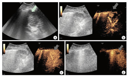
图 1 患者女,72岁,肝脏S8段eHCC
注:a,二维超声声像图示肝脏S8段低回声结节(箭头);b,注射造影剂16 s后病灶明显增强(箭头);c,注射造影剂26 s后病灶呈等回声(箭头);d, 注射造影剂1 min 8 s病灶呈低回声(箭头)。
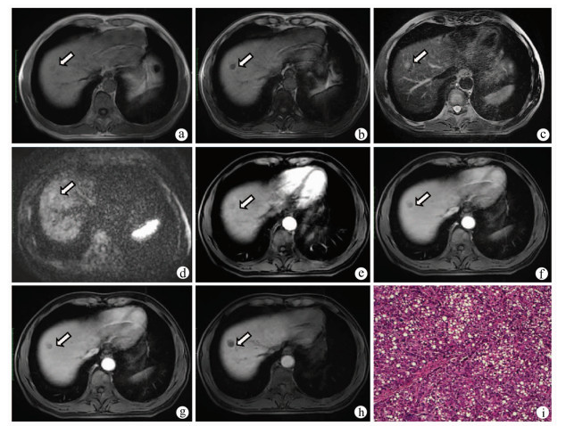
图 2 患者男,46岁,HBV感染者,肝硬化合并eHCC
注:a,同相位MR T1WI示肝脏S8段等或稍低信号(箭头);b,病灶反相位信号减低,提示其内存在脂肪变性(箭头);c,T2WI呈稍高信号;d,DWI呈稍高信号;e,增强扫描动脉晚期呈等信号(箭头);f、g,门静脉期及延迟期呈低信号(箭);h,肝胆期呈低信号;i,病理学诊断为高分化HCC,其内存在较多脂肪(HE染色,×100)。
-
[1] WANG M, WANG Y, FENG X, et al. Contribution of hepatitis B virus and hepatitis C virus to liver cancer in China north areas: Experience of the Chinese National Cancer Center[J]. Int J Infect Dis, 2017, 65: 15-21. DOI: 10.1016/j.ijid.2017.09.003. [2] National Health and Family Planning Commission of the People's Republic of China. Diagnosis, management, and treatment of hepatocellular carcinoma (V2017)[J]. J Clin Hepatol, 2017, 33(8): 1419-1431. DOI: 10.3969/j.issn.1001-5256.2017.08.003.中华人民共和国国家卫生和计划生育委员会. 原发性肝癌诊疗规范(2017年版)[J]. 临床肝胆病杂志, 2017, 33(8): 1419-1431. DOI: 10.3969/j.issn.1001-5256.2017.08.003. [3] International Consensus Group for Hepatocellular Neoplasia. Pathologic diagnosis of early hepatocellular carcinoma: A report of the international consensus group for hepatocellular neoplasia[J]. Hepatology, 2009, 49(2): 658-664. DOI: 10.1002/hep.22709. [4] HYTIROGLOU P. Well-differentiated hepatocellular nodule: Making a diagnosis on biopsy and resection specimens of patients with advanced stage chronic liver disease[J]. Semin Diagn Pathol, 2017, 34(2): 138-145. DOI: 10.1053/j.semdp.2016.12.009. [5] KOBAYASHI M, IKEDA K, HOSAKA T, et al. Dysplastic nodules frequently develop into hepatocellular carcinoma in patients with chronic viral hepatitis and cirrhosis[J]. Cancer, 2006, 106(3): 636-647. DOI: 10.1002/cncr.21607. [6] DIETRICH CF, KONO Y, COSGROVE D, et al. Contrast enhanced ultrasound: Liver imaging reporting and data system (CEUS LI-RADS)[J]. Ultrasound Med Biol, 2017, 43: s38-s39. [7] Abdominal Study Group, Chinese Society of Radiology, Chinese Medical Association. Standard guidelines for abdominal CT scan (trial version)[J]. Chin J Radiol, 2007, 41(9): 999-1004. DOI: 10.3760/j.issn:1005-1201.2007.09.031.中华医学会放射学分会腹部学组. 腹部CT扫描规范指南(试用稿)[J]. 中华放射学杂志, 2007, 41(9): 999-1004. DOI: 10.3760/j.issn:1005-1201.2007.09.031. [8] DING Y, RAO SX, CHEN C, et al. Assessing liver function in patients with HBV-related HCC: A comparison of T1 mapping on Gd-EOB-DTPA-enhanced MR imaging with DWI[J]. Eur Radiol, 2015, 25(5): 1392-1398. DOI: 10.1007/s00330-014-3542-x. [9] PIETRYGA JA, BURKE LM, MARIN D, et al. Respiratory motion artifact affecting hepatic arterial phase imaging with gadoxetate disodium: Examination recovery with a multiple arterial phase acquisition[J]. Radiology, 2014, 271(2): 426-434. DOI: 10.1148/radiol.13131988. [10] van KESSEL CS, VELDHUIS WB, van den BOSCH MA, et al. MR liver imaging with Gd-EOB-DTPA: A delay time of 10 minutes is sufficient for lesion characterisation[J]. Eur Radiol, 2012, 22(10): 2153-2160. DOI: 10.1007/s00330-012-2486-2. [11] ESTERSON YB, FLUSBERG M, OH S, et al. Improved parenchymal liver enhancement with extended delay on Gd-EOB-DTPA-enhanced MRI in patients with parenchymal liver disease: Associated clinical and imaging factors[J]. Clin Radiol, 2015, 70(7): 723-729. DOI: 10.1016/j.crad.2015.03.005. [12] BASHIR MR, MERKLE EM. Improved liver lesion conspicuity by increasing the flip angle during hepatocyte phase MR imaging[J]. Eur Radiol, 2011, 21(2): 291-294. DOI: 10.1007/s00330-010-1917-1. [13] YANG ZH, FENG F, WANG XY. A guide to technique of magnetic resonance imaging[M]. Beijing: People's Military Medical Press, 2007: 185-212.杨正汉, 冯逢, 王霄英. 磁共振成像技术指南[M]. 北京: 人民军医出版社, 2007: 185-212. [14] SATO T, KONDO F, EBARA M, et al. Natural history of large regenerative nodules and dysplastic nodules in liver cirrhosis: 28-year follow-up study[J]. Hepatol Int, 2015, 9(2): 330-336. DOI: 10.1007/s12072-015-9620-6. [15] NIU ZS, NIU XJ, WANG WH, et al. Latest developments in precancerous lesions of hepatocellular carcinoma[J]. World J Gastroenterol, 2016, 22(12): 3305-3314. DOI: 10.3748/wjg.v22.i12.3305. [16] European Association for the study of the liver; European Organisation for Research and Treatment of Cancer. EASL-EORTC clinical practice guidelines: Management of hepatocellular carcinoma[J]. J Hepatol, 2012, 56(4): 908-943. DOI: 10.1016/j.jhep.2011.12.001. [17] TERRAULT NA, LOK A, MCMAHON BJ, et al. Update on prevention, diagnosis, and treatment of chronic hepatitis B: AASLD 2018 hepatitis B guidance[J]. Hepatology, 2018, 67(4): 1560-1599. DOI: 10.1002/hep.29800. [18] OMATA M, CHENG AL, KOKUDO N, et al. Asia-Pacific clinical practice guidelines on the management of hepatocellular carcinoma: A 2017 update[J]. Hepatol Int, 2017, 11(4): 317-370. DOI: 10.1007/s12072-017-9799-9. [19] KIM BR, LEE JM, LEE DH, et al. Diagnostic performance of gadoxetic acid-enhanced liver MR imaging versus multidetector CT in the detection of dysplastic nodules and early hepatocellular carcinoma[J]. Radiology, 2017, 285(1): 134-146. DOI: 10.1148/radiol.2017162080. [20] SANO K, ICHIKAWA T, MOTOSUGI U, et al. Imaging study of early hepatocellular carcinoma: Usefulness of gadoxetic acid-enhanced MR imaging[J]. Radiology, 2011, 261(3): 834-844. DOI: 10.1148/radiol.11101840. -

 下载:
下载:

 本文二维码
本文二维码
图(3)
计量
- 文章访问数: 512
- HTML全文浏览量: 257
- PDF下载量: 187
- 被引次数: 0

