
 PDF下载 ( 2208 KB)

Research advances in Kupffer cells participating in the regulation of hepatocellular carcinoma
-
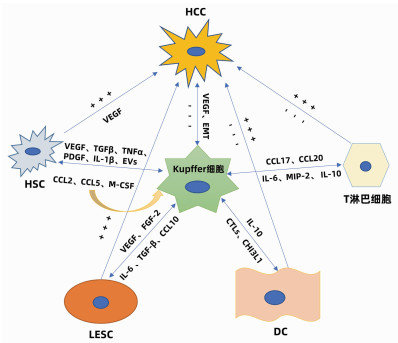
摘要: 肝细胞癌(HCC)是一种在全球范围内常见的高死亡率原发性肝癌。HCC在我国常见恶性肿瘤中发病率排第四位,死亡率排第二位,严重威胁着国民的健康和生命安全。主要介绍了Kupffer细胞在HCC中的双重作用,简述了其与肝实质细胞和非实质细胞的交互作用及其针对性靶点的治疗。分析表明深入研究Kupffer细胞对HCC的调控有助于为HCC的进一步治疗提供新的思路。Abstract: Hepatocellular carcinoma (HCC) is a common type of primary liver cancer with high mortality worldwide. Among the common malignant tumors in China, HCC ranks fourth in terms of incidence rate and ranks second in terms of mortality, which seriously threatens the health and life safety of the Chinese people. This article mainly introduces the dual role of Kupffer cells (KCs) in HCC and briefly describes its interaction with liver parenchymal cells and nonparenchymal cells and related targeted treatment methods. The analysis shows that in-depth research on KCs in the regulation of HCC helps to provide new ideas for further treatment of HCC.
-
Key words:
- Carcinoma, Hepatocellular /
- Kupffer Cells /
- Cytokines
-
[1] THANDRA KC, BARSOUK A, SAGINALA K, et al. Epidemiology of non-alcoholic fatty liver disease and risk of hepatocellular carcinoma progression[J]. Clin Exp Hepatol, 2020, 6(4): 289-294. DOI: 10.5114/ceh.2020.102153. [2] CHEN J, DENG X, LIU Y, et al. Kupffer cells in non-alcoholic fatty liver disease: Friend or foe?[J]. Int J Biol Sci, 2020, 16(13): 2367-2378. DOI: 10.7150/ijbs.47143. [3] TACKE F. Targeting hepatic macrophages to treat liver diseases[J]. J Hepatol, 2017, 66(6): 1300-1312. DOI: 10.1016/j.jhep.2017.02.026. [4] YANG YM, KIM SY, SEKI E. Inflammation and liver cancer: Molecular mechanisms and therapeutic targets[J]. Semin Liver Dis, 2019, 39(1): 26-42. DOI: 10.1055/s-0038-1676806. [5] FU XT, DAI Z, SONG K, et al. Macrophage-secreted IL-8 induces epithelial-mesenchymal transition in hepatocellular carcinoma cells by activating the JAK2/STAT3/Snail pathway[J]. Int J Oncol, 2015, 46(2): 587-596. DOI: 10.3892/ijo.2014.2761. [6] ABDULLAH Z, KNOLLE PA. Liver macrophages in healthy and diseased liver[J]. Pflugers Arch, 2017, 469(3-4): 553-560. DOI: 10.1007/s00424-017-1954-6. [7] LI P, HE K, LI J, et al. The role of Kupffer cells in hepatic diseases[J]. Mol Immunol, 2017, 85: 222-229. DOI: 10.1016/j.molimm.2017.02.018. [8] WANG Z, DABROSIN C, YIN X, et al. Broad targeting of angiogenesis for cancer prevention and therapy[J]. Semin Cancer Biol, 2015, 35(Suppl): s224-s243. DOI: 10.1016/j.semcancer.2015.01.001. [9] MURAKAMI K, KASAJIMA A, KAWAGISHI N, et al. The prognostic significance of vasohibin 1-associated angiogenesis in patients with hepatocellular carcinoma[J]. Hum Pathol, 2014, 45(3): 589-597. DOI: 10.1016/j.humpath.2013.10.028. [10] XUE X, GAO W, SUN B, et al. Vasohibin 2 is transcriptionally activated and promotes angiogenesis in hepatocellular carcinoma[J]. Oncogene, 2013, 32(13): 1724-1734. DOI: 10.1038/onc.2012.177. [11] LI XY, WU L, LI SW, et al. Effect of CD16a, the surface receptor of Kupffer cells, on the growth of hepatocellular carcinoma cells[J]. Int J Mol Med, 2016, 37(6): 1465-1474. DOI: 10.3892/ijmm.2016.2561. [12] AKIBA J, YANO H, OGASAWARA S, et al. Expression and function of interleukin-8 in human hepatocellular carcinoma[J]. Int J Oncol, 2001, 18(2): 257-264. DOI: 10.3892/ijo.18.2.257. [13] CHEN JA, SHI M, LI JQ, et al. Angiogenesis: Multiple masks in hepatocellular carcinoma and liver regeneration[J]. Hepatol Int, 2010, 4(3): 537-547. DOI: 10.1007/s12072-010-9192-4. [14] JOO YY, JANG JW, LEE SW, et al. Circulating pro- and anti-angiogenic factors in multi-stage liver disease and hepatocellular carcinoma progression[J]. Sci Rep, 2019, 9(1): 9137. DOI: 10.1038/s41598-019-45537-w. [15] MONNIER J, PIQUET-PELLORCE C, FEIGE JJ, et al. Prokineticin 2/Bv8 is expressed in Kupffer cells in liver and is down regulated in human hepatocellular carcinoma[J]. World J Gastroenterol, 2008, 14(8): 1182-1191. DOI: 10.3748/wjg.14.1182. [16] AKAZAWA Y, KONO H, HARA M, et al. M-CSF receptor antagonists inhibit the initiation and progression of hepatocellular carcinoma in mice[J]. Anticancer Res, 2019, 39(9): 4787-4794. DOI: 10.21873/anticanres.13663. [17] MALEHMIR M, PFISTER D, GALLAGE S, et al. Platelet GPIbα is a mediator and potential interventional target for NASH and subsequent liver cancer[J]. Nat Med, 2019, 25(4): 641-655. DOI: 10.1038/s41591-019-0379-5. [18] YAN L, XU F, DAI CL. Relationship between epithelial-to-mesenchymal transition and the inflammatory microenvironment of hepatocellular carcinoma[J]. J Exp Clin Cancer Res, 2018, 37(1): 203. DOI: 10.1186/s13046-018-0887-z. [19] WANG D, LUO L, CHEN W, et al. Significance of the vascular endothelial growth factor and the macrophage migration inhibitory factor in the progression of hepatocellular carcinoma[J]. Oncol Rep, 2014, 31(3): 1199-1204. DOI: 10.3892/or.2013.2946. [20] KLEMKE L, DE OLIVEIRA T, WITT D, et al. Hsp90-stabilized MIF supports tumor progression via macrophage recruitment and angiogenesis in colorectal cancer[J]. Cell Death Dis, 2021, 12(2): 155. DOI: 10.1038/s41419-021-03426-z. [21] BRODT P. Role of the microenvironment in liver metastasis: From pre- to prometastatic niches[J]. Clin Cancer Res, 2016, 22(24): 5971-5982. DOI: 10.1158/1078-0432.CCR-16-0460. [22] MIYAZOE Y, MIUMA S, MIYAAKI H, et al. Extracellular vesicles from senescent hepatic stellate cells promote cell viability of hepatoma cells through increasing EGF secretion from differentiated THP-1 cells[J]. Biomed Rep, 2020, 12(4): 163-170. DOI: 10.3892/br.2020.1279. [23] LI H, ZHOU Y, WANG H, et al. Crosstalk between liver macrophages and surrounding cells in nonalcoholic steatohepatitis[J]. Front Immunol, 2020, 11: 1169. DOI: 10.3389/fimmu.2020.01169. [24] RAMIREZ-PEDRAZA M, FERNÁNDEZ M. Interplay between macrophages and angiogenesis: A double-edged sword in liver disease[J]. Front Immunol, 2019, 10: 2882. DOI: 10.3389/fimmu.2019.02882. [25] POISSON J, LEMOINNE S, BOULANGER C, et al. Liver sinusoidal endothelial cells: Physiology and role in liver diseases[J]. J Hepatol, 2017, 66(1): 212-227. DOI: 10.1016/j.jhep.2016.07.009. [26] MANZI M, BACIGALUPO ML, CARABIAS P, et al. Galectin-1 controls the proliferation and migration of liver sinusoidal endothelial cells and their interaction with hepatocarcinoma cells[J]. J Cell Physiol, 2016, 231(7): 1522-1533. DOI: 10.1002/jcp.25244. [27] DAI S, LIU F, QIN Z, et al. Kupffer cells promote T-cell hepatitis by producing CXCL10 and limiting liver sinusoidal endothelial cell permeability[J]. Theranostics, 2020, 10(16): 7163-7177. DOI: 10.7150/thno.44960. [28] UWATOKU R, SUEMATSU M, EZAKI T, et al. Kupffer cell-mediated recruitment of rat dendritic cells to the liver: Roles of N-acetylgalactosamine-specific sugar receptors[J]. Gastroenterology, 2001, 121(6): 1460-1472. DOI: 10.1053/gast.2001.29594. [29] LO TH, SILVEIRA PA, FROMM PD, et al. Characterization of the expression and function of the C-type lectin receptor CD302 in mice and humans reveals a role in dendritic cell migration[J]. J Immunol, 2016, 197(3): 885-898. DOI: 10.4049/jimmunol.1600259. [30] DI ROSA M, TIBULLO D, SACCONE S, et al. CHI3L1 nuclear localization in monocyte derived dendritic cells[J]. Immunobiology, 2016, 221(2): 347-356. DOI: 10.1016/j.imbio.2015.09.023. [31] BAMBOAT ZM, OCUIN LM, BALACHANDRAN VP, et al. Conventional DCs reduce liver ischemia/reperfusion injury in mice via IL-10 secretion[J]. J Clin Invest, 2010, 120(2): 559-69. DOI: 10.1172/JCI40008. [32] HEFETZ-SELA S, STEIN I, KLIEGER Y, et al. Acquisition of an immunosuppressive protumorigenic macrophage phenotype depending on c-Jun phosphorylation[J]. Proc Natl Acad Sci U S A, 2014, 111(49): 17582-17587. DOI: 10.1073/pnas.1409700111. [33] DOU L, SHI X, HE X, et al. Macrophage phenotype and function in liver disorder[J]. Front Immunol, 2019, 10: 3112. DOI: 10.3389/fimmu.2019.03112. [34] NOONAN A, PAWLIK TM. Hepatocellular carcinoma: An update on investigational drugs in phase I and Ⅱ clinical trials[J]. Expert Opin Investig Drugs, 2019, 28(11): 941-949. DOI: 10.1080/13543784.2019.1677606. [35] HUANG A, YANG XR, CHUNG WY, et al. Targeted therapy for hepatocellular carcinoma[J]. Signal Transduct Target Ther, 2020, 5(1): 146. DOI: 10.1038/s41392-020-00264-x. [36] RIZZO A, RICCI AD, BRANDI G. Immune-based combinations for advanced hepatocellular carcinoma: Shaping the direction of first-line therapy[J]. Future Oncol, 2021, 17(7): 755-757. DOI: 10.2217/fon-2020-0986. [37] ABDELGALIL AA, ALKAHTANI HM, AL-JENOOBI FI. Sorafenib[J]. Profiles Drug Subst Excip Relat Methodol, 2019, 44: 239-266. DOI: 10.1016/bs.podrm.2018.11.003. [38] PICCIONI F, FIORE E, BAYO J, et al. 4-methylumbelliferone inhibits hepatocellular carcinoma growth by decreasing IL-6 production and angiogenesis[J]. Glycobiology, 2015, 25(8): 825-835. DOI: 10.1093/glycob/cwv023. [39] FUKUMURA D, KLOEPPER J, AMOOZGAR Z, et al. Enhancing cancer immunotherapy using antiangiogenics: Opportunities and challenges[J]. Nat Rev Clin Oncol, 2018, 15(5): 325-340. DOI: 10.1038/nrclinonc.2018.29. [40] KIM CG, JANG M, KIM Y, et al. VEGF-A drives TOX-dependent T cell exhaustion in anti-PD-1-resistant microsatellite stable colorectal cancers[J]. Sci Immunol, 2019, 4(41): eaay0555. DOI: 10.1126/sciimmunol.aay0555. [41] DE GRAMONT A, FAIVRE S, RAYMOND E. Novel TGF-β inhibitors ready for prime time in onco-immunology[J]. Oncoimmunology, 2017, 6(1): e1257453. DOI: 10.1080/2162402X.2016.1257453. [42] GRETEN TF, LAI CW, LI G, et al. Targeted and immune-based therapies for hepatocellular carcinoma[J]. Gastroenterology, 2019, 156(2): 510-524. DOI: 10.1053/j.gastro.2018.09.051. -

 下载:
下载:
 本文二维码
本文二维码
图(1)
计量
- 文章访问数: 793
- HTML全文浏览量: 122
- PDF下载量: 83
- 被引次数: 0

