
 PDF下载 ( 2289 KB)

肝窦内皮细胞与肝纤维化的关系
DOI: 10.3969/j.issn.1001-5256.2023.02.027
利益冲突声明:所有作者均声明不存在利益冲突。
作者贡献声明:戴伟明负责文章结构设计,撰写文章;陆伦根负责论文修改;蔡晓波负责拟定写作思路,修改文章。
-
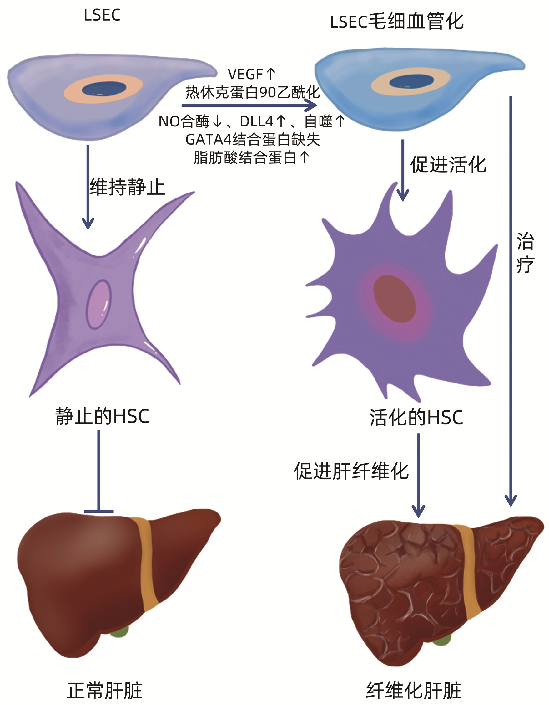
摘要: 生理情况下,肝窦内皮细胞(LSEC)对肝脏稳态的维持起重要作用。而在肝脏病理损伤条件下,LSEC可通过改变其自身结构即毛细血管化对损伤做出反应,并加重肝损伤。此外,LSEC与肝脏中其他细胞的相互作用对肝纤维化的发生发展也有一定作用,其中与肝纤维化的主要效应细胞——肝星状细胞的交互作用占主体地位。本文主要阐述慢性肝损伤时LSEC在肝纤维化发生发展中的作用。Abstract: Liver sinusoidal endothelial cells (LSECs) are crucial to the maintenance of hepatic homeostasis under physiologic conditions, while under the conditions of pathological liver damage, LSEC can respond to the damage by changing their structure through the process called capillarization, thereby aggravating liver damage. In addition, the interaction between LSEC and other cells in the liver plays a certain role in the development and progression of liver fibrosis, especially the interaction between LSEC and hepatic stellate cells, which are the primary effector cells of liver fibrosis. This article mainly elaborates on the role of LSEC in the development and progression of liver fibrosis during chronic liver injury.
-
[1] LI H. Intercellular crosstalk of liver sinusoidal endothelial cells in liver fibrosis, cirrhosis and hepatocellular carcinoma[J]. Dig Liver Dis, 2022, 54(5): 598-613. DOI: 10.1016/j.dld.2021.07.006. [2] DELEVE LD, WANG X, HU L, et al. Rat liver sinusoidal endothelial cell phenotype is maintained by paracrine and autocrine regulation[J]. Am J Physiol Gastrointest Liver Physiol, 2004, 287(4): G757-G763. DOI: 10.1152/ajpgi.00017.2004. [3] POISSON J, LEMOINNE S, BOULANGER C, et al. Liver sinusoidal endothelial cells: Physiology and role in liver diseases[J]. J Hepatol, 2017, 66(1): 212-227. DOI: 10.1016/j.jhep.2016.07.009. [4] SUN X, HARRIS EN. New aspects of hepatic endothelial cells in physiology and nonalcoholic fatty liver disease[J]. Am J Physiol Cell Physiol, 2020, 318(6): C1200-C1213. DOI: 10.1152/ajpcell.00062.2020. [5] ZAPOTOCZNY B, SZAFRANSKA K, KUS E, et al. Tracking fenestrae dynamics in live murine liver sinusoidal endothelial cells[J]. Hepatology, 2019, 69(2): 876-888. DOI: 10.1002/hep.30232. [6] XIE G, WANG L, WANG X, et al. Isolation of periportal, midlobular, and centrilobular rat liver sinusoidal endothelial cells enables study of zonated drug toxicity[J]. Am J Physiol Gastrointest Liver Physiol, 2010, 299(5): G1204-G1210. DOI: 10.1152/ajpgi.00302.2010. [7] DELEVE LD. Liver sinusoidal endothelial cells in hepatic fibrosis[J]. Hepatology, 2015, 61(5): 1740-1746. DOI: 10.1002/hep.27376. [8] SIMON-SANTAMARIA J, RINALDO CH, KARDAS P, et al. Efficient uptake of blood-borne BK and JC polyomavirus-like particles in endothelial cells of liver sinusoids and renal vasa recta[J]. PLoS One, 2014, 9(11): e111762. DOI: 10.1371/journal.pone.0111762. [9] BHANDARI S, LARSEN AK, MCCOURT P, et al. The scavenger function of liver sinusoidal endothelial cells in health and disease[J]. Front Physiol, 2021, 12: 757469. DOI: 10.3389/fphys.2021.757469. [10] LI TP, GUAN SH, WANG Q, et al. Soluble mannose receptor as a predictor of prognosis of hepatitis B virus-related acute-on-chronic liver failure[J]. World J Gastroenterol, 2019, 25(37): 5667-5675. DOI: 10.3748/wjg.v25.i37.5667. [11] TURMAN JM, CHEPLOWITZ AM, TIWARI C, et al. Accelerated clearance and degradation of cell-free HIV by neutralizing antibodies occurs via FcγRⅡb on liver sinusoidal endothelial cells by endocytosis[J]. J Immunol, 2021, 206(6): 1284-1296. DOI: 10.4049/jimmunol.2000772. [12] HAMMOUTENE A, RAUTOU PE. Role of liver sinusoidal endothelial cells in non-alcoholic fatty liver disease[J]. J Hepatol, 2019, 70(6): 1278-1291. DOI: 10.1016/j.jhep.2019.02.012. [13] LAFOZ E, RUART M, ANTON A, et al. The endothelium as a driver of liver fibrosis and regeneration[J]. Cells, 2020, 9(4). DOI: 10.3390/cells9040929. [14] MCGUIRE RF, BISSELL DM, BOYLES J, et al. Role of extracellular matrix in regulating fenestrations of sinusoidal endothelial cells isolated from normal rat liver[J]. Hepatology, 1992, 15(6): 989-997. DOI: 10.1002/hep.1840150603. [15] MOHAMAD M, MITCHELL SJ, WU LE, et al. Ultrastructure of the liver microcirculation influences hepatic and systemic insulin activity and provides a mechanism for age-related insulin resistance[J]. Aging Cell, 2016, 15(4): 706-715. DOI: 10.1111/acel.12481. [16] DI MARTINO J, MASCALCHI P, LEGROS P, et al. Actin depolymerization in dedifferentiated liver sinusoidal endothelial cells promotes fenestrae re-formation[J]. Hepatol Commun, 2019, 3(2): 213-219. DOI: 10.1002/hep4.1301. [17] GRACIA-SANCHO J, CAPARRÓS E, FERNÁNDEZ-IGLESIAS A, et al. Role of liver sinusoidal endothelial cells in liver diseases[J]. Nat Rev Gastroenterol Hepatol, 2021, 18(6): 411-431. DOI: 10.1038/s41575-020-00411-3. [18] MAK KM, KEE D, SHIN DW. Alcohol-associated capillarization of sinusoids: A critique since the discovery by Schaffner and Popper in 1963[J]. Anat Rec (Hoboken), 2022, 305(7): 1592-1610. DOI: 10.1002/ar.24829. [19] DU W, WANG L. The crosstalk between liver sinusoidal endothelial cells and hepatic microenvironment in nash related liver fibrosis[J]. Front Immunol, 2022, 13: 936196. DOI: 10.3389/fimmu.2022.936196. [20] DELEVE LD, WANG X, GUO Y. Sinusoidal endothelial cells prevent rat stellate cell activation and promote reversion to quiescence[J]. Hepatology, 2008, 48(3): 920-930. DOI: 10.1002/hep.22351. [21] JARNAGIN WR, ROCKEY DC, KOTELIANSKY VE, et al. Expression of variant fibronectins in wound healing: cellular source and biological activity of the EⅢA segment in rat hepatic fibrogenesis[J]. J Cell Biol, 1994, 127(6 Pt 2): 2037-2048. DOI: 10.1083/jcb.127.6.2037. [22] MARRONE G, RUSSO L, ROSADO E, et al. The transcription factor KLF2 mediates hepatic endothelial protection and paracrine endothelial-stellate cell deactivation induced by statins[J]. J Hepatol, 2013, 58(1): 98-103. DOI: 10.1016/j.jhep.2012.08.026. [23] LEMOINNE S, CADORET A, RAUTOU PE, et al. Portal myofibroblasts promote vascular remodeling underlying cirrhosis formation through the release of microparticles[J]. Hepatology, 2015, 61(3): 1041-1055. DOI: 10.1002/hep.27318. [24] DESROCHES-CASTAN A, TILLET E, RICARD N, et al. Bone morphogenetic protein 9 is a paracrine factor controlling liver sinusoidal endothelial cell fenestration and protecting against hepatic fibrosis[J]. Hepatology, 2019, 70(4): 1392-1408. DOI: 10.1002/hep.30655. [25] TATEYA S, RIZZO NO, HANDA P, et al. Endothelial NO/cGMP/VASP signaling attenuates Kupffer cell activation and hepatic insulin resistance induced by high-fat feeding[J]. Diabetes, 2011, 60(11): 2792-2801. DOI: 10.2337/db11-0255. [26] ROH YS, SEKI E. Chemokines and chemokine receptors in the development of NAFLD[J]. Adv Exp Med Biol, 2018, 1061: 45-53. DOI: 10.1007/978-981-10-8684-7_4. [27] YANG Y, SANGWUNG P, KONDO R, et al. Alcohol-induced Hsp90 acetylation is a novel driver of liver sinusoidal endothelial dysfunction and alcohol-related liver disease[J]. J Hepatol, 2021, 75(2): 377-386. DOI: 10.1016/j.jhep.2021.02.028. [28] FANG ZQ, RUAN B, LIU JJ, et al. Notch-triggered maladaptation of liver sinusoidal endothelium aggravates nonalcoholic steatohepatitis through endothelial nitric oxide synthase[J]. Hepatology, 2022, 76(3): 742-758. DOI: 10.1002/hep.32332. [29] CHEN L, GU T, LI B, et al. Delta-like ligand 4/DLL4 regulates the capillarization of liver sinusoidal endothelial cell and liver fibrogenesis[J]. Biochim Biophys Acta Mol Cell Res, 2019, 1866(10): 1663-1675. DOI: 10.1016/j.bbamcr.2019.06.011. [30] RUART M, CHAVARRIA L, CAMPRECIÓS G, et al. Impaired endothelial autophagy promotes liver fibrosis by aggravating the oxidative stress response during acute liver injury[J]. J Hepatol, 2019, 70(3): 458-469. DOI: 10.1016/j.jhep.2018.10.015. [31] WINKLER M, STANICZEK T, KVRSCHNER SW, et al. Endothelial GATA4 controls liver fibrosis and regeneration by preventing a pathogenic switch in angiocrine signaling[J]. J Hepatol, 2021, 74(2): 380-393. DOI: 10.1016/j.jhep.2020.08.033. [32] WU X, SHU L, ZHANG Z, et al. Adipocyte fatty acid binding protein promotes the onset and progression of liver fibrosis via mediating the crosstalk between liver sinusoidal endothelial cells and hepatic stellate cells[J]. Adv Sci (Weinh), 2021, 8(11): e2003721. DOI: 10.1002/advs.202003721. [33] WU Y, LI Z, XIU AY, et al. Carvedilol attenuates carbon tetrachloride-induced liver fibrosis and hepatic sinusoidal capillarization in mice[J]. Drug Des Devel Ther, 2019, 13: 2667-2676. DOI: 10.2147/DDDT.S210797. [34] ABRALDES JG, RODRÍGUEZ-VILARRUPLA A, GRAUPERA M, et al. Simvastatin treatment improves liver sinusoidal endothelial dysfunction in CCl4 cirrhotic rats[J]. J Hepatol, 2007, 46(6): 1040-1046. DOI: 10.1016/j.jhep.2007.01.020. [35] MARRONE G, MAESO-DÍAZ R, GARCÍA-CARDENA G, et al. KLF2 exerts antifibrotic and vasoprotective effects in cirrhotic rat livers: behind the molecular mechanisms of statins[J]. Gut, 2015, 64(9): 1434-1443. DOI: 10.1136/gutjnl-2014-308338. [36] BRAVO M, RAURELL I, HIDE D, et al. Restoration of liver sinusoidal cell phenotypes by statins improves portal hypertension and histology in rats with NASH[J]. Sci Rep, 2019, 9(1): 20183. DOI: 10.1038/s41598-019-56366-2. [37] HU L, SU L, DONG Z, et al. AMPK agonist AICAR ameliorates portal hypertension and liver cirrhosis via NO pathway in the BDL rat model[J]. J Mol Med (Berl), 2019, 97(3): 423-434. DOI: 10.1007/s00109-019-01746-4. [38] HUNT NJ, LOCKWOOD GP, KANG S, et al. The effects of metformin on age-related changes in the liver sinusoidal endothelial cell[J]. J Gerontol A Biol Sci Med Sci, 2020, 75(2): 278-285. DOI: 10.1093/gerona/glz153. [39] TALAMINI L, PICCHETTI P, FERREIRA LM, et al. Organosilica cages target hepatic sinusoidal endothelial cells avoiding macrophage filtering[J]. ACS Nano, 2021, 15(6): 9701-9716. DOI: 10.1021/acsnano.1c00316. [40] BAE CR, ZHANG H, KWON YG. The endothelial dysfunction blocker CU06-1004 ameliorates choline-deficient L-amino acid diet-induced non-alcoholic steatohepatitis in mice[J]. PLoS One, 2020, 15(12): e0243497. DOI: 10.1371/journal.pone.0243497. -

 下载:
下载:
 本文二维码
本文二维码
图(1)
计量
- 文章访问数: 865
- HTML全文浏览量: 523
- PDF下载量: 95
- 被引次数: 0

