
 PDF下载 ( 5713 KB)

多房棘球蚴源性外泌体对巨噬细胞极化的影响
DOI: 10.3969/j.issn.1001-5256.2023.04.019
Effect of exosomes derived from Echinococcus multilocularis on macrophage polarization: A preliminary study
-
摘要:
目的 探究不同时间及浓度多房棘球蚴源性外泌体对巨噬细胞极化的影响。 方法 从60只造模BALB/c小鼠中随机选取4只,应用7.0T MRI观察腹腔病灶生长情况;解剖造模小鼠,通过腹腔病灶提取原头节进行体外培养,超速离心法从培养上清中提取外泌体,透射电镜及蛋白质免疫印迹法鉴定外泌体表征。将未加外泌体处理的巨噬细胞单独培养组设为对照组,不同浓度多房棘球蚴来源外泌体与巨噬细胞共培养组设为实验组(10 μg/mL组、50 μg/mL组),分别共培养48 h和72 h,显微镜下观察巨噬细胞形态变化。通过流式细胞术和酶联免疫吸附实验(ELISA)检测极化状态。符合正态分布的计量资料多组间比较采用单因素方差分析,进一步两两比较采用LSD-t检验。 结果 7.0T MRI显示小鼠腹腔内弥漫分布、大小不等的病灶形成;多房棘球蚴源性外泌体直径100 nm左右,呈杯型或茶托型,其表面标志物CD9、TSG101和CD63表达阳性。共培养后,实验组大部分细胞拉长,形态不规则,主要呈多角形;流式细胞术检测发现,共培养48 h,对照组CD16/32、CD206、CD369阳性率分别为(99.53±0.06)%、(90.27±0.21)%、(2.40±0.20)%;与对照组相比,除10 μg/mL外泌体组CD369阳性率[(0.80±0.00)%]降低(P<0.05),其余组别CD16/32、CD206、CD369阳性率均明显升高(P值均<0.000 1);共培养72 h,对照组CD16/32、CD206、CD369阳性率分别为(99.67±0.06)%、(85.47±0.55)%、(6.60±0.20)%,实验组CD16/32、CD206、CD369阳性率较对照组均明显升高(P值均<0.05)。ELISA结果示:共培养48 h,对照组IL-6及TNFα水平分别为(58.53±15.52) pg/mL、(320.70±5.30)pg/mL,实验组外泌体浓度为50 μg/mL时IL-6[(98.81±15.55) pg/mL]较对照组升高(P<0.05);共培养72 h,对照组IL-6及TNFα水平分别为(76.22±9.68)pg/mL、(323.90±87.37)pg/mL,当外泌体浓度为10 μg/mL时TNFα水平[(164.20±14.17)pg/mL]较对照组明显下降(P<0.05);当外泌体浓度为50 μg/mL时IL-6水平[(99.52±8.35)pg/mL]较对照组升高(P<0.05)。 结论 多房棘球蚴源性外泌体可调控巨噬细胞极化,且在浓度为10 μg/mL,共培养72 h后,可导致巨噬细胞M2样极化,具体方式有待进一步研究。 -
关键词:
- 棘球蚴病, 肝 /
- 外泌体 /
- 巨噬细胞 /
- 小鼠,近交BALB C
Abstract:Objective To investigate the effect of exosomes derived from Echinococcus multilocularis on macrophage polarization after treatment for different durations and concentrations. Methods A total of 60 BALB/c mice were used for modeling, among which 4 mice were selected to observe the growth of abdominal lesions on 7.0T MRI. The mice for modeling were dissected, and the protoscoleces was taken from the abdominal lesion and cultured in vitro; ultracentrifugation was used to extract the exosomes from the supernatant, and transmission electron microscopy and Western blotting were used for the characterization of exosomes. The macrophages without exosome treatment were established as control group, and the macrophages co-cultured with different concentrations of exosomes derived from Echinococcus multilocularis were established as experimental group (10 μg/mL group and 50 μg/mL group) and were cultured for 48 and 72 hours. The morphological changes of macrophages were observed under a microscope, and flow cytometry and ELISA were used to observe polarization state. A one-way analysis of variance was used for comparison of normally distributed continuous data between multiple groups, and the least significant difference t-test was used for further comparison between two groups. Results The results of 7.0T MRI showed the formation of diffuse lesions with different sizes in the abdominal cavity of mice, and the exosomes derived from Echinococcus multilocularis were approximately 100 nm in diameter and were cup-shaped or saucer-shaped, with the positive expression of the surface markers CD9, TSG101, and CD63. After co-culture, most of the cells in the experimental group were elongated with an irregular and polygonal shape. Flow cytometry showed that after 48 hours of co-culture, the positive rates of CD16/32, CD206, and CD369 in the control group were 99.53%±0.06%, 90.27%±0.21%, and 2.40%±0.20%, respectively; compared with the control group, except that the 10 μg/mL exosome group had a significant reduction in the positive rate of CD369 (0.80%±0.00%) (P < 0.05), all the other groups had a significant increase in the positive rates of CD16/32, CD206, and CD369 (all P < 0.000 1); after 72 hours of co-culture, the positive rates of CD16/32, CD206, and CD369 in the control group were 99.67%±0.06%, 85.47%±0.55%, and 6.60%±0.20%, respectively, and compared with the control group, the experimental group had significant increases in the positive rates of CD16/32, CD206, and CD369 (all P < 0.05). ELISA showed that after 48 hours of co-culture, the levels of IL-6 and TNFα in the control group were 58.53±15.52 pg/mL and 320.70±5.30 pg/mL, respectively, and when the exosome concentration was 50 μg/mL, the level of IL-6 in the experimental group was 98.81±15.55 pg/mL, which was higher than that in the control group (P < 0.05); after 72 hours of co-culture, the levels of IL-6 and TNFα in the control group were 76.22±9.68 pg/mL and 323.90±87.37 pg/mL, respectively, and when the exosome concentration was 10 μg/mL, the level of TNFα was 164.20±14.17 pg/mL, which was significantly lower than that in the control group (P < 0.05); when the exosome concentration was 50 μg/mL, the level of IL-6 was 99.52±8.35 pg/mL, which was significantly higher than that in the control group (P < 0.05). Conclusion Exosomes derived from Echinococcus multilocularis can regulate macrophage polarization and induce M2-like polarization of macrophages after co-culture at a concentration of 10 μg /mL for 72 hours, and further studies are needed to clarify the specific method. -
Key words:
- Echinococcosis, Hepatic /
- Exosomes /
- Macrophages /
- Mice, Inbred BALB C
-
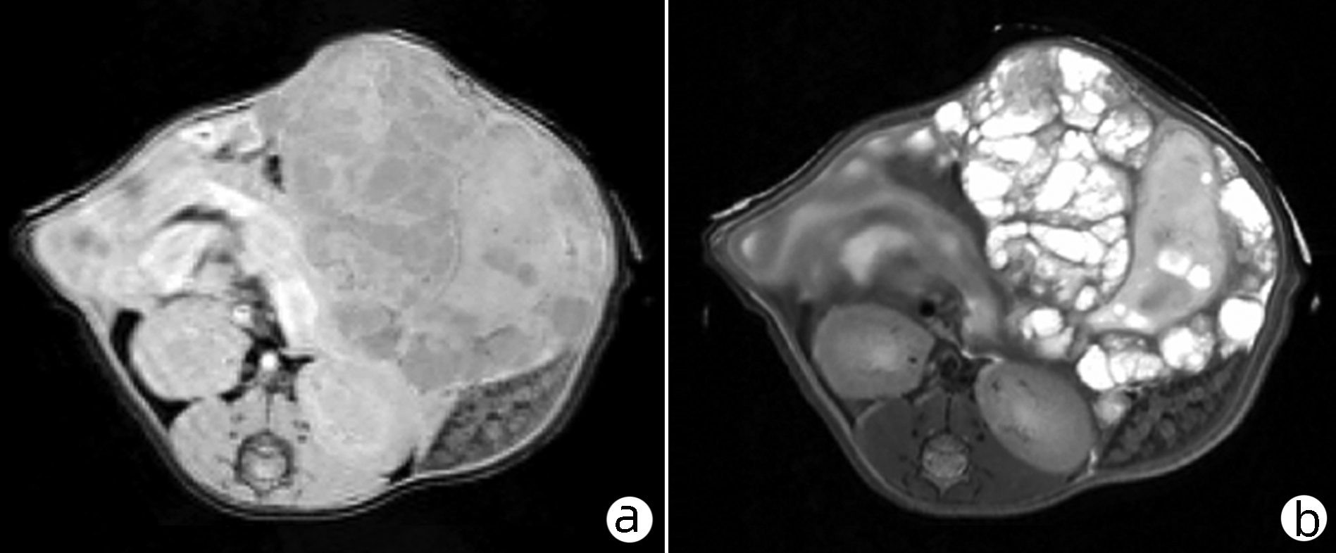
图 1 7.0T MRI在模型小鼠中的应用
注:a, T1WI像;b, T2WI像。
Figure 1. Application of 7.0T MRI in mouse model
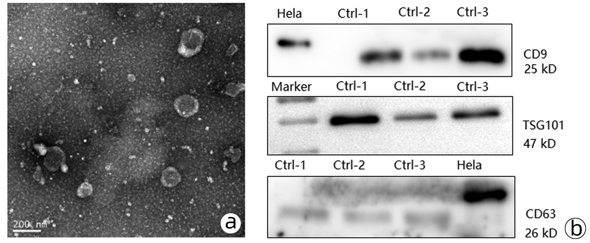
图 2 多房棘球蚴源性外泌体的鉴定
注:a, TEM观察多房棘球蚴形态;b, Western Blot检测相关标志物表达。
Figure 2. Identification of exosomes derived from echinococcus multilocularis

图 3 不同浓度外泌体与J774A.1细胞共培养的形态变化(×200)
Figure 3. Morphological changes of J774A.1 cells co-cultured with different concentrations of exosomes (×200)

图 4 流式细胞术检测不同时间及浓度外泌体刺激巨噬细胞CD16/32、CD206、CD369表达量
Figure 4. The expressions of CD16/32, CD206 and CD369 in macrophages stimulated by exosomes at different time and concentration were detected by flow cytometry
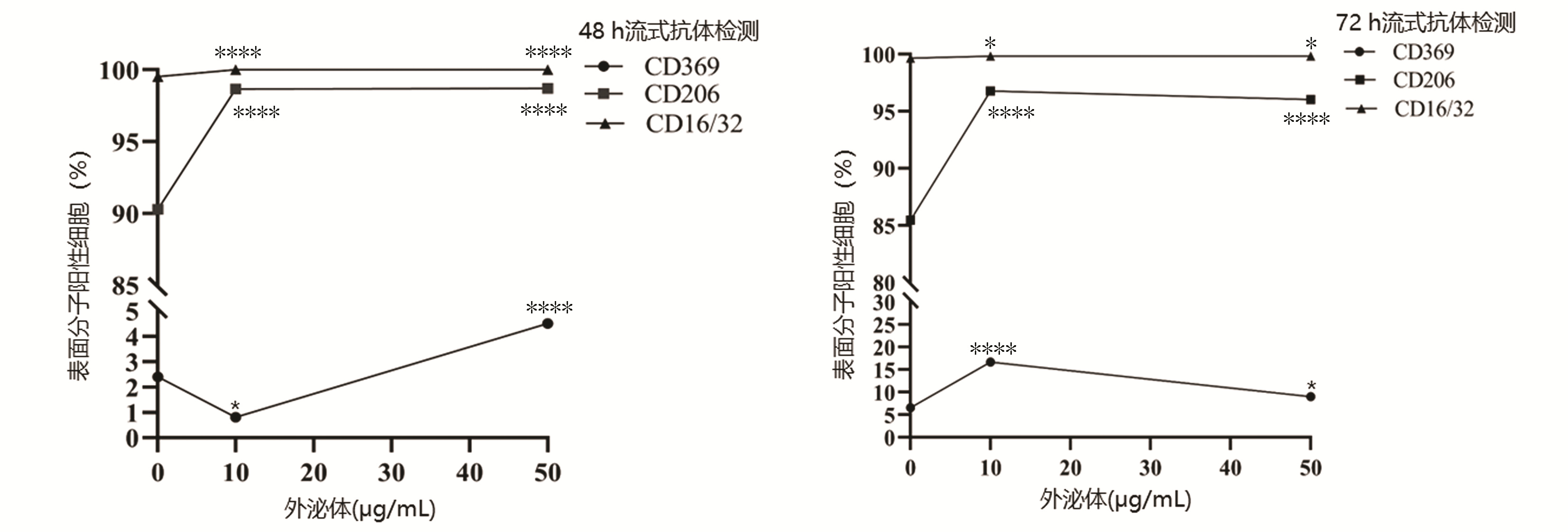
图 5 共培养后CD16/32、CD206、CD369变化趋势
注:与对照组比较,*<0.05, ****P<0.000 1。
Figure 5. The change trend of CD16/32, CD206 and CD369 after co-culture
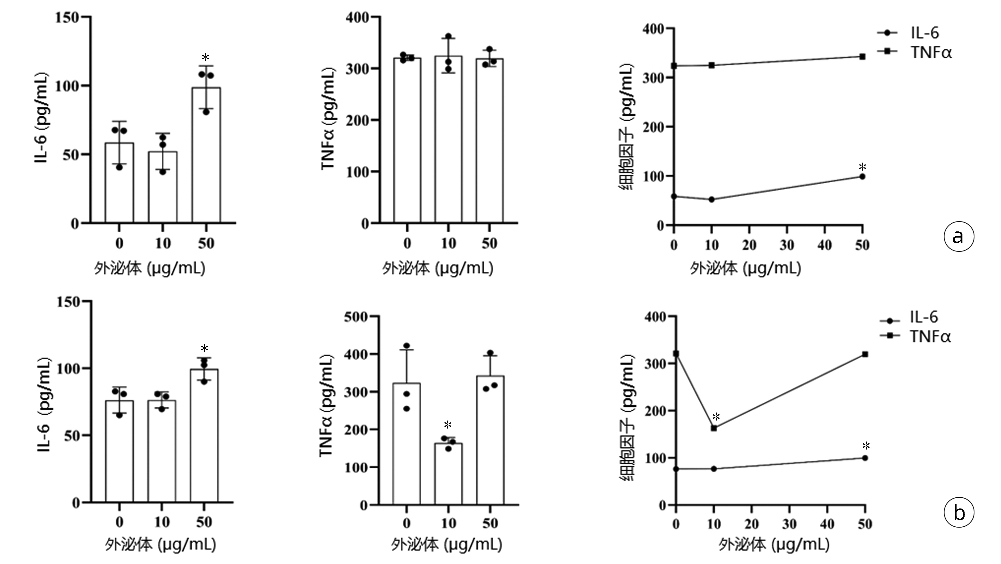
图 6 ELISA检测不同浓度的外泌体刺激巨噬细胞后相关分子表达情况
注:a, 48 h; b, 72 h。与对照组比较,*P<0.05。
Figure 6. ELISA was used to detect the expression of related molecules in macrophages stimulated by exosomes at different concentrations
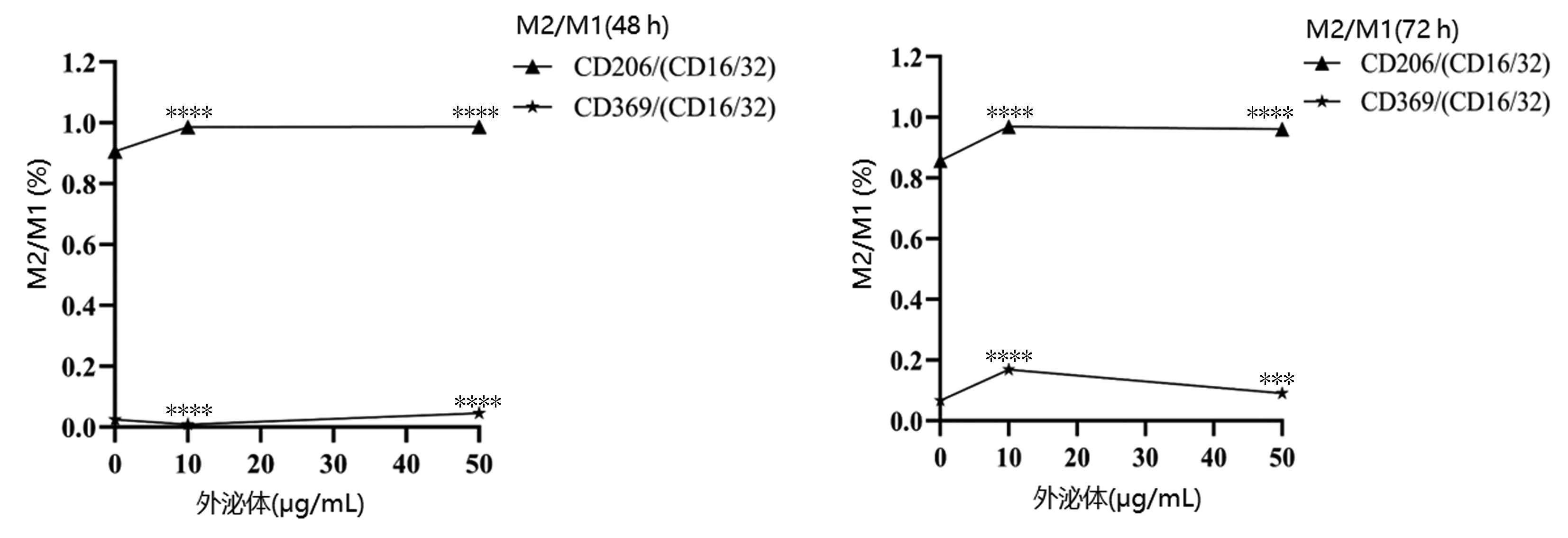
图 7 M2/M1型巨噬细胞相关分子比值变化趋势
注:与对照组比较,***P<0.001,****P<0.000 1。
Figure 7. The trends in the molecular ratio of M2/M1 macrophages
表 1 外泌体与巨噬细胞共培养48 h流式结果
Table 1. Flow cytometry results of exosomes co-cultured with macrophages for 48 hours
分组 CD16/32(%) CD206(%) CD369(%) 48 h 72 h 48 h 72 h 48 h 72 h 对照组 99.53±0.06 99.67±0.06 90.27±0.21 85.47±0.55 2.40±0.20 6.60±0.20 10 μg/mL组 100.00±0.002) 99.83±0.061) 98.67±0.212) 96.77±0.152) 0.80±0.001) 16.67±0.452) 50 μg/mL组 100.00±0.002) 99.83±0.061) 98.70±0.202) 96.03±0.232) 4.50±0.302) 9.03±0.231) F值 196.00 8.33 1 678.00 946.90 238.40 836.90 P值 <0.000 1 <0.05 <0.000 1 <0.000 1 <0.000 1 <0.000 1 注:与对照组比较,1)P<0.05, 2)P<0.000 1。  下载: 导出CSV
表 2 外泌体与巨噬细胞共培养ELISA结果
Table 2. ELISA results of exosomes co-cultured with macrophages
组别 IL-6(pg/mL) TNFα(pg/mL) 48 h 72 h 48 h 72 h 对照组 58.53±15.52 76.22±9.68 320.70±5.30 323.90±87.37 10 μg/mL组 52.19±13.16 76.46±6.03 324.70±33.48 164.20±14.171) 50 μg/mL组 98.81±15.551) 99.52±8.351) 319.60±15.91 342.70±52.79 F值 8.77 8.07 0.05 8.15 P值 <0.05 <0.05 0.95 <0.05 注:与对照组比较,1)P<0.05。
下载: 导出CSV
表 3 M2/M1型巨噬细胞相关分子比值
Table 3. The ratio of M2/M1 macrophage-associated molecules
组别 CD206/(CD16/32) CD369/(CD16/32) 48 h 72 h 48 h 72 h 对照组 0.907±0.002 0.858±0.005 0.024±0.002 0.066±0.002 10 μg/mL组 0.987±0.0021) 0.969±0.0021) 0.008±0.0001) 0.169±0.0081) 50 μg/mL组 0.987±0.0021) 0.962±0.0031) 0.045±0.0031) 0.090±0.0022) F值 1 767.0 953.6 237.5 378.9 P值 <0.000 1 <0.000 1 <0.000 1 <0.000 1 注:与对照组比较,1)P<0.000 1,2)P<0.001。
下载: 导出CSV
-
[1] Sichuan Hydatid Disease Clinical Medical Research Center, Hydatid Disease Professional Committee of Sichuan Medical Association. Expert consensus on diagnosis and treatment of alveolar hepatic echinococcosis(2020 version)[J]. Chin J Bases Clin Gen Surg, 2020, 27(1): 13-17. DOI: 10.7507/1007-9424.201911105.四川省包虫病临床医学研究中心, 四川省医师协会包虫病专业委员会. 泡型肝包虫病诊疗专家共识(2020版)[J]. 中国普外基础与临床杂志, 2020, 27(1): 13-17. DOI: 10.7507/1007-9424.201911105. [2] SCHULTZE JL, SCHMIDT SV. Molecular features of macrophage activation[J]. Semin Immunol, 2015, 27(6): 416-423. DOI: 10.1016/j.smim.2016.03.009. [3] SCHULTZE JL, FREEMAN T, HUME DA, et al. A transcriptional perspective on human macrophage biology[J]. Semin Immunol, 2015, 27(1): 44-50. DOI: 10.1016/j.smim.2015.02.001. [4] LI WH, ZHANG YX, ZHAO D, et al. Dectin-1 affects heart remodeling after myocardial infarction by regulating macrophage polarization[J]. Immunol J, 2021, 37(8): 692-697. DOI: 10.13431/j.cnki.immunol.j.20210096.李文华, 张一馨, 赵迪, 等. Dectin-1通过调节巨噬细胞极化影响心肌梗死后的心脏重塑[J]. 免疫学杂志, 2021, 37(8): 692-697. DOI: 10.13431/j.cnki.immunol.j.20210096. [5] MOSSER DM, EDWARDS JP. Exploring the full spectrum of macrophage activation[J]. Nat Rev Immunol, 2008, 8(12): 958-969. DOI: 10.1038/nri2448. [6] YAO T, XU ZH, YAO JY, et al. Effect of hepatocellular carcinoma cell-derived exosomes on M2 polarization of tumor-associated macrophages[J]. J Clin Hepatol, 2022, 38(3): 558-562. DOI: 10.3969/j.issn.1001-5256.2022.03.013.姚涛, 徐植红, 姚纪友, 等. 肝癌细胞来源外泌体对肿瘤相关M2型巨噬细胞极化的影响[J]. 临床肝胆病杂志, 2022, 38(3): 558-562. DOI: 10.3969/j.issn.1001-5256.2022.03.013. [7] van NIEL G, D'ANGELO G, RAPOSO G. Shedding light on the cell biology of extracellular vesicles[J]. Nat Rev Mol Cell Biol, 2018, 19(4): 213-228. DOI: 10.1038/nrm.2017.125. [8] GRUBOR NM, JOVANOVA-NESIC KD, SHOENFELD Y. Liver cystic echinococcosis and human host immune and autoimmune follow-up: A review[J]. World J Hepatol, 2017, 9(30): 1176-1189. DOI: 10.4254/wjh.v9.i30.1176. [9] WU Z, WANG L, LI J, et al. Extracellular vesicle-mediated communication within host-parasite interactions[J]. Front Immunol, 2018, 9: 3066. DOI: 10.3389/fimmu.2018.03066. [10] WANG L, LI Z, SHEN J, et al. Exosome-like vesicles derived by Schistosoma japonicum adult worms mediates M1 type immune- activity of macrophage[J]. Parasitol Res, 2015, 114(5): 1865-1873. DOI: 10.1007/s00436-015-4373-7. [11] ZAMANIAN M, FRASER LM, AGBEDANU PN, et al. Release of small RNA-containing exosome-like vesicles from the human filarial parasite brugia malayi[J]. PLoS Negl Trop Dis, 2015, 9(9): e0004069. DOI: 10.1371/journal.pntd.0004069. [12] LI Y, LIU Y, XIU F, et al. Characterization of exosomes derived from Toxoplasma gondii and their functions in modulating immune responses[J]. Int J Nanomedicine, 2018, 13: 467-477. DOI: 10.2147/IJN.S151110. [13] PAN BT, JOHNSTONE RM. Fate of the transferrin receptor during maturation of sheep reticulocytes in vitro: selective externalization of the receptor[J]. Cell, 1983, 33(3): 967-978. DOI: 10.1016/0092-8674(83)90040-5. [14] HESSVIK NP, LLORENTE A. Current knowledge on exosome biogenesis and release[J]. Cell Mol Life Sci, 2018, 75(2): 193-208. DOI: 10.1007/s00018-017-2595-9. [15] RAPOSO G, NIJMAN HW, STOORVOGEL W, et al. B lymphocytes secrete antigen-presenting vesicles[J]. J Exp Med, 1996, 183(3): 1161-1172. DOI: 10.1084/jem.183.3.1161. [16] COAKLEY G, MAIZELS RM, BUCK AH. Exosomes and other extracellular vesicles: The new communicators in parasite infections[J]. Trends Parasitol, 2015, 31(10): 477-489. DOI: 10.1016/j.pt.2015.06.009. [17] COAKLEY G, BUCK AH, MAIZELS RM. Host parasite communications-Messages from helminths for the immune system: Parasite communication and cell-cell interactions[J]. Mol Biochem Parasitol, 2016, 208(1): 33-40. DOI: 10.1016/j.molbiopara.2016.06.003. [18] SHAPOURI-MOGHADDAM A, MOHAMMADIAN S, VAZINI H, et al. Macrophage plasticity, polarization, and function in health and disease[J]. J Cell Physiol, 2018, 233(9): 6425-6440. DOI: 10.1002/jcp.26429. [19] ZHANG C, LIN R, LI Z, et al. Immune exhaustion of T cells in alveolar echinococcosis patients and its reversal by blocking checkpoint receptor TIGIT in a murine model[J]. Hepatology, 2020, 71(4): 1297-1315. DOI: 10.1002/hep.30896. [20] GAO YS, ZHU MB, GUO YZ, et al. Clinical analysis on hepatic hydatid disease in Yili River Valley[J]. Chin J Parasitol Parasitic Dis, 2005, 23(1): 3-13. DOI: 10.3969/j.issn.1000-7423.2005.01.003.高永盛, 朱马拜, 郭永忠, 等. 新疆伊犁河谷肝棘球蚴病临床资料分析[J]. 中国寄生虫学与寄生虫病杂志, 2005, 23(1): 3-13. DOI: 10.3969/j.issn.1000-7423.2005.01.003. [21] WANG DX, WANG H, FAN HN, et al. Study on the role of macrophage polarization during E. multilocularis-infection in mice[J]. Chin High Altitude Med Biology, 2018, 39(2): 118-122. DOI: 10.13452/j.cnki.jqmc.2018.02.010.王东旭, 王虎, 樊海宁, 等. 巨噬细胞极化在小鼠泡型包虫病中的作用[J]. 中国高原医学与生物学杂志, 2018, 39(2): 118-122. DOI: 10.13452/j.cnki.jqmc.2018.02.010. -

 本文二维码
本文二维码
计量
- 文章访问数: 231
- HTML全文浏览量: 54
- PDF下载量: 29
- 被引次数: 0

