
 PDF下载 ( 2850 KB)

内镜逆行胰胆管造影联合经口子母胆道镜治疗胆总管结石合并胆囊颈部结石及胆囊息肉1例报告
DOI: 10.3969/j.issn.1001-5256.2023.05.024
Endoscopic retrograde cholangiopancreatography combined with peroral choledochoscopy in treatment of common bile duct stones with gallbladder neck stones and gallbladder polyps: A case report
-
-
关键词:
- 胆总管结石病 /
- 胆囊结石病 /
- 息肉 /
- 胰胆管造影术, 内窥镜逆行 /
- 经口子母胆道镜
-

注:a,胆总管下段腔内可见多个直径6 mm以下小圆形低信号充盈缺损影(白色圆圈),其上方胆总管扩张,最宽约达13 mm。胆囊体积不大,壁厚,其内可见小类圆形短T2信号充盈缺损影;胆囊管低位汇合,胆囊颈部可见多发小圆形低信号充盈缺损影(红色箭头);b,胆囊壁增厚,呈双层结构(黄色箭头)。
图 1 MRCP影像(2022年8月10日)
Figure 1. MRCP image (August 10, 2022)

注:a,胆囊颈部可见强回声光团,后伴声影(红色箭头),可见其上方并行的扩张胆总管(蓝色箭头);b,胆总管下段可见多发强回声光团,后伴声影(黄色圆圈)。
图 2 EUS影像(2022年8月11日)
Figure 2. EUS image (August 11, 2022)
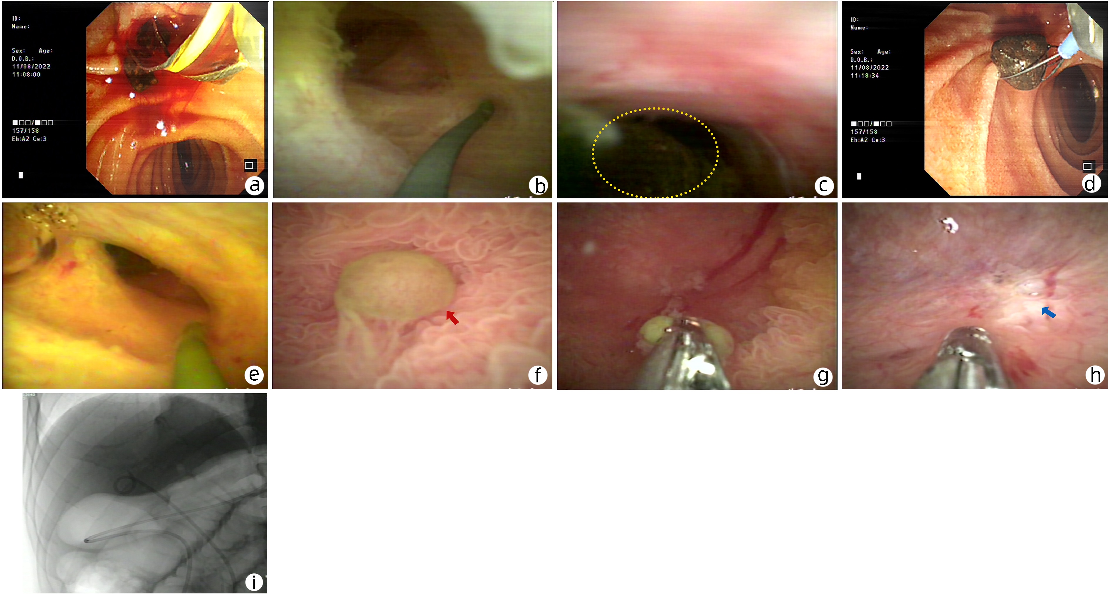
注:a,常规ERCP行胆总管结石网篮取石;b,eyeMax经口子母胆道镜进入胆道内,直视找到胆囊管开口,沿着导丝进入胆囊颈管;c,eyeMax经口子母胆道镜进入胆囊颈部可见胆囊颈部结石(黄色虚线内);d,应用经口子母胆道镜的子母网篮套取胆囊颈部结石,将其拖拽至肠道;e,再次进入eyeMax经口子母胆道镜,可见胆囊颈部螺旋状结构;f,进入胆囊腔内,应用甲硝唑溶液冲洗后可见胆囊壁网格状结构,并清晰可见息肉样病变,略呈黄色(红色箭头);g,将子母热活检钳通过子母胆道镜工作孔道进入胆囊腔内,直视下钳取息肉样隆起;h,直视下电凝切除息肉样隆起,可见切除后的创面(蓝色箭头);i,术后留置鼻胆囊引流管,X线示鼻胆管位置良好。
图 3 ERCP联合eyeMax经口子母胆道镜治疗过程(2022年8月11日)
Figure 3. ERCP combined with eyeMax via oral choledochoscopy (August 11, 2022)
-
[1] Gallbladder-Preserving Committee, Endoscopes Doctor Branch of Chinese Medical Doctor Association. Expert consensus on choledochoscopic gallbladder-preserving surgery for benign gallbladder diseases (2018 edition)[J]. China J Endosc, 2018, 24(9): 106-112. DOI: 10.3969/j.issn.1007-1989.2018.09.022.中国医师协会内镜医师分会内镜微创保胆专业委员会. 内镜微创保胆手术治疗胆囊良性疾病专家共识(2018版)[J]. 中国内镜杂志, 2018, 24(9): 106-112. DOI: 10.3969/j.issn.1007-1989.2018.09.022. [2] TSAI TJ, CHAN HH, LAI KH, et al. Gallbladder function predicts subsequent biliary complications in patients with common bile duct stones after endoscopic treatment?[J]. BMC Gastroenterol, 2018, 18(1): 32. DOI: 10.1186/s12876-018-0762-6. [3] MYERS RP, SHAFFER EA, BECK PL. Gallbladder polyps: epidemiology, natural history and management[J]. Can J Gastroenterol, 2002, 16(3): 187-194. DOI: 10.1155/2002/787598. [4] YANG HL, SUN YG, WANG Z. Polypoid lesions of the gallbladder: diagnosis and indications for surgery[J]. Br J Surg, 1992, 79(3): 227-229. DOI: 10.1002/bjs.1800790312. [5] European Association for the Study of the Liver (EASL). EASL Clinical Practice Guidelines on the prevention, diagnosis and treatment of gallstones[J]. J Hepatol, 2016, 65(1): 146-181. DOI: 10.1016/j.jhep.2016.03.005. [6] ZHANG LD, LIU DQ. Endoscopic technology boosts the development of precision biliary and pancreatic surgery[J]. Chin J Dig Surg, 2022, 21(1): 66-68. DOI: 10.3760/cma.j.cn115610-20211222-00674.张雷达, 刘丹青. 内镜技术助力精准胆胰外科的发展[J]. 中华消化外科杂志, 2022, 21(1): 66-68. DOI: 10.3760/cma.j.cn115610-20211222-00674. [7] DONG WF, PANG EJ, DAI ZL. Clinical efficacy of ERCP combined with LC in treatment of patients with cholecystolithiasis combined with choledocholithiasis and influencing factors for recurrence of choledocholithiasis after surgery[J]. Clin Misdiagn Misther, 2021, 34(5): 85-90. DOI: 10.3969/j.issn.1002-3429.2021.05.017.董维峰, 庞尔君, 代镇岭. ERCP联合LC治疗胆囊结石合并胆总管结石临床效果及术后胆总管结石复发影响因素分析[J]. 临床误诊误治, 2021, 34(5): 85-90. DOI: 10.3969/j.issn.1002-3429.2021.05.017. [8] HAN W, YUE Q, LIU K, et al. Endoscopic nasogallbladder drainage combined with laparoscopic surgery for type Ⅰ mirizzi syndrome with acute cholecystitis: A case series report[J]. Gastroenterol Res Pract, 2020, 2020: 2417539. DOI: 10.1155/2020/2417539. [9] BACA-ARZAGA AA, NAVARRO-CHÁVEZ A, GALINDO-JIMÉNEZ A, et al. Gallstone lithotripsy with SpyGlassTM system through a cholecystoduodenal fistula in a patient with type Ⅲa Mirizzi syndrome[J]. Rev Gastroenterol Mex (Engl Ed), 2021, 86(1): 99-101. DOI: 10.1016/j.rgmx.2020.01.003. [10] MA SR, ZHANG YC, YANG Z, et al. Application of endoscopic retrograde cholangiopancreatography for patients with acute cholecystitis[J]. Chin J Dig Endosc, 2013, 30(5): 269-272. DOI: 10.3760/cma.j.issn.1007-5232.2013.05.008.麻树人, 张迎春, 杨卓, 等. 内镜逆行胰胆管造影技术在急性胆囊炎患者中的临床应用价值[J]. 中华消化内镜杂志, 2013, 30(5): 269-272. DOI: 10.3760/cma.j.issn.1007-5232.2013.05.008. [11] ZHANG H, LIU DQ, XIAO L, et al. ERCP combined with SpyGlass system for the treatment of concomitant gallbladder stones and secondary common bile duct stones in high-risky surgical patients[J]. Chin J Pract Surg, 2018, 38(11): 1310-1313. DOI: 10.19538/j.cjps.issn1005-2208.2018.11.21.张航, 刘丹青, 肖乐, 等. ERCP联合SpyGlass系统治疗高危胆囊结石合并继发胆总管结石可行性研究[J]. 中国实用外科杂志, 2018, 38(11): 1310-1313. DOI: 10.19538/j.cjps.issn1005-2208.2018.11.21. [12] WANG HG, TAO LY, GUO QM. Endoscopic retrograde cholangiopancreatography combined with SpyGlass digital system choledochoscope in treatment of gallbladder neck stones and acute cholecystitis[J]. J Surg Concepts Pract, 2020, 25(6): 481-485. DOI: 10.16139/j.1007-9610.2020.06.008.王宏光, 陶丽莹, 郭庆梅. 内镜逆行胰胆管造影联合SpyGlass DS胆道镜治疗胆囊颈结石和急性胆囊炎[J]. 外科理论与实践, 2020, 25(6): 481-485. DOI: 10.16139/j.1007-9610.2020.06.008. [13] TAO LY, WANG HG, GUO X, et al. Efficacy of endoscopic retrograde cholangiopancreatography combined with SpyGlass system in treatment of acute cholecystitis secondary to choledocholithiasis[J]. J Clin Hepatol, 2022, 38(8): 1854-1858. DOI: 10.3969/j.issn.1001-5256.2022.08.025.陶丽莹, 王宏光, 郭享, 等. 内镜逆行胰胆管造影联合SpyGlass系统治疗胆总管结石继发急性胆囊炎的效果观察[J]. 临床肝胆病杂志, 2022, 38(8): 1854-1858. DOI: 10.3969/j.issn.1001-5256.2022.08.025. [14] FOLEY KG, LAHAYE MJ, THOENI RF, et al. Management and follow-up of gallbladder polyps: updated joint guidelines between the ESGAR, EAES, EFISDS and ESGE[J]. Eur Radiol, 2022, 32(5): 3358-3368. DOI: 10.1007/s00330-021-08384-w. [15] CHAVAN S, RATHI P. Gallbladder polyp: Review and proposed algorithm for management[J]. J Assoc Physicians India, 2022, 70(1): 11-12. [16] KAMADA H, KOBARA H, YAMANA H, et al. Endoscopic direct visualization of gallbladder polypoid lesion using peroral digital single-operator cholangioscopy[J]. Endoscopy, 2021, 53(7): E263-E264. DOI: 10.1055/a-1252-2704. [17] CAO J, DING X, WU H, et al. Classification of the cystic duct patterns and endoscopic transpapillary cannulation of the gallbladder to prevent post-ERCP cholecystitis[J]. BMC Gastroenterol, 2019, 19(1): 139. DOI: 10.1186/s12876-019-1053-6. -

 下载:
下载:

 本文二维码
本文二维码
计量
- 文章访问数: 1185
- HTML全文浏览量: 472
- PDF下载量: 69
- 被引次数: 0

