
 PDF下载 ( 1547 KB)

CCl4诱导的肝纤维化大鼠模型肝小叶内卵圆细胞总体积与轮廓数密度变化的体视学分析
DOI: 10.12449/JCH240113
Changes in the total volume and contour density of oval cells in hepatic lobules of rats with carbon tetrachloride-induced hepatic fibrosis: A stereological study
-
摘要:
目的 定量研究CCl4诱导的肝纤维化大鼠肝小叶内卵圆细胞(HOC)总体积和轮廓数密度的变化。 方法 将11只雄性健康SD大鼠随机分为对照组(n=5)、肝纤维化组(n=6),每周2次皮下注射CCl4与橄榄油混悬液,每次3 mL/kg。在肝纤维化造模5周后取材,从每只大鼠肝脏随机抽选5个大小约1 mm3的肝组织块分别制作1张Epon812环氧树脂包埋超薄切片,运用体视学方法结合透射电子显微镜技术,对大鼠肝小叶内的HOC总体积和轮廓数密度进行定量研究,另从每只大鼠剩余肝脏等距随机抽选4个2 mm厚的肝组织块并分别制作2张石蜡包埋的Masson染色切片,按照肝纤维化Metavir分期标准定性评估每只大鼠的肝纤维化程度。计量资料两组间比较采用成组t检验。 结果 体视学定量研究显示,对照组肝小叶内HOC总体积为(15.40±7.63) mm3,肝纤维化组肝小叶内HOC总体积为(146.80±114.00) mm3,与对照组比较,肝纤维化组大鼠肝小叶内HOC总体积显著增加了8.53倍(t=-2.551,P=0.031);对照组肝小叶内HOC轮廓数密度为(56.20±40.40),肝纤维化组肝小叶内HOC轮廓数密度为(566.50±317.00),与对照组比较,肝纤维化组大鼠肝小叶内轮廓数密度显著增加了9.08倍(t=-3.539,P=0.006);定性观察研究结果显示,肝纤维化大鼠肝纤维化分期按照Metavir评分标准达到Ⅱ~Ⅲ期,大鼠窦周隙内肝星状细胞增生部位周围伴随着HOC的大量增生。 结论 CCl4诱导肝纤维化大鼠肝小叶内HOC显著增生。 -
关键词:
- 肝纤维化 /
- 大鼠, Sprague-Dawley /
- 肝卵圆细胞 /
- 肝星状细胞
Abstract:Objective To quantitatively investigate the changes in the total volume and contour density of hepatic oval cells (HOC) in hepatic lobules of rats with carbon tetrachloride (CCl4)-induced hepatic fibrosis. Methods A total of 11 healthy male Sprague-Dawley rats were randomly divided into control group with 5 rats and hepatic fibrosis group with 6 rats, and CCl4 and olive oil suspension were injected subcutaneously twice a week, 3 mL/kg each time. After five weeks of hepatic fibrosis modeling, five liver tissue blocks with a size of about 1 mm3 were randomly selected from the liver of each rat to prepare one Epon812 epoxy resin-embedded ultrathin section, and the stereological method and transmission electron microscopy were used for the quantitative analysis of the total volume and contour density of HOC in the hepatic lobules of rats. In addition, four liver tissue blocks with a thickness of 2 mm were randomly selected from the remaining liver of each rat to prepare two paraffin-embedded Masson staining sections, and the degree of liver fibrosis in each rat was qualitatively evaluated according to the Metavir staging criteria for liver fibrosis. The independent-samples t test was used for comparison of continuous data between groups. Results The quantitative stereological analysis showed that the total volume of HOC in hepatic lobules was 15.40±7.63 mm3 in the control group and 146.80±114.00 mm3 in the liver fibrosis group, and compared with the control group, the total volume of HOC in hepatic lobules of rats in the liver fibrosis group was significantly increased by 8.53 times (t=-2.551, P=0.031); the contour density of HOC in hepatic lobules was 56.20±40.40 in the control group and 566.50±317.00 in the liver fibrosis group, and compared with the control group, the contour density of HOC in hepatic lobules of rats in the liver fibrosis group was significantly increased by 9.08 times (t=-3.539, P=0.006). Qualitative observation showed that liver fibrosis stage of rats reached stage Ⅱ-Ⅲ according to the Metavir scoring criteria, and massive proliferation of HOC was observed around the proliferation site of hepatic stellate cells in the perisinusoidal space of rats. Conclusion CCl4 induces significant proliferation of HOC in hepatic lobules of rats with liver fibrosis. -
Key words:
- Hepatic Fibrosis /
- Rats, Sprague-Dawley /
- Hepatic Oval Cells /
- Hepatic Stellate Cells
-
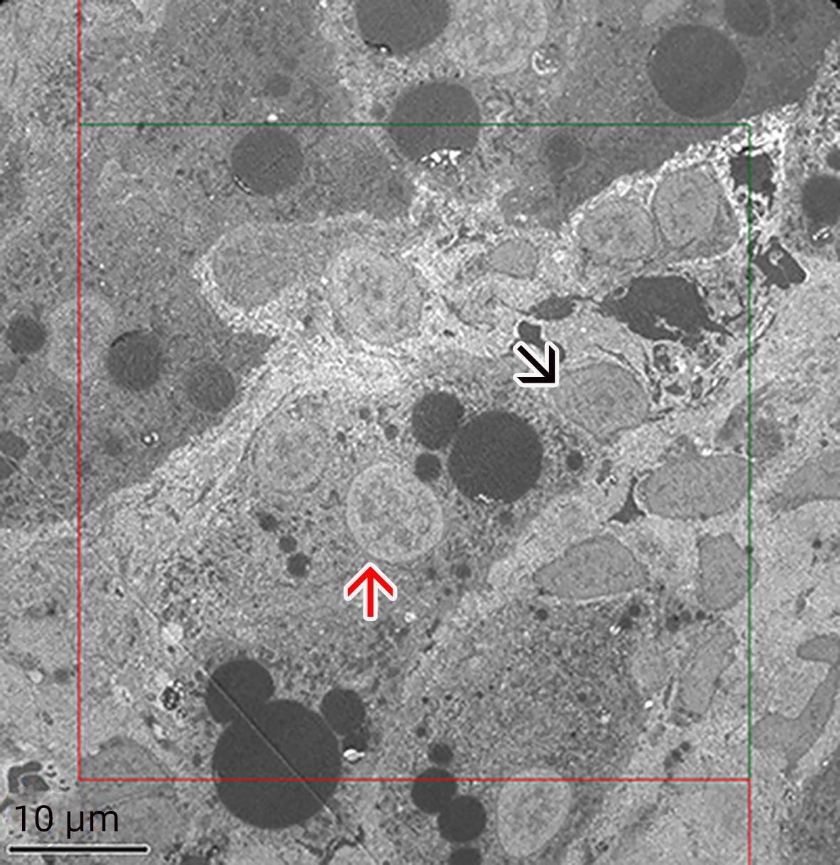
图 1 体视框计数肝卵圆细胞数密度
注: 黑色箭头示肝HOC;红色箭头示肝细胞。体视框面积为2 354.45 μm2,红线为禁线,绿线为计数线,计数落在体视框内、或与绿线相交的肝HOC,落在红线上的肝HOC不计数。
Figure 1. Contour number density of hepatic oval cells counted by stereoscopic frame
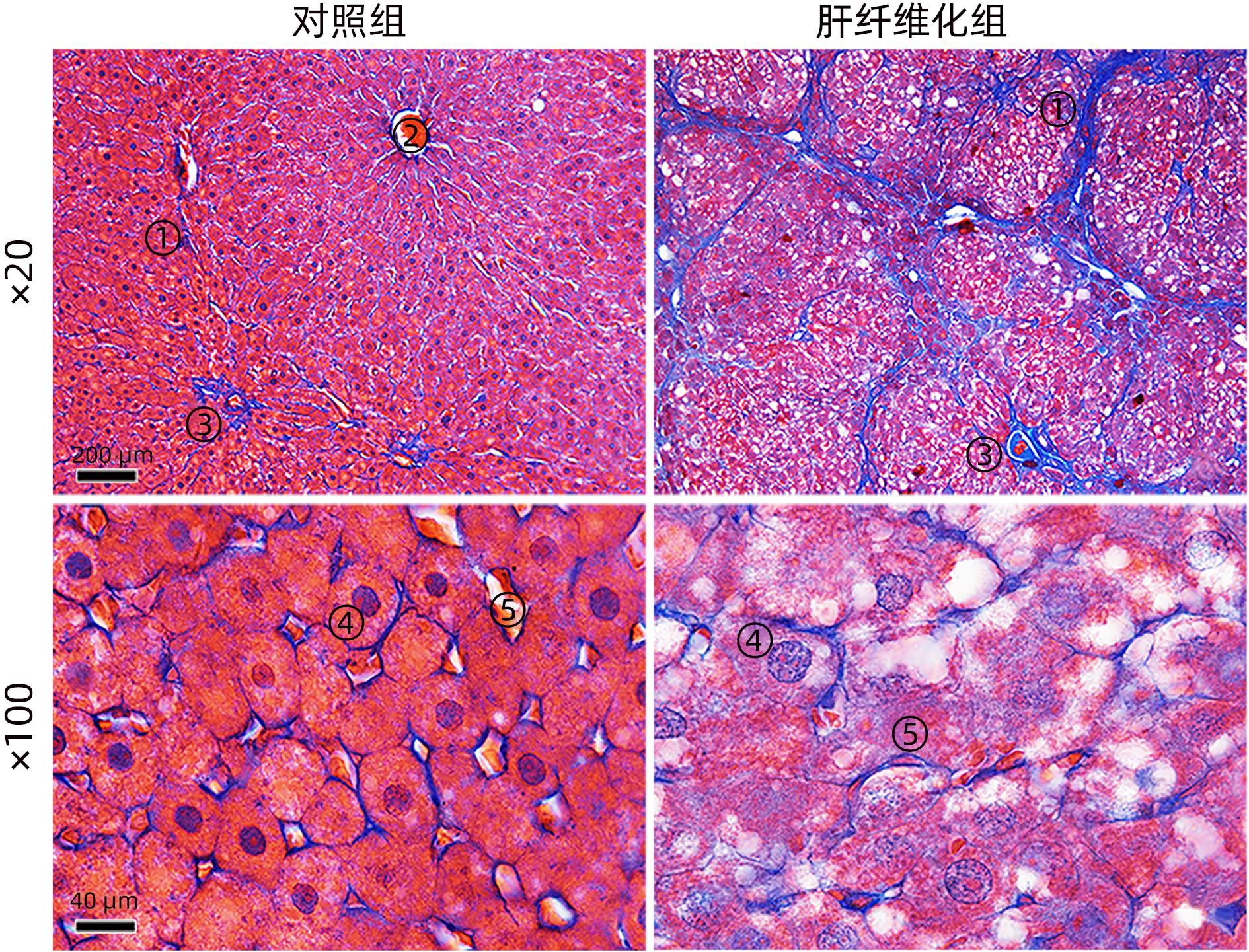
图 2 Masson染色的肝组织结构
注: ①胶原纤维;②中央静脉;③汇管区;④肝细胞;⑤肝血窦。
Figure 2. Histological structure of liver by Masson staining
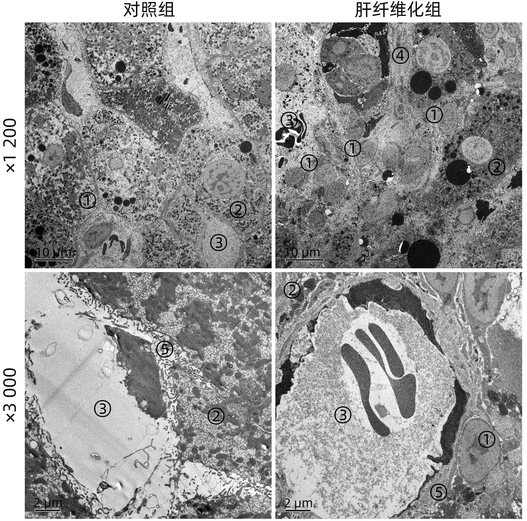
图 3 电镜下肝组织超微结构
注: ①HOC;②肝细胞;③肝血窦;④肝星状细胞;⑤窦周隙。
Figure 3. Ultrastructure of liver tissue under electron microscope
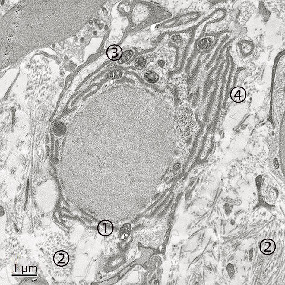
图 4 电镜下HOC超微结构(×7 000)
注: ①肝卵圆细胞;②胶原纤维;③线粒体;④粗面内质网。
Figure 4. Ultrastructure of hepatic oval cell under electron microscope(×7 000)
表 1 各组大鼠肝小叶内肝HOC总体积与轮廓数密度(每mm2细胞数量)变化
Table 1. Changes in total volume (mm3) and contour number density (number of cells per mm2) of hepatic oval cells in hepatic lobules of rats in each group
项目 对照组(n=5) 肝纤维化组(n=6) t值 P值 肝卵圆细胞轮廓数密度 56.20±40.40 566.50±317.00 -3.539 0.006 肝卵圆细胞总体积(mm3) 15.40±7.63 146.80±114.00 -2.551 0.031  下载: 导出CSV
-
[1] ROEHLEN N, CROUCHET E, BAUMERT T F. Liver fibrosis: mechanistic concepts and therapeutic perspectives[J]. Cells, 2020, 9( 4): 875- 918. DOI: 10.3390/cells9040875. [2] PAROLA M, PINZANI M. Liver fibrosis: Pathophysiology, pathogenetic targets and clinical issues[J]. Mol Aspects Med, 2019, 65: 37- 55. DOI: 10.1016/j.mam.2018.09.002. [3] LI T, LIU HB, HU WY, et al. Role of inflammation hepatic fibrosis[J]. J Clin Hepatol, 2022, 38( 10): 2368- 2372. DOI: 10.3969/j.issn.1001-5256.2022.10.032.李婷, 刘华宝, 胡文艳, 等. 炎症在肝纤维化中的作用[J]. 临床肝胆病杂志, 2022, 38( 10): 2368- 2372. DOI: 10.3969/j.issn.1001-5256.2022.10.032. [4] WANG CL. Effects of the changes in the number and volume of hepatic stellate cells on the volume of collagen fibers in rat fibrotic liver and the effects of magnesium isoglycetate on them——a stereological study[D]. Nanchong: North Sichuan Medical Colleage, 2019.王川林. 肝星状细胞数量和体积变化对肝纤维化大鼠肝组织胶原纤维体积的影响及异甘草酸镁的作用——体视学研究[D]. 南充: 川北医学院, 2019. [5] LOTOWSKA JM, SOBANIEC-LOTOWSKA ME, LEBENSZTEJN DM, et al. Ultrastructural characteristics of rat hepatic oval cells and their intercellular contacts in the model of biliary fibrosis: new insights into experimental liver fibrogenesis[J]. Gastroenterol Res Pract, 2017, 2017: 2721547. DOI: 10.1155/2017/2721547. [6] de VOS R, DESMET V. Ultrastructural characteristics of novel epithelial cell types identified in human pathologic liver specimens with chronic ductular reaction[J]. Am J Pathol, 1992, 140( 6): 1441- 1450. [7] DING ZY, JIN GN, WANG W, et al. Activin a-smad signaling mediates connective tissue growth factor synthesis in liver progenitor cells[J]. Int J Mol Sci, 2016, 17( 3): 408. DOI: 10.3390/ijms17030408. [8] MANDAL A, RAJU S, VISWANATHAN C. Cryopreserved hepatic progenitor cells derived from human embryonic stem cells can arrest progression of liver fibrosis in rats[J]. Cell Biol Int, 2016, 40( 10): 1107- 1115. DOI: 10.1002/cbin.10649. [9] HE Z, FENG M. Activation, isolation, identification and culture of hepatic stem cells from porcine liver tissues[J]. Cell Prolif, 2011, 44( 6): 558- 566. DOI: 10.1111/j.1365-2184.2011.00781.x. [10] DORRELL C, ERKER L, SCHUG J, et al. Prospective isolation of a bipotential clonogenic liver progenitor cell in adult mice[J]. Genes Dev, 2011, 25( 11): 1193- 1203. DOI: 10.1101/gad.2029411. [11] WANG CL, YANG X, CUI QL, et al. Effect of magnesium isoglycyrrhizinate on the pathological changes of liver tissue in CCl4 induced hepatic fibrosis rats[J]. J North Sichuan Med Coll, 2018, 33( 3): 360- 363. DOI: 10.3969/j.issn.1005-3697.2018.03.017.王川林, 杨霞, 崔秋林, 等. 异甘草酸镁对四氯化碳肝纤维化大鼠肝组织病理改变的影响[J]. 川北医学院学报, 2018, 33( 3): 360- 363. DOI: 10.3969/j.issn.1005-3697.2018.03.017. [12] OH SH, HATCH HM, PETERSEN BE. Hepatic oval‘stem’ cell in liver regeneration[J]. Semin Cell Dev Biol, 2002, 13( 6): 405- 409. DOI: 10.1016/s1084952102001271. [13] LIN Y, DONG MQ, LIU ZM, et al. A strategy of vascular-targeted therapy for liver fibrosis[J]. Hepatology, 2022, 76( 3): 660- 675. DOI: 10.1002/hep.32299. [14] DHAR D, BAGLIERI J, KISSELEVA T, et al. Mechanisms of liver fibrosis and its role in liver cancer[J]. Exp Biol Med(Maywood), 2020, 245( 2): 96- 108. DOI: 10.1177/1535370219898141. [15] AWAN SJ, BAIG MT, YAQUB F, et al. In vitro differentiated hepatic oval-like cells enhance hepatic regeneration in CCl(4)-induced hepatic injury[J]. Cell Biol Int, 2017, 41( 1): 51- 61. DOI: 10.1002/cbin.10699. [16] YANG AT, HU DD, WANG P, et al. TGF-β1 induces the dual regulation of hepatic progenitor cells with both anti- and proliver fibrosis[J]. Stem Cells Int, 2016, 2016: 1492694. DOI: 10.1155/2016/1492694. [17] KÖHN-GAONE J, GOGOI-TIWARI J, RAMM GA, et al. The role of liver progenitor cells during liver regeneration, fibrogenesis, and carcinogenesis[J]. Am J Physiol Gastrointest Liver Physiol, 2016, 310( 3): G143- 154. DOI: 10.1152/ajpgi.00215.2015. [18] QIU DK, MA X, PENG YS, et al. Locational and quantitative study of hepatic oval cells in chronic liver diseases——Pathologic analysis of 29 liver samples from patients with chronic liver diseases[J]. Chin J Dig, 2000, 20( 5): 301- 303. DOI: 10.3760/j.issn:0254-1432.2000.05.004.邱德凯, 马雄, 彭延申, 等. 慢性肝病患者肝脏卵圆细胞的定位和定量研究——29例慢性肝病患者肝组织病理学分析[J]. 中华消化杂志, 2000, 20( 5): 301- 303. DOI: 10.3760/j.issn:0254-1432.2000.05.004. [19] EZHILARASAN D, SOKAL E, NAJIMI M. Hepatic fibrosis: It is time to go with hepatic stellate cell-specific therapeutic targets[J]. Hepatobiliary Pancreat Dis Int, 2018, 17( 3): 192- 197. DOI: 10.1016/j.hbpd.2018.04.003. [20] KAUR S, SIDDIQUI H, BHAT MH. Hepatic progenitor cells in action: liver regeneration or fibrosis?[J]. Am J Pathol, 2015, 185( 9): 2342- 2350. DOI: 10.1016/j.ajpath.2015.06.004. [21] STRAZZABOSCO M, FABRIS L. Development of the bile ducts: essentials for the clinical hepatologist[J]. J Hepatol, 2012, 56( 5): 1159- 1170. DOI: 10.1016/j.jhep.2011.09.022. [22] WANG P, LIU T, CONG M, et al. Expression of extracellular matrix genes in cultured hepatic oval cells: an origin of hepatic stellate cells through transforming growth factor beta?[J]. Liver Int, 2009, 29( 4): 575- 584. DOI: 10.1111/j.1478-3231. [23] FAKTOR VM, RADAEVA SA. The formation of oval-cell ducts during hepatic carcinogenesis in mice. Its relationship to the pre-existing canals of Hering[J]. Ontogenez, 1992, 23( 4): 407- 418. [24] BURKE ZD, SHEN CN, RALPHS KL, et al. Characterization of liver function in transdifferentiated hepatocytes[J]. J Cell Physiol, 2006, 206( 1): 147- 159. DOI: 10.1002/jcp.20438. -

 本文二维码
本文二维码
计量
- 文章访问数: 123
- HTML全文浏览量: 50
- PDF下载量: 25
- 被引次数: 0

