
 PDF下载 ( 915 KB)

肝硬化营养不良患者肠道菌群的组成特征分析
DOI: 10.12449/JCH240115
Characteristics of the composition of intestinal flora in cirrhotic patients with malnutrition
-
摘要:
目的 研究肝硬化营养不良患者肠道菌群的组成特征和血清内毒素水平,旨在为提高肝硬化患者的营养状况提供新的诊疗思路。 方法 收集2021年3月—2022年11月在郑州人民医院消化内科住院的58例肝硬化患者作为试验组(LC组),根据皇家自由医院营养优先排序工具分为低营养不良风险组(LC-A组,n=28)和中/高营养不良风险组(LC-B组,n=30),并选择同期25例健康体检者作为对照组(HC组)。采集所有受试对象的外周血和粪便样本,用鲎试剂凝胶法检测外周血内毒素浓度,并用高通量测序技术及生物信息学分析肠道菌群间的特征。正态分布的计量资料两组间比较采用成组t检验,多组间比较采用单因素方差分析,进一步两两比较采用LSD-t检验;非正态分布的计量资料两组间比较采用Mann-Whitney U检验,多组间比较采用Kruskal-Wallis H检验。计数资料组间比较采用 结果 三组ALT(H=7.054)、GGT(H=9.644)、Alb(F=32.768)、TBil(H=20.980)及血清内毒素水平(F=108.672)差异均有统计学意义(P值均<0.05)。三组间Chao1指数比较差异有统计学意义(F=5.110,P=0.008),HC组与LC-B组Chao1指数比较差异有统计学意义(P<0.05)。与HC组比较,LC-A组、LC-B组的Chao1指数和Shannon指数均下降,HC组与LC-B组Chao1指数比较差异有统计学意义(P<0.05)。在门水平上,各组主要由拟杆菌门(Bacteroidota)、厚壁菌门(Firmicutes)、变形菌门(Proteobacteria)和放线菌门(Actinobacteriota)组成,并占总体门类的95%以上,HC组与LC-B组厚壁门相对丰度差异有统计学意义(P<0.05)。血清内毒素与瘤胃菌科呈显著负相关(r=-0.420,P=0.007)。螺旋体与TBil呈显著正相关(r=0.419,P=0.007),与Alb呈显著负相关(r=-0.492,P=0.001)。 结论 肝硬化营养不良患者中存在独特的肠道菌群改变,并且差异菌群与内毒素血症存在相关性,改善肝硬化肠道微生态可能有助于改善营养状况。 Abstract:Objective To investigate the characteristics of the composition of intestinal flora and the serum level of endotoxin in cirrhotic patients with malnutrition, and to provide new diagnosis and treatment ideas for improving the nutritional status of patients with liver cirrhosis. Methods A total of 58 patients with liver cirrhosis who were hospitalized in Department of Gastroenterology, People’s Hospital of Zhengzhou, from March 2021 to November 2022 were enrolled as experimental group (LC group), and according to the Royal Free Hospital-Nutritional Prioritizing Tool, they were divided into low malnutrition risk group (LC-A group with 28 patients) and moderate/high malnutrition risk group (LC-B group with 30 patients); 25 individuals who underwent physical examination during the same period of time were enrolled as control group (HC group). Peripheral blood and feces samples were collected from all subjects. The limulus amebocyte lysate gel method was used to measure the concentration of endotoxin in peripheral blood, and high-throughput sequencing and bioinformatics analysis were used to investigate the characteristics of intestinal flora. The independent-samples t test was used for comparison of normally distributed continuous data between two groups; a one-way analysis of variance was used for comparison between multiple groups, and the least significant difference t-test were used for further comparison between two groups. The Mann-Whitney U test was used for comparison of non-normally distributed continuous data between two groups, and the Kruskal-Wallis H test was used for comparison between multiple groups. The chi-square test was used for comparison of categorical data between groups. The Spearman test was used for correlation analysis. Results There were significant differences between the three groups in the levels of alanine aminotransferase (H=7.054, P<0.05), gamma-glutamyl transpeptidase (H=9.644, P<0.05), albumin (Alb) (F=32.768, P<0.05), total bilirubin (TBil) (H=20.980, P<0.05), and serum endotoxin (F=108.672, P<0.05). There was a significant difference in Chao1 index between the three groups (F=5.110, P=0.008) and between the HC group and the LC-B group (P<0.05). Compared with the HC group, the LC-A group and the LC-B group had significant reductions in Chao1 index and Shannon index, and there was a significant difference in Chao1 index between the HC group and the LC-B group (P<0.05). At the phylum level, the intestinal flora in each group was mainly composed of Bacteroidota, Firmicutes, Proteobacteria, and Actinobacteriota, accounting for more than 95% of all phyla, and there was a significant difference in the relative abundance of Firmicutes between the HC group and the LC-B group (P<0.05). Serum endotoxin was significantly negatively correlated with Ruminococcaceae (r=-0.420, P=0.007), and spirochete was significantly positively correlated with TBil (r=0.419, P=0.007) and was significantly negatively correlated with Alb (r=-0.492, P=0.001). Conclusion There are unique changes in intestinal flora in cirrhotic patients with malnutrition, and differentially expressed flora are associated with endotoxemia. Improving intestinal microecology in liver cirrhosis may help to improve nutritional status. -
Key words:
- Liver Cirrhosis /
- Gastrointestinal Microbiome /
- Malnutrition
-

图 1 肠道菌群主要属(前20位)相对丰度百分比
Figure 1. Relative abundance percentage of major genera (top 20) of intestinal flora
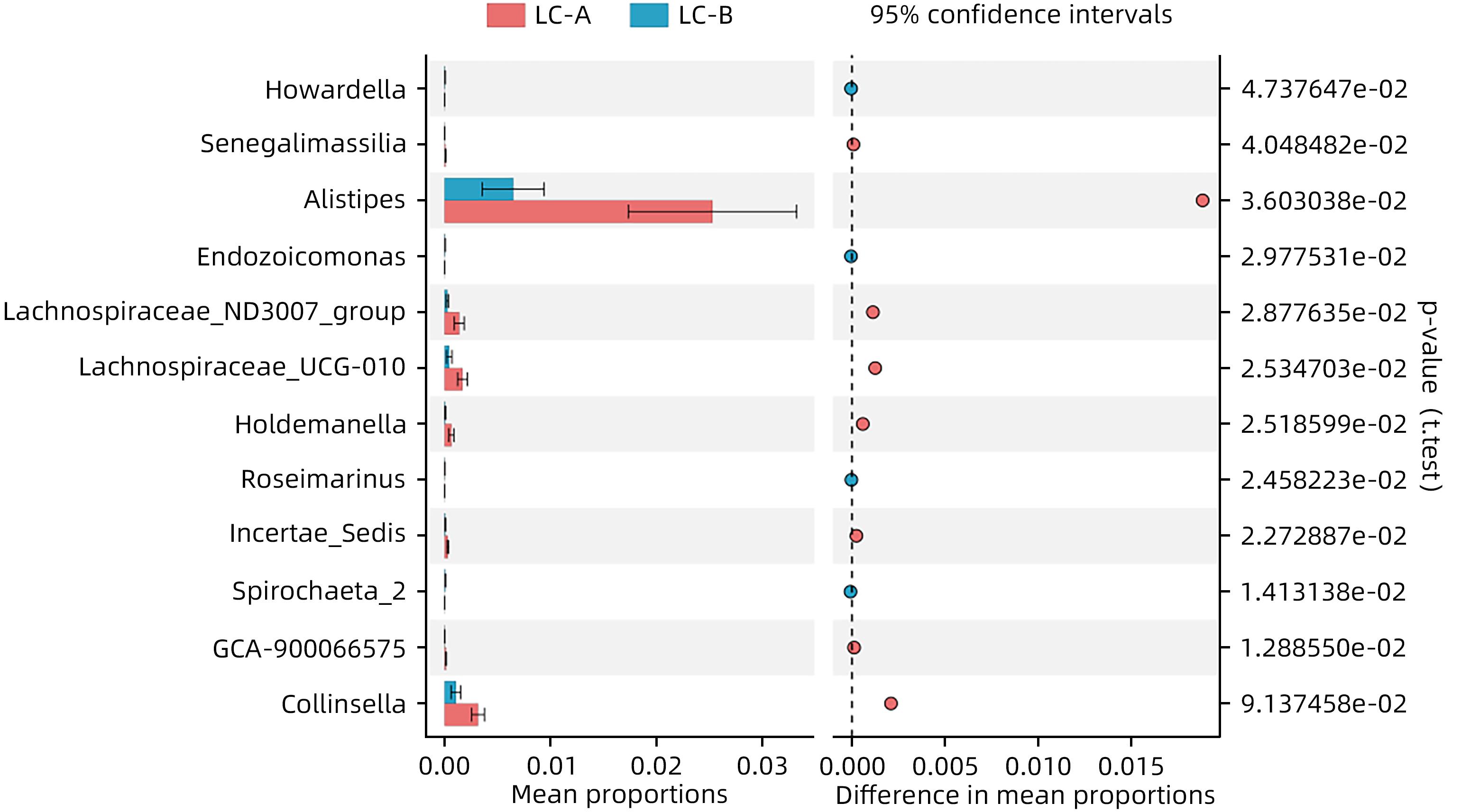
图 2 肠道菌群属水平物种相对丰度比较
Figure 2. Comparison of relative abundance of species at genus level of gut microbiota
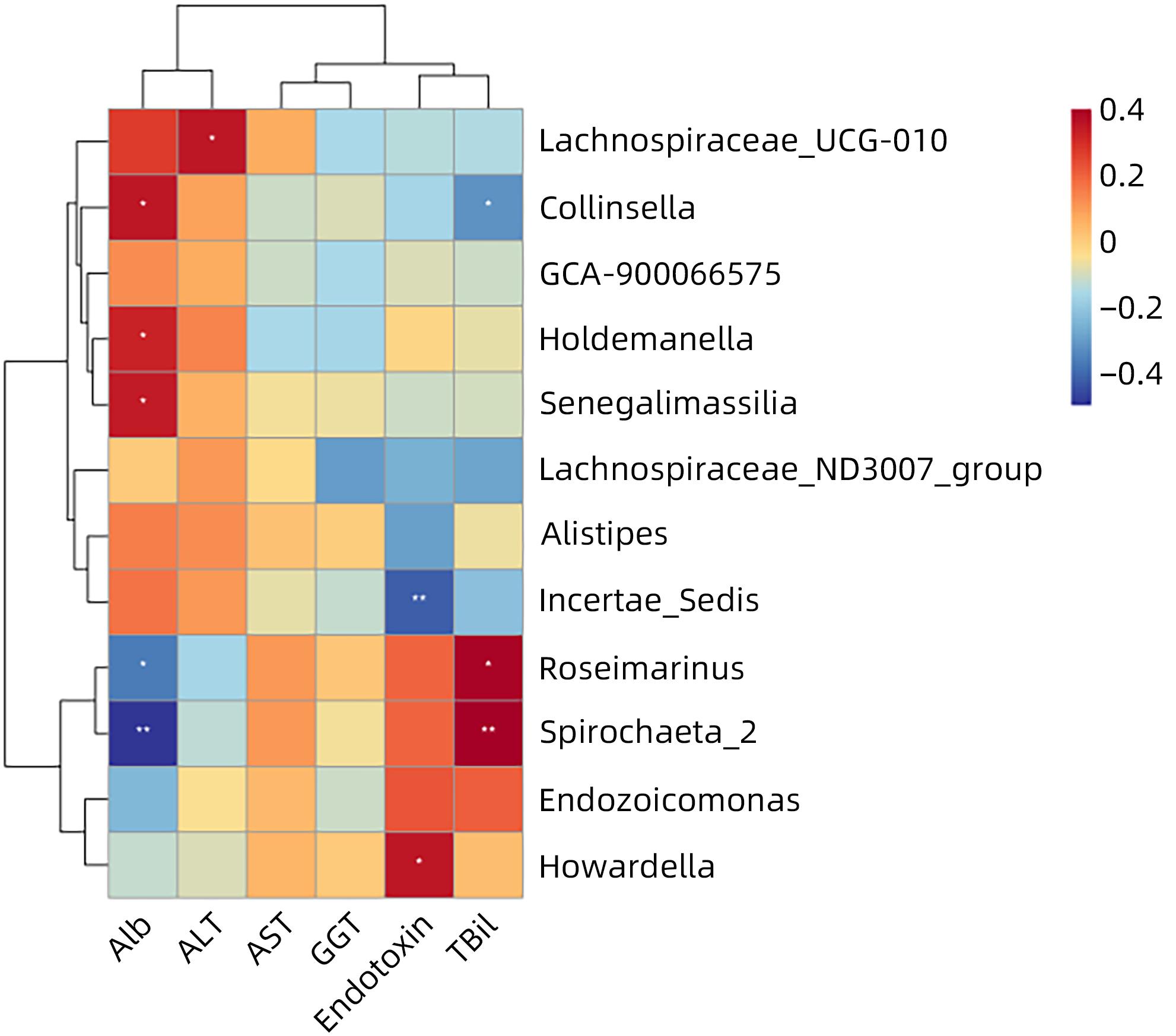
图 3 肠道差异菌群与临床指标的关系
Figure 3. Relationship between gut differential flora and clinical indicators
表 1 各组基本资料
Table 1. Basic information of each group
项目 HC组(n=25) LC-A组(n=28) LC-B组(n=30) 统计值 P值 年龄(岁) 54.68±8.56 57.19±11.21 59.10±9.67 F=2.004 0.139 男性[例(%)] 12(48.00) 19(67.86) 20(66.67) 0.184 ALT(U/L) 20.00(17.70~26.62) 29.00(29.34~74.45)1) 26.00(24.66~103.34)1) H=7.054 0.029 AST(U/L) 35.00(29.14~39.98) 35.00(35.04~69.38) 46.00(40.55~112.74) H=2.929 0.231 GGT(U/L) 26.00(22.42~50.30) 61.00(41.29~139.71)1) 49.00(67.09~134.06)1) H=9.644 0.008 Alb(g/L) 41.80±4.54 37.54±6.021) 31.23±6.461)2) F=32.768 <0.001 TBil(μmol/L) 20.20(19.33~29.32) 20.20(17.56~33.34) 41.30(38.35~61.79)1)2) H=20.98 <0.001 内毒素(EU/mL) 0.04±0.02 0.19±0.071) 0.32±0.111)2) F=108.672 <0.001 注:与HC组比较,1)P<0.05;与LC-A组比较,2)P<0.05。  下载: 导出CSV
表 2 各组肠道菌群丰度和多样性比较
Table 2. Comparison of the abundance and diversity of intestinal flora in each group
项目 HC组(n=25) LC-A组(n=28) LC-B组(n=30) F值 P值 Chao1指数 215.75±74.18 191.76±70.41 159.25±53.311) 5.110 0.008 Shannon指数 5.15±0.89 5.02±0.86 4.59±1.23 2.271 0.110 注:与HC组比较,1)P<0.05。
下载: 导出CSV
表 3 各组肠道菌群门水平相对丰度组成比较
Table 3. Composition comparison of relative abundance of intestinal flora at phylum level in each group
项目 HC组(n=25) LC-A组(n=28) LC-B组(n=30) H值 P值 拟杆菌门 65.24(34.99~73.87) 47.89(36.85~58.42) 60.30(44.50~65.76) 1.357 0.507 厚壁菌门 9.94(7.04~44.30) 12.19(11.13~32.08) 4.02(3.85~14.93)1) 6.217 0.045 变形菌门 15.85(9.91~26.62) 26.30(20.19~35.21) 26.10(22.25~37.01) 4.230 0.121 放线菌门 0.58(0.23~1.67) 0.73(0.66~3.24) 0.55(0.41~5.53) 1.739 0.419 注:与LC-A组比较,1)P<0.05。
下载: 导出CSV
-
[1] TRAUB J, REISS L, ALIWA B, et al. Malnutrition in patients with liver cirrhosis[J]. Nutrients, 2021, 13( 2): 540. DOI: 10.3390/nu13020540. [2] PASHAYEE-KHAMENE F, HAJIMOHAMMADEBRAHIM-KETABFOROUSH M, SHAHRBAF MA, et al. Malnutrition and its association with the mortality in liver cirrhosis; a prospective nutritional assessment in two referral centers in Iran[J]. Clin Nutr ESPEN, 2023, 54: 453- 458. DOI: 10.1016/j.clnesp.2023.02.021. [3] MERLI M, APRILE F. The European Association for the Study of Liver(EASL) nutrition guidelines[J]. Recenti Prog Med, 2021, 112( 2): 103- 109. DOI: 10.1701/3559.35370. [4] XIAO HJ, HAN T. Prevention and treatment of malnutrition, sarcopenia, and osteoporosis in patients with liver cirrhosis[J]. J Clin Hepatol, 2021, 37( 1): 26- 30. DOI: 10.3969/j.issn.1001-5256.2021.01.006.肖慧娟, 韩涛. 肝硬化患者营养不良、肌肉减少症及骨质疏松的防治[J]. 临床肝胆病杂志, 2021, 37( 1): 26- 30. DOI: 10.3969/j.issn.1001-5256.2021.01.006. [5] STADLBAUER V, KOMAROVA I, KLYMIUK I, et al. Disease severity and proton pump inhibitor use impact strongest on faecal microbiome composition in liver cirrhosis[J]. Liver Int, 2020, 40( 4): 866- 877. DOI: 10.1111/liv.14382. [6] PLAUTH M, BERNAL W, DASARATHY S, et al. ESPEN guideline on clinical nutrition in liver disease[J]. Clin Nutr, 2019, 38( 2): 485- 521. DOI: 10.1016/j.clnu.2018.12.022. [7] Chinese Society of Hepatology, Chinese Medical Association. Chinese guidelines on the management of liver cirrhosis[J]. J Clin Hepatol, 2019, 35( 11): 2408- 2425. DOI: 10.3969/j.issn.1001-5256.2019.11.006.中华医学会肝病学分会. 肝硬化诊治指南[J]. 临床肝胆病杂志, 2019, 35( 11): 2408- 2425. DOI: 10.3969/j.issn.1001-5256.2019.11.006. [8] MEYER F, BANNERT K, WIESE M, et al. Molecular mechanism contributing to malnutrition and sarcopenia in patients with liver cirrhosis[J]. Int J Mol Sci, 2020, 21( 15): 5357. DOI: 10.3390/ijms21155357. [9] CHAPMAN B, SINCLAIR M, GOW PJ, et al. Malnutrition in cirrhosis: More food for thought[J]. World J Hepatol, 2020, 12( 11): 883- 896. DOI: 10.4254/wjh.v12.i11.883. [10] RACHAKONDA V, BORHANI AA, DUNN MA, et al. Serum leptin is a biomarker of malnutrition in decompensated cirrhosis[J]. PLoS One, 2016, 11( 9): e0159142. DOI: 10.1371/journal.pone.0159142. [11] BUNCHORNTAVAKUL C. Sarcopenia and frailty in cirrhosis: Assessment and management[J]. Med Clin North Am, 2023, 107( 3): 589- 604. DOI: 10.1016/j.mcna.2022.12.007. [12] RUIGROK RAAA, WEERSMA RK, VICH VILA A. The emerging role of the small intestinal microbiota in human health and disease[J]. Gut Microbes, 2023, 15( 1): 2201155. DOI: 10.1080/19490976.2023.2201155. [13] GHOSH G, JESUDIAN AB. Small intestinal bacterial overgrowth in patients with cirrhosis[J]. J Clin Exp Hepatol, 2019, 9( 2): 257- 267. DOI: 10.1016/j.jceh.2018.08.006. [14] BOJKO M. Causes of sarcopenia in liver cirrhosis[J]. Clin Liver Dis, 2019, 14( 5): 167- 170. DOI: 10.1002/cld.851. [15] PALMER LB, KUFTINEC G, PEARLMAN M, et al. Nutrition in cirrhosis[J]. Curr Gastroenterol Rep, 2019, 21( 8): 38. DOI: 10.1007/s11894-019-0706-5. [16] ZENG YB, CHEN SJ, FU Y, et al. Gut microbiota dysbiosis in patients with hepatitis B virus-induced chronic liver disease covering chronic hepatitis, liver cirrhosis and hepatocellular carcinoma[J]. J Viral Hepat, 2020, 27( 2): 143- 155. DOI: 10.1111/jvh.13216. [17] GUO XX, HU N, LIAN XX, et al. Features of intestinal flora imbalance in patients with liver cirrhosis and related driving factors[J]. J Clin Hepatol, 2020, 36( 7): 1527- 1533. DOI: 10.3969/j.issn.1001-5256.2020.07.016.郭晓霞, 胡娜, 廉晓晓, 等. 肝硬化患者肠道菌群失调的特征及驱动因子分析[J]. 临床肝胆病杂志, 2020, 36( 7): 1527- 1533. DOI: 10.3969/j.issn.1001-5256.2020.07.016. [18] KRISS M, HAZLETON KZ, NUSBACHER NM, et al. Low diversity gut microbiota dysbiosis: Drivers, functional implications and recovery[J]. Curr Opin Microbiol, 2018, 44: 34- 40. DOI: 10.1016/j.mib.2018.07.003. [19] LAI JC, TANDON P, BERNAL W, et al. Malnutrition, frailty, and sarcopenia in patients with cirrhosis: 2021 practice guidance by the American association for the study of liver diseases[J]. Hepatology, 2021, 74( 3): 1611- 1644. DOI: 10.1002/hep.32049. [20] HAO SS, REN XJ, YUAN LL, et al. Correlation of intestinal microecology to muscle mass loss in patients with liver cirrhosis[J]. J Pract Hepatol, 2020, 23( 4): 462- 466. DOI: 10.3969/j.issn.1672-5069.2020.04.003.郝莎莎, 任晓静, 原丽莉, 等. 肝硬化患者肠道微生态与肌量减少的相关性研究[J]. 实用肝脏病杂志, 2020, 23( 4): 462- 466. DOI: 10.3969/j.issn.1672-5069.2020.04.003. [21] IEBBA V, GUERRIERI F, di GREGORIO V, et al. Combining amplicon sequencing and metabolomics in cirrhotic patients highlights distinctive microbiota features involved in bacterial translocation, systemic inflammation and hepatic encephalopathy[J]. Sci Rep, 2018, 8( 1): 8210. DOI: 10.1038/s41598-018-26509-y. [22] SHAO L, LING ZX, CHEN DY, et al. Disorganized gut microbiome contributed to liver cirrhosis progression: A meta-omics-based study[J]. Front Microbiol, 2018, 9: 3166. DOI: 10.3389/fmicb.2018.03166. [23] SUNG CM, LIN YF, CHEN KF, et al. Predicting clinical outcomes of cirrhosis patients with hepatic encephalopathy from the fecal microbiome[J]. Cell Mol Gastroenterol Hepatol, 2019, 8( 2): 301- 318.e2. DOI: 10.1016/j.jcmgh.2019.04.008. [24] PARKER BJ, WEARSCH PA, VELOO ACM, et al. The genus Alistipes: Gut bacteria with emerging implications to inflammation, cancer, and mental health[J]. Front Immunol, 2020, 11: 906. DOI: 10.3389/fimmu.2020.00906. [25] BLAAK EE, CANFORA EE, THEIS S, et al. Short chain fatty acids in human gut and metabolic health[J]. Benef Microbes, 2020, 11( 5): 411- 455. DOI: 10.3920/BM2020.0057. [26] MARTIN-GALLAUSIAUX C, MARINELLI L, BLOTTIÈRE HM, et al. SCFA: Mechanisms and functional importance in the gut[J]. Proc Nutr Soc, 2021, 80( 1): 37- 49. DOI: 10.1017/S0029665120006916. [27] TYAGI AM, YU MC, DARBY TM, et al. The microbial metabolite butyrate stimulates bone formation via T regulatory cell-mediated regulation of WNT10B expression[J]. Immunity, 2018, 49( 6): 1116- 1131.e7. DOI: 10.1016/j.immuni.2018.10.013. [28] TANDON P, MONTANO-LOZA AJ, LAI JC, et al. Sarcopenia and frailty in decompensated cirrhosis[J]. J Hepatol, 2021, 75( Suppl 1): S147- S162. DOI: 10.1016/j.jhep.2021.01.025. [29] YAO YQ, YANG YQ, CHEN XR, et al. Visual analysis of knowledge map correlation between cirrhosis and gut microbiota[J]. Chin Med Herald, 2023, 20( 20): 17- 22. DOI: 10.20047/j.issn1673-7210.2023.20.03.姚倚琦, 杨雅钦, 陈宣睿, 等. 肝硬化与肠道菌群相关性的知识图谱可视化分析[J]. 中国医药导报, 2023, 20( 20): 17- 22. DOI: 10.20047/j.issn1673-7210.2023.20.03. -

 本文二维码
本文二维码
计量
- 文章访问数: 152
- HTML全文浏览量: 28
- PDF下载量: 32
- 被引次数: 0

