
 PDF下载 ( 4389 KB)

骨髓巨噬细胞M2亚型共培养后的骨髓间充质干细胞移植治疗肝硬化大鼠模型的效果分析
DOI: 10.12449/JCH240117
Therapeutic effect of transplantation of bone marrow mesenchymal stem cells co-cultured with bone marrow M2 macrophages on a rat model of liver cirrhosis
-
摘要:
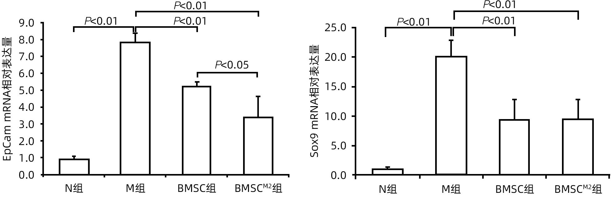
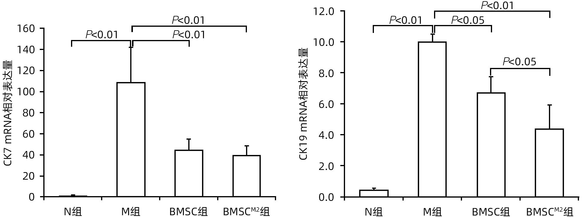
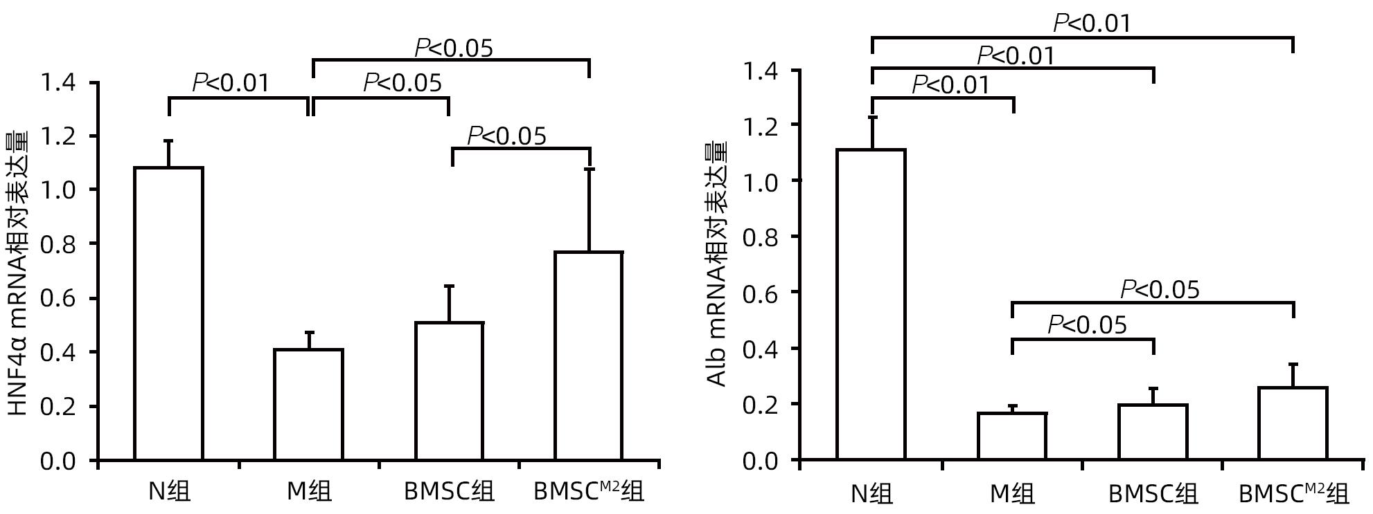
目的 探讨骨髓巨噬细胞M2亚型(M2-BMDM)共培养后的骨髓间充质干细胞(BMSCM2)移植对四氯化碳/2-乙酰氨基芴(CCl4/2-AAF)诱导肝硬化大鼠模型进展的影响。 方法 分离大鼠BMDM并极化为M2表型;分离大鼠BMSC,培养至第3代时与M2-BMDM共培养后获取BMSCM2。CCl4皮下注射6周建立大鼠肝硬化模型。将模型大鼠随机分为模型组(M组)、BMSC组、BMSCM2组,同时设有正常组(N组),每组6只。第7周开始,模型大鼠于CCl4皮下注射的同时予以2-AAF灌胃,分组干预,10周末取材,观察肝功能、肝组织病理、肝组织羟脯氨酸(Hyp)含量,以及肝星状细胞、肝祖细胞、胆管细胞、肝细胞标志物的变化情况。计量资料多组间比较采用单因素方差分析,进一步两两比较采用LSD-t检验。 结果 与N组比较,M组大鼠血清ALT、AST活性均显著升高(P值均<0.01);与M组比较,BMSC组和BMSCM2组大鼠ALT、AST均显著降低(P值均<0.01),且BMSCM2组显著优于BMSC组(P值均<0.05)。与N组比较,M组大鼠肝脏Hyp含量、α-SMA mRNA及蛋白表达均显著升高(P值均<0.01);与M组比较,BMSC组和BMSCM2组Hyp含量、α-SMA表达均显著降低(P值均<0.05),且BMSCM2组α-SMA水平显著低于BMSC组(P<0.01)。与N组比较,M组大鼠肝祖细胞标志物EpCam、Sox9以及胆管细胞标志物CK7、CK19 mRNA表达均显著增加(P值均<0.01),肝细胞标志物HNF-4α和Alb表达均显著降低(P值均<0.01);与M组比较,BMSC组和BMSCM2组EpCam、Sox9、CK7和CK19 mRNA表达均显著降低(P值均<0.05),HNF-4α和Alb mRNA表达均显著增加(P值均<0.05);且与BMSC组比较,BMSCM2组EpCam和CK19 mRNA表达均显著降低(P值均<0.05),而HNF-4α mRNA表达显著增加(P<0.05)。 结论 M2-BMDM可提高BMSC对CCl4/2-AAF诱导大鼠肝硬化的治疗效应,为进一步提高BMSC治疗肝硬化的作用提供了新思路。 Abstract:Objective To investigate the effect of transplantation of bone marrow mesenchymal stem cells (BMSCs) co-cultured with bone marrow-derived M2 macrophages (M2-BMDMs), named as BMSCM2, on a rat model of liver cirrhosis induced by carbon tetrachloride (CCl4)/2-acetaminofluorene (2-AAF). Methods Rat BMDMs were isolated and polarized into M2 phenotype, and rat BMSCs were isolated and co-cultured with M2-BMDMs at the third generation to obtain BMSCM2. The rats were given subcutaneous injection of CCl4 for 6 weeks to establish a model of liver cirrhosis, and then they were randomly divided into model group (M group), BMSC group, and BMSCM2 group, with 6 rats in each group. A normal group (N group) with 6 rats was also established. Since week 7, the model rats were given 2-AAF by gavage in addition to the subcutaneous injection of CCl4. Samples were collected at the end of week 10 to observe liver function, liver histopathology, and hydroxyproline (Hyp) content in liver tissue, as well as changes in the markers for hepatic stellate cells, hepatic progenitor cells, cholangiocytes, and hepatocytes. A one-way analysis of variance was used for comparison of continuous data between multiple groups, and the least significant difference t-test was used for further comparison between two groups. Results Compared with the N group, the M group had significant increases in the activities of serum alanine aminotransferase (ALT) and aspartate aminotransferase (AST) (P<0.01); compared with the M group, the BMSC and BMSCM2 groups had significant reductions in ALT and AST (P<0.01), and the BMSCM2 group had significantly better activities than the BMSC group (P<0.05). Compared with the N group, the M group had significant increases in Hyp content and the mRNA and protein expression levels of alpha-smooth muscle actin (α-SMA) in the liver (P<0.01); compared with the M group, the BMSC and BMSCM2 groups had significant reductions in Hyp content and the expression of α-SMA (P<0.05), and the BMSCM2 group had a significantly lower level of α-SMA than the BMSC group (P<0.01). Compared with the N group, the M group had significant increases in the mRNA expression levels of the hepatic progenitor cell markers EpCam and Sox9 and the cholangiocyte markers CK7 and CK19 (P<0.01) and significant reductions in the expression levels of the hepatocyte markers HNF-4α and Alb (P<0.01); compared with the M group, the BMSC and BMSCM2 groups had significant reductions in the mRNA expression levels of EpCam, Sox9, CK7, and CK19 (P<0.05) and significant increases in the mRNA expression levels of HNF-4α and Alb (P<0.05), and compared with the BMSC group, the BMSCM2 group had significant reductions in the mRNA expression levels of EpCam and CK19 (P<0.05) and significant increase in the expression level of HNF-4α (P<0.05). Conclusion M2-BMDMs can enhance the therapeutic effect of BMSCs on CCl4/2-AAF-induced liver cirrhosis in rats, which provides new ideas for further improving the therapeutic effect of BMSCs on liver cirrhosis. -
Key words:
- Liver Cirrhosis /
- Mesenchymal Stem Cells /
- Macrophages /
- Rats, Wistar
-

图 1 BMDM和BMSC鉴定
注: a,BMSC镜下形态学观察(×100);b,BMSC成骨诱导(茜素红染色,×100);c,BMSC成脂诱导(油红O染色,×100);d,M2-BMDM流式细胞鉴定;e,BMSC细胞周期检测;f,BMSC流式细胞鉴定。
Figure 1. BMDM and BMSC identification

图 2 BMSCM2抑制肝脏炎症反应
注: a,HE染色(×200);b,CD68免疫组化染色(×200);c,血清ALT、AST活性;d,肝组织TNF-α、TGF-β1及CD68 mRNA表达水平;e,肝组织CD68免疫印迹;f,肝组织CD68免疫印迹灰度积分比值。
Figure 2. BMSCM2 inhibits hepatic inflammatory response
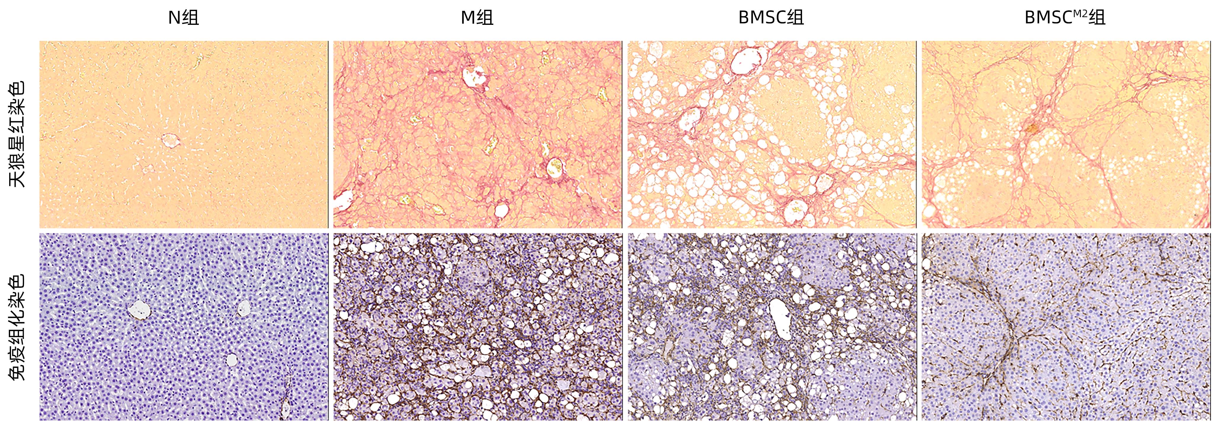
图 3 天狼星红染色和α-SMA免疫组化染色结果(×200)
Figure 3. Sirius red staining and α-SMA immunohistochemical staining results(×200)
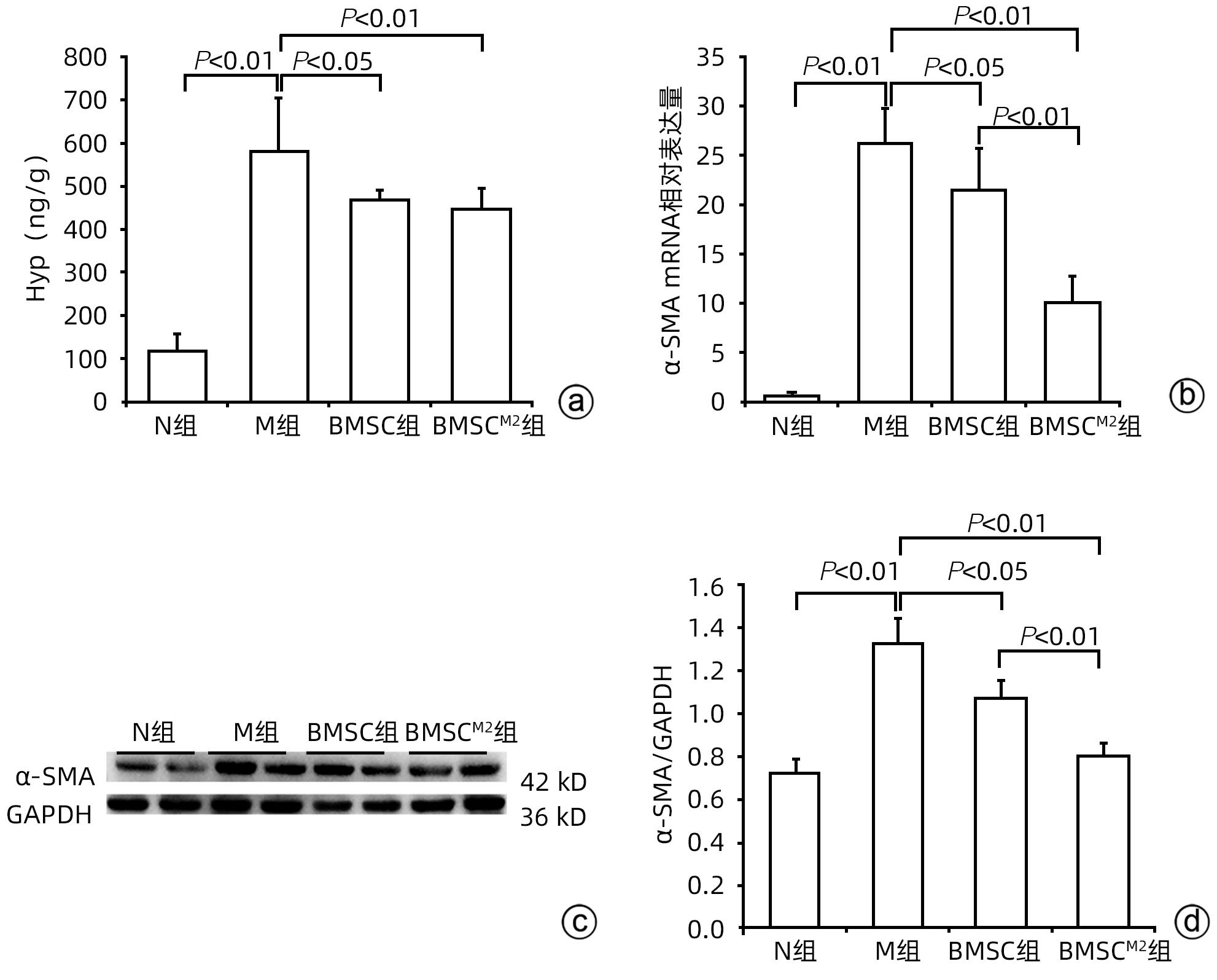
图 4 BMSCM2抑制肝硬化进展
注: a,肝组织Hyp含量;b,肝组织α-SMA mRNA表达水平;c,肝组织α-SMA免疫印迹;d,肝组织α-SMA免疫印迹灰度积分比值。
Figure 4. BMSCM2 inhibits the progression of liver cirrhosis

图 5 EpCam和Sox9免疫组化染色结果(×200)
Figure 5. Immunohistochemical staining results of EpCam and Sox9 (×200)

图 7 CK7和CK19免疫组化染色结果(×200)
Figure 7. Immunohistochemical staining results of CK7 and CK19 (×200)
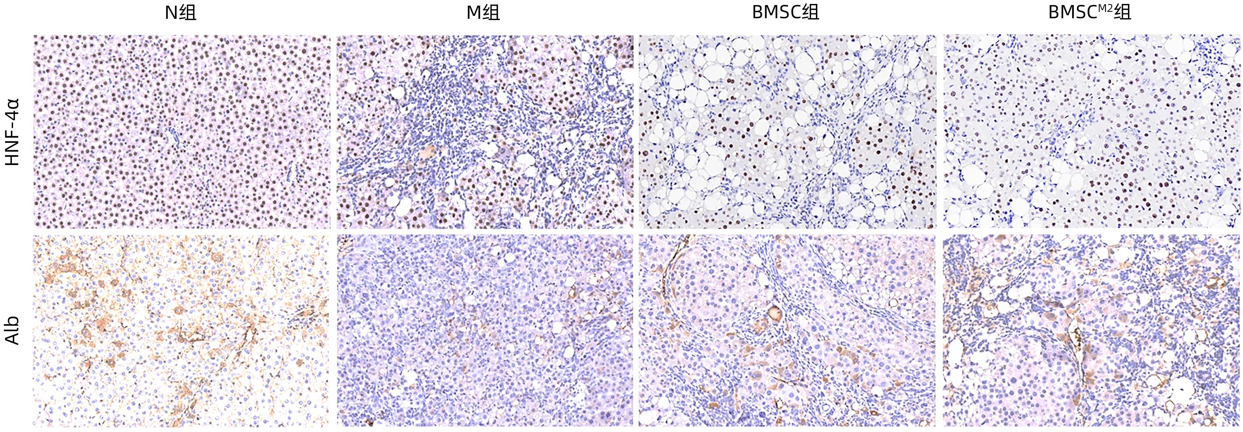
图 9 HNF-4α和Alb免疫组化染色结果(×200)
Figure 9. Immunohistochemical staining results of HNF-4α and Alb (×200)
-
[1] FREEMAN RB Jr, STEFFICK DE, GUIDINGER MK, et al. Liver and intestine transplantation in the United States, 1997-2006[J]. Am J Transplant, 2008, 8( 4 Pt 2): 958- 976. DOI: 10.1111/j.1600-6143.2008.02174.x. [2] ZHANG YT, LI YW, ZHANG LL, et al. Mesenchymal stem cells: Potential application for the treatment of hepatic cirrhosis[J]. Stem Cell Res Ther, 2018, 9( 1): 59. DOI: 10.1186/s13287-018-0814-4. [3] XIE RP, GU MQ, ZHANG FB, et al. Current status and prospect of surgical technique of liver transplantation[J]. Ogran Transplant, 2022, 13( 1): 105- 110. DOI: 10.3969/j.issn.1674-7445.2022.01.016.谢闰鹏, 谷明旗, 张凤博, 等. 肝移植手术技术的现状和展望[J]. 器官移植, 2022, 13( 1): 105- 110. DOI: 10.3969/j.issn.1674-7445.2022.01.016. [4] XIA Q, SHA M. Progress and prospect of living donor liver transplantation[J]. Chin J Dig Surg, 2022, 21( 1): 39- 42. DOI: 10.3760/cma.j.cn115610-20211205-00622.夏强, 沙朦. 活体肝移植的进展与展望[J]. 中华消化外科杂志, 2022, 21( 1): 39- 42. DOI: 10.3760/cma.j.cn115610-20211205-00622. [5] HUANG W, BHADURI A, VELMESHEV D, et al. Origins and proliferative states of human oligodendrocyte precursor cells[J]. Cell, 2020, 182( 3): 594- 608. DOI: 10.1016/j.cell.2020.06.027. [6] KHARAZIHA P, HELLSTRÖM PM, NOORINAYER B, et al. Improvement of liver function in liver cirrhosis patients after autologous mesenchymal stem cell injection: A phase I-II clinical trial[J]. Eur J Gastroenterol Hepatol, 2009, 21( 10): 1199- 1205. DOI: 10.1097/MEG.0b013e32832a1f6c. [7] SUK KT, YOON JH, KIM MY, et al. Transplantation with autologous bone marrow-derived mesenchymal stem cells for alcoholic cirrhosis: Phase 2 trial[J]. Hepatology, 2016, 64( 6): 2185- 2197. DOI: 10.1002/hep.28693. [8] ESMAEILZADEH A, OMMATI H, KOOSHYAR MM, et al. Autologous bone marrow stem cell transplantation in liver cirrhosis after correcting nutritional anomalies, A controlled clinical study[J]. Cell J, 2019, 21( 3): 268- 273. DOI: 10.22074/cellj.2019.6108. [9] JIA SS, LIU X, LI WY, et al. Peroxisome proliferator-activated receptor gamma negatively regulates the differentiation of bone marrow-derived mesenchymal stem cells toward myofibroblasts in liver fibrogenesis[J]. Cell Physiol Biochem, 2015, 37( 6): 2085- 2100. DOI: 10.1159/000438567. [10] JIAN X, WANG DY, XU YN, et al. Effect of polarized bone marrow-derived macrophage transplantation on the progression of CCl4-induced liver fibrosis in rats[J]. J Clin Hepatol, 2021, 37( 12): 2830- 2837. DOI: 10.3969/j.issn.1001-5256.2021.12.020.简迅, 王丹阳, 许燕楠, 等. 极化骨髓巨噬细胞移植对CCl4诱导的肝纤维化大鼠模型的影响[J]. 临床肝胆病杂志, 2021, 37( 12): 2830- 2837. DOI: 10.3969/j.issn.1001-5256.2021.12.020. [11] XU YN, XU W, ZHANG X, et al. BM-MSCs overexpressing the Numb enhance the therapeutic effect on cholestatic liver fibrosis by inhibiting the ductular reaction[J]. Stem Cell Res Ther, 2023, 14( 1): 45. DOI: 10.1186/s13287-023-03276-w. [12] JAMALL IS, FINELLI VN, QUE HEE SS. A simple method to determine nanogram levels of 4-hydroxyproline in biological tissues[J]. Anal Biochem, 1981, 112( 1): 70- 75. DOI: 10.1016/0003-2697(81)90261-x. [13] MU YP, OGAWA T, KAWADA N. Reversibility of fibrosis, inflammation, and endoplasmic reticulum stress in the liver of rats fed a methionine-choline-deficient diet[J]. Lab Invest, 2010, 90( 2): 245- 256. DOI: 10.1038/labinvest.2009.123. [14] PRADERE JP, KLUWE J, DE MINICIS S, et al. Hepatic macrophages but not dendritic cells contribute to liver fibrosis by promoting the survival of activated hepatic stellate cells in mice[J]. Hepatology, 2013, 58( 4): 1461- 1473. DOI: 10.1002/hep.26429. [15] KARLMARK KR, WEISKIRCHEN R, ZIMMERMANN HW, et al. Hepatic recruitment of the inflammatory Gr1+ monocyte subset upon liver injury promotes hepatic fibrosis[J]. Hepatology, 2009, 50( 1): 261- 274. DOI: 10.1002/hep.22950. [16] VANNELLA KM, WYNN TA. Mechanisms of organ injury and repair by macrophages[J]. Annu Rev Physiol, 2017, 79: 593- 617. DOI: 10.1146/annurev-physiol-022516-034356. [17] ORECCHIONI M, GHOSHEH Y, PRAMOD AB, et al. Macrophage polarization: Different gene signatures in M1(LPS+) vs. classically and M2(LPS-) vs. alternatively activated macrophages[J]. Front Immunol, 2019, 10: 1084. DOI: 10.3389/fimmu.2019.01084. [18] ZHOU T, YUAN ZN, WENG JY, et al. Challenges and advances in clinical applications of mesenchymal stromal cells[J]. J Hematol Oncol, 2021, 14( 1): 24. DOI: 10.1186/s13045-021-01037-x. [20] NISHINA T, HOSHIKAWA KT, UENO Y. Current cell-based therapies in the chronic liver diseases[J]. Adv Exp Med Biol, 2018, 1103: 243- 253. DOI: 10.1007/978-4-431-56847-6_13. [21] SAITO Y, IKEMOTO T, TOKUDA K, et al. Effective three-dimensional culture of hepatocyte-like cells generated from human adipose-derived mesenchymal stem cells[J]. J Hepatobiliary Pancreat Sci, 2021, 28( 9): 705- 715. DOI: 10.1002/jhbp.1024. [22] LUO XY, MENG XJ, CAO DC, et al. Transplantation of bone marrow mesenchymal stromal cells attenuates liver fibrosis in mice by regulating macrophage subtypes[J]. Stem Cell Res Ther, 2019, 10( 1): 16. DOI: 10.1186/s13287-018-1122-8. [23] CHAI NL, ZHANG XB, CHEN SW, et al. Umbilical cord-derived mesenchymal stem cells alleviate liver fibrosis in rats[J]. World J Gastroenterol, 2016, 22( 26): 6036- 6048. DOI: 10.3748/wjg.v22.i26.6036. [24] GHAFOURI-FARD S, NIAZI V, HUSSEN BM, et al. The emerging role of exosomes in the treatment of human disorders with a special focus on mesenchymal stem cells-derived exosomes[J]. Front Cell Dev Biol, 2021, 9: 653296. DOI: 10.3389/fcell.2021.653296. [25] FONDEVILA MF, FERNANDEZ U, HERAS V, et al. Inhibition of carnitine palmitoyltransferase 1A in hepatic stellate cells protects against fibrosis[J]. J Hepatol, 2022, 77( 1): 15- 28. DOI: 10.1016/j.jhep.2022.02.003. [26] NOVO E, MARRA F, ZAMARA E, et al. Dose dependent and divergent effects of superoxide anion on cell death, proliferation, and migration of activated human hepatic stellate cells[J]. Gut, 2006, 55( 1): 90- 97. DOI: 10.1136/gut.2005.069633. [27] WU XP, SHU LL, ZHANG ZX, et al. Adipocyte fatty acid binding protein promotes the onset and progression of liver fibrosis via mediating the crosstalk between liver sinusoidal endothelial cells and hepatic stellate cells[J]. Adv Sci, 2021, 8( 11): e2003721. DOI: 10.1002/advs.202003721. [28] PENG JY, LI F, WANG J, et al. Identification of a rare Gli1+ progenitor cell population contributing to liver regeneration during chronic injury[J]. Cell Discov, 2022, 8( 1): 118. DOI: 10.1038/s41421-022-00474-3. [29] TARLOW BD, FINEGOLD MJ, GROMPE M. Clonal tracing of Sox9+ liver progenitors in mouse oval cell injury[J]. Hepatology, 2014, 60( 1): 278- 289. DOI: 10.1002/hep.27084. [30] MISHRA L, BANKER T, MURRAY J, et al. Liver stem cells and hepatocellular carcinoma[J]. Hepatology, 2009, 49( 1): 318- 329. DOI: 10.1002/hep.22704. [31] CHEN JM, ZHANG X, XU Y, et al. Hepatic progenitor cells contribute to the progression of 2-acetylaminofluorene/carbon tetrachloride-induced cirrhosis via the non-canonical Wnt pathway[J]. PLoS One, 2015, 10( 6): e0130310. DOI: 10.1371/journal.pone.0130310. [32] YAGI K, KOJIMA M, OYAGI S, et al. Application of mesenchymal stem cells to liver regenerative medicine[J]. Yakugaku Zasshi, 2008, 128( 1): 3- 9. DOI: 10.1248/yakushi.128.3. -

 下载:
下载:
 本文二维码
本文二维码
计量
- 文章访问数: 158
- HTML全文浏览量: 40
- PDF下载量: 21
- 被引次数: 0

