
 PDF下载 ( 2417 KB)

DynaCT在原发性肝癌患者经肝动脉化疗栓塞术中的指导作用及对疗效的评估价值
DOI: 10.3969/j.issn.1001-5256.2022.04.021
Guiding role of cone beam CT with DynaCT in transcatheter arterial chemoembolization for patients with primary liver cancer and its value in assessing treatment outcome
-
摘要:
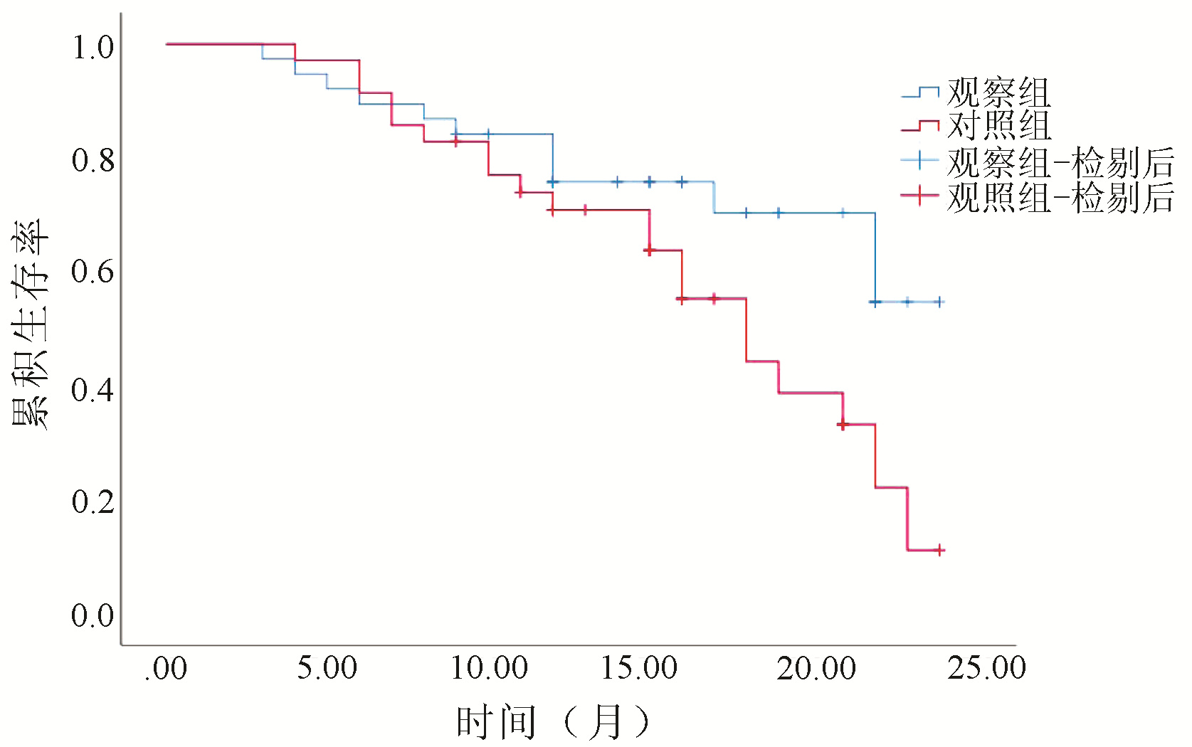
目的 探讨分析肝癌经肝动脉化疗栓塞术(TACE)治疗患者采用DynaCT的效果及对患者预后的影响。 方法 选择2017年5月-2019年5月在川北医学院附属第二医院就诊的原发性肝癌患者73例, 将患者随机分为观察组(n=38)和对照组(n=35)。对照组患者仅行2D-DSA造影下TACE治疗, 观察组患者在2D-DSA造影后再行DynaCT造影。比较两组患者手术时间、X线曝光量以及造影剂用量, 2D-DSA造影与DynaCT造影对肝内肿瘤病灶检出情况及供血动脉显示情况, 二维X线透视与平扫DynaCT对肿瘤病灶内碘化油沉积的评估。两组间计量资料比较采用独立样本t检验, 计数资料两组间比较采用χ2检验; 绘制Kaplan-Meier生存曲线分析生存情况, 两组间比较采用log-rank检验。 结果 两组患者手术时间、X线曝光量以及造影剂用量比较差异均无统计学意义(P值均>0.05)。观察组患者中共检出肿瘤病灶93个, 病灶供血动脉阳性占比为84.95%(79/93), 对照组患者中共检出肿瘤病灶61个, 病灶供血动脉阳性占比为55.74%(34/61), 观察组患者病灶中供血动脉检出阳性占比显著高于对照组(χ2=16.088, P < 0.05)。术后对两组患者的113个病灶进行碘化油沉积分析, 二维X线透视显示89个病灶碘油均匀沉积, 24个病灶碘油部分或全部缺失; 而平扫DynaCT显示78个病灶碘油均匀沉积, 35个病灶碘油部分或全部缺失。观察组患者术后总体生存情况显著优于对照组(χ2=4.347, P=0.037)。 结论 DynaCT在不增加术中X线曝光量与术中造影剂使用量的同时, 能够提高对肝内乏血病灶和重叠病灶的检出率, 从而提高插管准确性, 减少患者血管损伤, 同时也可应用于栓塞术后碘化油沉积评价, 在肝癌TACE术中具有重要的应用价值, 有助于改善患者术后生存。 -
关键词:
- 肝肿瘤 /
- 化学栓塞, 治疗性 /
- 血管造影三维软组织成像技术
Abstract:Objective To investigate the effect of cone beam CT with DynaCT on liver cancer patients undergoing transcatheter arterial chemoembolization (TACE) and its influence on the prognosis of patients. Methods A total of 73 patients with primary liver cancer who attended The Second Affiliated Hospital of North Sichuan Medical College from May 2017 to May 2019 were enrolled and randomly divided into observation group with 38 patients and control group with 35 patients. The patients in the control group underwent TACE under 2D-DSA angiography, and those in the observation group underwent DynaCT angiography after 2D-DSA angiography. The two groups were compared in terms of time of operation, X-ray exposure, amount of contrast agent used, intrahepatic tumor lesions detected and blood supplying arteries displayed by 2D-DSA angiography versus DynaCT angiography, and lipiodol deposition in tumor lesions evaluated by postoperative two-dimensional X-ray fluoroscopy versus plain DynaCT scan. The two-independent-samples t test was used for comparison between two groups, and the chi-square test was used for comparison of categorical data between two groups; the Kaplan-Meier survival curve was plotted for survival analysis, and the log-rank test was used for comparison between two groups. Results There were no significant differences between the two groups in time of operation, X-ray exposure, and amount of contrast agent used (all P>0.05). For the observation group, a total of 93 tumor lesions were detected, among which 79 (84.95%) were positive for blood supplying arteries, while in the control group, a total of 61 tumor lesions were detected, among which 34 (55.74%) were positive for blood supplying arteries, suggesting that the proportion of lesions positive for blood supplying arteries in the observation group was significantly higher than that in the control group (χ2=16.088, P < 0.05). After surgery, 113 lesions of the two groups were analyzed for lipiodol deposition; two-dimensional X-ray fluoroscopy showed that lipiodol was evenly deposited in 89 lesions and was partially or completely missing in 24 lesions, while plain DynaCT scan showed that lipiodol was evenly deposited in 78 lesions and was partially or completely missing in 35 lesions. The observation group had significantly better overall survival than the control group (χ2=4.347, P=0.037). Conclusion DynaCT can increase the detection rate of ischemic lesions and overlapping lesions in the liver without increasing the amount of intraoperative X-ray exposure and contrast agent used, thereby improving the accuracy of intubation and reducing the patient's vascular injury, and at the same time, it can be used to evaluate the deposition of lipiodol after embolization. It has an important application value in TACE for liver cancer and can help to improve the survival of patients after surgery. -
Key words:
- Liver Neoplasms /
- Chemoembolization, Therapeutic /
- DynaCT
-
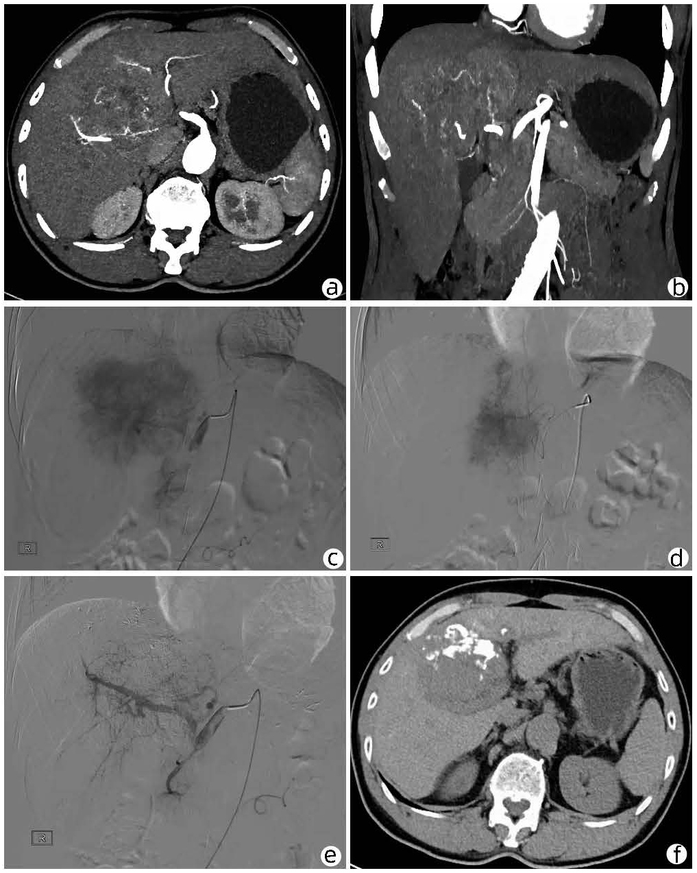
图 1 典型患者影像学检查情况
注:a,术前椎型CT动脉期显示供血动脉与肿瘤结构关系;b,肿瘤实质期显示,肿瘤实质轻度强化;c,术前DSA显示肿瘤供血动脉及肿块染色;d,术中栓塞,碘油沉积明显;e,术后碘油栓塞治疗后,肿瘤微循环血管大部分消失;f,术后CT示碘油沉积。
Figure 1. Typical patient imaging findings
表 1 两组患者临床资料比较
Table 1. Comparison of clinical data of two groups of patients
临床资料 观察组(n=38) 对照组(n=35) 统计值 P值 性别(例) χ2=0.434 0.510 男 21 22 女 17 13 年龄(岁) 56.28±9.22 57.10±10.52 t=0.355 0.724 BCLC分期(例) χ2=0.096 0.757 B期 27 26 C期 11 9 Child-Pugh分级(例) χ2=1.051 0.305 A级 15 18 B级 23 17  下载: 导出CSV
表 2 两组患者手术时间、X线曝光量以及造影剂用量比较
Table 2. Comparison of operation time, X-ray exposure and contrast agent consumption between the two groups
组别 例数 手术时间(min) X线曝光量(mGy) 造影剂用量(mL) 观察组 38 30.47±6.29 879.28±110.94 57.28±6.44 对照组 35 29.38±7.22 860.33±102.32 55.20±7.21 t值 0.689 0.757 1.302 P值 0.493 0.452 0.197
下载: 导出CSV
表 3 两组患者二维X线透视与平扫DynaCT造影评估肿瘤病灶内碘化油沉积对比
Table 3. Comparison of two-dimensional X-ray fluoroscopy and plain DynaCT angiography in evaluating the deposition of lipiodol in tumor lesions
检查方式 碘油沉积类型 Ⅰ型 Ⅱ型 Ⅲ型 Ⅳ型 二维X线透视(个) 89 9 4 11 平扫DynaCT造影(个) 78 15 13 7
下载: 导出CSV
-
[1] SIA D, VILLANUEVA A, FRIEDMAN SL, et al. Liver cancer cell of origin, molecular class, and effects on patient prognosis[J]. Gastroenterology, 2017, 152(4): 745-761. DOI: 10.1053/j.gastro.2016.11.048. [2] GAO YX, YANG TW, YIN JM, et al. Progress and prospects of biomarkers in primary liver cancer (Review)[J]. Int J Oncol, 2020, 57(1): 54-66. DOI: 10.3892/ijo.2020.5035. [3] LI CH, LUO HY, WANG JJ, et al. The value of using contrast-enhanced ultrasound in combination with blood γ-GT level to evaluate the efficacy of transcatheter arterial chemoembolization therapy for patients with primary hepatic carcinoma[J]. Chin Hepatol, 2021, 26(3): 266-269. DOI: 10.3969/j.issn.1008-1704.2021.03.014.李丛辉, 罗红缨, 王建钧, 等. 超声造影联合血γ-GT水平在原发性肝癌患者TACE术后疗效评估中的价值[J]. 肝脏, 2021, 26(3): 266-269. DOI: 10.3969/j.issn.1008-1704.2021.03.014. [4] ZHAO RG, KANG JB, ZHU Q. Curative effect analysis of TACE combined with SRT in the treatment of unresectable primary liver cancer[J]. China Med Equip, 2021, 18(1): 71-74. DOI: 10.3969/J.ISSN.1672-8270.2021.01.018.赵儒钢, 康静波, 朱奇. 经导管肝动脉化疗栓塞联合立体定向放射治疗对不可切除原发性肝癌的疗效分析[J]. 中国医学装备, 2021, 18(1): 71-74. DOI: 10.3969/J.ISSN.1672-8270.2021.01.018. [5] WEI JT. Efficacy and safety of intracavitary catheter radiofrequency ablation combined with TACE in portal vein tumor thrombus in patients with primary liver cancer[J]. J Hepatobiliary Surg, 2021, 29(2): 132-135. DOI: 10.3969/j.issn.1006-4761.2021.02.015.魏健体. 腔内导管射频消融联合TACE对原发性肝癌患者门静脉癌栓的疗效及安全性分析[J]. 肝胆外科杂志, 2021, 29(2): 132-135. DOI: 10.3969/j.issn.1006-4761.2021.02.015. [6] HUANG SS, ZHANG W, XIE ZP, et al. Diagnostic effect of contrast-enhanced ultrasound combined with microvascular imaging and Gd-EOB-DTPA-enhanced MRI for hepatocellular carcinoma recurred after TACE[J]. Prog Mod Biomed, 2021, 21(17): 3289-3294. DOI: 10.13241/j.cnki.pmb.2021.17.020.黄珊珊, 张维, 谢昭鹏, 等. 超声造影联合微血管成像技术与钆塞酸二钠增强MRI评价原发性肝癌TACE术后复发的诊断效能对照分析[J]. 现代生物医学进展, 2021, 21(17): 3289-3294. DOI: 10.13241/j.cnki.pmb.2021.17.020. [7] PEISEN F, MAURER M, GROSSE U, et al. Intraprocedural cone-beam CT with parenchymal blood volume assessment for transarterial chemoembolization guidance: Impact on the effectiveness of the individual TACE sessions compared to DSA guidance alone[J]. Eur J Radiol, 2021, 140: 109768. DOI: 10.1016/j.ejrad.2021.109768. [8] YAO H, LIANG CR, HE XL, et al. The value of cone beam computed tomography in the transcatheter arterial chemoembolization therapy for primary and metastatic liver cancers[J]. Chin Hepatol, 2020, 25(9): 926-929. DOI: 10.3969/j.issn.1008-1704.2020.09.010.姚欢, 梁春蕊, 何响玲, 等. 锥形束CT用于肝转移瘤患者TACE术的价值观察[J]. 肝脏, 2020, 25(9): 926-929. DOI: 10.3969/j.issn.1008-1704.2020.09.010. [9] LI Z, JIAO D, HAN X, et al. Transcatheter arterial chemoembolization combined with simultaneous DynaCT-guided microwave ablation in the treatment of small hepatocellular carcinoma[J]. Cancer Imaging, 2020, 20(1): 13. DOI: 10.1186/s40644-020-0294-5. [10] YANG J, YIN Y, NI CF, et al. Value of ABCR scoring system in assessing the prognosis of hepatocellular carcinoma after transcatheter arterial che-moembolization[J]. J Clin Hepatol, 2020, 36(9): 1980-1984. DOI: 10.3969/j.issn.1001-5256.2020.09.014.杨俊, 印于, 倪才方, 等. ABCR评分系统对经肝动脉化疗栓塞术治疗肝细胞癌预后的评估价值[J]. 临床肝胆病杂志, 2020, 36(9): 1980-1984. DOI: 10.3969/j.issn.1001-5256.2020.09.014. [11] LI QG, TANG Y, LONG Y. Clinical observation on application of raltitrexed combined with cisplatin in transcatheter arterial chemoem-bolization for primary liver cancer[J]. Med Pharm J Chin PLA, 2021, 33(2): 29-32. DOI: 10.3969/j.issn.2095-140X.2021.02.007.李清桂, 唐瑛, 龙禹. 雷替曲塞联合顺铂在原发性肝癌肝动脉化疗栓塞术中应用的临床观察[J]. 解放军医药杂志, 2021, 33(2): 29-32. DOI: 10.3969/j.issn.2095-140X.2021.02.007. [12] RAOUL JL, FORNER A, BOLONDI L, et al. Updated use of TACE for hepatocellular carcinoma treatment: How and when to use it based on clinical evidence[J]. Cancer Treat Rev, 2019, 72: 28-36. DOI: 10.1016/j.ctrv.2018.11.002. [13] GALLE PR, TOVOLI F, FOERSTER F, et al. The treatment of intermediate stage tumours beyond TACE: From surgery to systemic therapy[J]. J Hepatol, 2017, 67(1): 173-183. DOI: 10.1016/j.jhep.2017.03.007. [14] DAI CM, JIN S, ZHANG JZ. Effect of Dahuang Zhechong Pills combined with TACE on VEGF, MMP-2, TGF-β1 and immune function of patients with primary liver cancer (blood stasis and collaterals blocking type)[J]. China J Chin Mater Med, 2021, 46(3): 722-729. DOI: 10.19540/j.cnki.cjcmm.20200716.501.戴朝明, 靳松, 张济周. 大黄蛰虫丸联合TACE术对原发性肝癌患者(瘀血阻络型)VEGF, MMP-2, TGF-β1及免疫功能的影响[J]. 中国中药杂志, 2021, 46(3): 722-729. DOI: 10.19540/j.cnki.cjcmm.20200716.501. [15] GONG SH, LI SD, QIN X, et al. Evaluation value of liver specific contrast agent MRI and contrast-enhanced ultrasound in the treatment of hepatocellular carcinoma after TACE[J]. J Pract Radiol, 2021, 37(2): 309-312. DOI: 10.3969/j.issn.1002-1671.2021.02.033.龚姝卉, 李绍东, 秦响, 等. 普美显MRI与超声造影对肝细胞癌经肝动脉化疗栓塞治疗后的疗效评估价值[J]. 实用放射学杂志, 2021, 37(2): 309-312. DOI: 10.3969/j.issn.1002-1671.2021.02.033. [16] YUAN H, LIU F, LI X, et al. Transcatheter arterial chemoembolization combined with simultaneous DynaCT-guided radiofrequency ablation in the treatment of solitary large hepatocellular carcinoma[J]. Radiol Med, 2019, 124(1): 1-7. DOI: 10.1007/s11547-018-0932-1. [17] YANG L, GU YM, XU H, et al. Contrast-enhanced ultrasound and MRI in post-treatment evaluation of hepatocellular carcinoma after TACE[J]. Chin J Hepatobiliary Surg, 2020, 26(9): 683-686. DOI: 10.3760/cma.j.cn113884-20191207-00401.杨亮, 顾玉明, 徐浩, 等. 超声造影与增强MRI在肝细胞癌TACE疗效评估中的应用价值[J]. 中华肝胆外科杂志, 2020, 26(9): 683-686. DOI: 10.3760/cma.j.cn113884-20191207-00401. [18] YANG L, GU YM, LU J, et al. Contrast-enhanced ultrasonography versus contrast-enhanced MRI in the evaluation of therapeutic effect of TACE for HCC: Comparison of application value[J]. J Intervent Radiol, 2019, 28(7): 682-686. DOI: 10.3969/j.issn.1008-794X.2019.07.015.杨亮, 顾玉明, 鹿皎, 等. 超声造影与增强MRI在评价肝癌TACE术后疗效的应用比较[J]. 介入放射学杂志, 2019, 28(7): 682-686. DOI: 10.3969/j.issn.1008-794X.2019.07.015. [19] LIU M, XU M, LI XJ, et al. Hepatocellular carcinoma treated with transcatheter arterial chemoembolization: influence factors of local efficacy analyzed by parametric contrast enhanced ultrasound[J]. J Chin Physician, 2019, 21(8): 1129-1132, 1135. DOI: 10.3760/cma.j.issn.1008-1372.2019.08.003.刘明, 徐明, 李晓菊, 等. 超声造影定量分析评价HCC行TACE术后局部疗效的影响因素[J]. 中国医师杂志, 2019, 21(8): 1129-1132, 1135. DOI: 10.3760/cma.j.issn.1008-1372.2019.08.003. [20] HU JG, WANG XD, ZHU X, et al. Evaluation of cone-beam CT hepatic angiography in detecting the tumor-feeding arteries during the performance of TACE for HCC[J]. J Intervent Radiol, 2015, 6: 481-487. DOI: 10.3969/j.issn.1008-794X.2015.06.005.胡俊刚, 王晓东, 朱旭, 等. 锥体束CT三维肝动脉造影在TACE术中判断原发性肝癌肿瘤供血动脉的临床价值[J]. 介入放射学杂志, 2015, 6: 481-487. DOI: 10.3969/j.issn.1008-794X.2015.06.005. [21] WANG HJ, CHENG RH, WANG CH, et al. Application of contrast-enhanced ultrasonography combined with microwave ablation in the evaluation of hepatocellular carcinoma effect of transcatheter arterial chemoembolization[J]. J Beihua Univ (Natural Science), 2017, 18(5): 645-648. DOI: 10.11713/j.issn.1009-4822.2017.05.019.王海军, 程瑞洪, 王朝晖, 等. 超声造影在肝动脉化疗栓塞术联合微波消融治疗肝癌效果评估中的应用[J]. 北华大学学报(自然科学版), 2017, 18(5): 645-648. DOI:10.11713/j.issn. 1009-4822.2017.05.019. -

 本文二维码
本文二维码
计量
- 文章访问数: 352
- HTML全文浏览量: 103
- PDF下载量: 44
- 被引次数: 0

