
 PDF下载 ( 2023 KB)

东西方国家急性坏死性胰腺炎影响因素的Meta分析
DOI: 10.3969/j.issn.1001-5256.2023.07.019
Influencing factors for acute necrotizing pancreatitis in Eastern and Western countries: A meta-analysis
-
摘要:
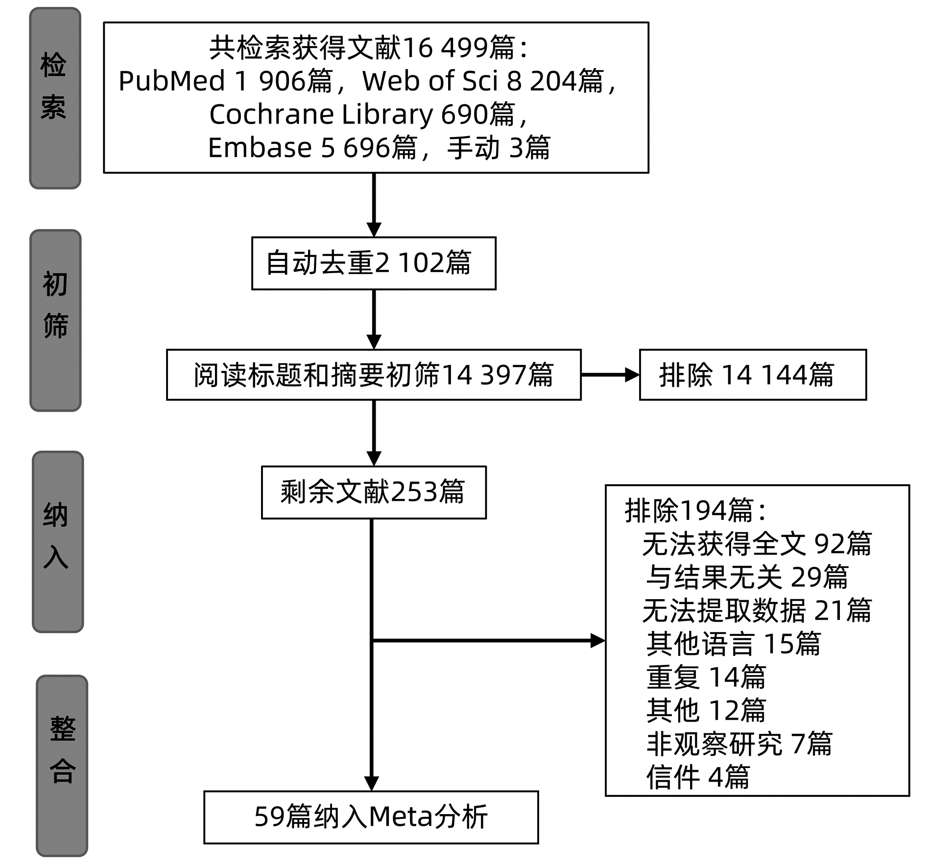
目的 探讨东西方国家急性坏死性胰腺炎(ANP)及感染性坏死性急性胰腺炎(IPN)影响因素的区别, 为预测和预防ANP的发生提供理论依据。 方法 在PubMed、Embase、Cochrane library、Web of Science等数据库检索公开发表的有关ANP、IPN的影响因素, 检索期限为建库至2021年1月21日, 运用Meta分析方法进行整合分析。 结果 共纳入59项研究, 其中22项来自东方国家, 37项来自西方国家。 结果 显示, ANP的影响因素: 在东方为男性(OR=1.51, 95%CI: 1.18~1.91, P < 0.01)、CRP (SMD=1.39, 95%CI: 1.06~1.71, P < 0.01)、D-二聚体(SMD=0.44, 95%CI: 0.07~0.81, P=0.02)、APACHE-Ⅱ评分(MD=3.51, 95%CI: 1.38~5.64, P < 0.01)、酒精性病因(OR=3.57, 95%CI: 2.68~4.75, P < 0.01)、胆源性病因(OR=0.60, 95%CI: 0.46~0.77, P < 0.01);在西方为男性(OR=1.63, 95%CI: 1.30~2.05, P < 0.01)、CRP (SMD=2.09, 95%CI: 1.12~3.05, P < 0.01)、APACHE-Ⅱ评分(MD=4.28, 95%CI: 2.73~5.83, P < 0.01)、Ranson评分(MD=2.99, 95%CI: 2.50~3.47, P < 0.01)和器官衰竭(OR=10.87, 95%CI: 2.62~45.04, P < 0.01)。IPN的影响因素: 在东方为年龄(MD=2.16, 95%CI: 0.43~3.89, P=0.01)、BMI (MD=1.74, 95%CI: 1.23~2.25, P < 0.01)、白蛋白水平(SMD=-0.43, 95%CI: -0.75~-0.12, P < 0.01)、CRP (SMD=0.58, 95%CI: 0.04~1.11, P=0.03)、PCT (SMD=0.80, 95%CI: 0.56~1.04, P < 0.01)、D-二聚体(MD=0.23, 95%CI: 0.15~0.31, P < 0.01)、APACHE-Ⅱ评分(MD=2.47, 95%CI: 0.73~4.22, P < 0.01)、Ranson评分(MD=1.60, 95%CI: 1.46~1.73, P<0.01)和坏死范围≥30%(OR=2.52, 95%CI: 1.26~5.06, P < 0.01);在西方为年龄(MD=4.07, 95%CI: 1.82~6.31, P < 0.01)、APACHE-Ⅱ评分(MD=3.28, 95%CI: 1.39~5.17, P < 0.01)、Ranson评分(MD=2.18, 95%CI: 1.75~2.62, P < 0.01)、SIRS评分(OR=3.88, 95%CI: 1.58~9.51, P < 0.01)、酒精性病因(OR=0.61, 95%CI: 0.42~0.87, P < 0.01)和器官衰竭(OR=3.63, 95%CI: 1.11~11.92, P=0.03)。 结论 当前证据显示, 东方人群ANP的特异性影响因素为胆源性病因及酒精性病因, 而Ranson评分是西方人群ANP的特异性影响因素; BMI和坏死范围≥30%是东方人群IPN特异性影响因素, 酒精性病因是西方人群IPN特异性影响因素。 -
关键词:
- 胰腺炎, 急性坏死性 /
- 影响因素 /
- Meta分析
Abstract:Objective To investigate the differences in the influencing factors for acute necrotizing pancreatitis (ANP) and infectious pancreatic necrosis (IPN) between Eastern and Western countries, and to provide a theoretical basis for the prediction and prevention of ANP. Methods Databases including PubMed, Embase, the Cochrane Library, and Web of Science were searched for articles on the influencing factors for ANP and IPN published up to January 21, 2021, and a Meta-analysis was performed. Results A total of 59 studies were included, with 22 studies from Eastern countries and 37 studies from Western countries.The Meta-analysis showed that in Eastern countries, male sex (odds ratio[OR]=1.51, 95% confidence interval[CI]: 1.18-1.91, P < 0.01), C-reactive protein (CRP)(standardized mean difference[SMD]=1.39, 95%CI: 1.06-1.71, P < 0.01), D-dimer (SMD=0.44, 95%CI: 0.07-0.81, P=0.02), Acute Physiology and Chronic Health Evaluation Ⅱ (APACHE-Ⅱ) score (mean difference[MD]=3.51, 95%CI: 1.38-5.64, P < 0.01), alcoholic etiology (OR=3.57, 95%CI: 2.68-4.75, P < 0.01), and biliary etiology (OR=0.60, 95%CI: 0.46-0.77, P < 0.01) were the influencing factors for ANP, and in Western countries, male sex (OR=1.63, 95%CI: 1.30-2.05, P < 0.01), CRP (SMD=2.09, 95%CI: 1.12-3.05, P < 0.01), APACHE-Ⅱ score (MD=4.28, 95%CI: 2.73-5.83, P < 0.01), Ranson score (MD=2.99, 95%CI: 2.50-3.47, P < 0.01), and organ failure (OR=10.87, 95%CI: 2.62-45.04, P < 0.01) were the influencing factors for ANP.In Eastern countries, age (MD=2.16, 95%CI: 0.43-3.89, P=0.01), body mass index (BMI)(MD=1.74, 95%CI: 1.23-2.25, P < 0.01), albumin level (SMD=-0.43, 95%CI: -0.75 to-0.12, P < 0.01), CRP (SMD=0.58, 95%CI: 0.04-1.11, P=0.03), procalcitonin (SMD=0.80, 95%CI: 0.56-1.04, P < 0.01), D-dimer (MD=0.23, 95%CI: 0.15-0.31, P < 0.01), APACHE-Ⅱ score (MD=2.47, 95%CI: 0.73-4.22, P < 0.01), Ranson score (MD=1.60, 95%CI: 1.46-1.73, P < 0.01), and extent of necrosis ≥30%(OR=2.52, 95%CI: 1.26-5.06, P < 0.01) were the influencing factors for IPN, while in Western countries, age (MD=4.07, 95%CI: 1.82-6.31, P < 0.01), APACHE-Ⅱ score (MD=3.28, 95%CI: 1.39-5.17, P < 0.01), Ranson score (MD=2.18, 95%CI: 1.75-2.62, P < 0.01), SIRS score (OR=3.88, 95%CI: 1.58-9.51, P < 0.01), alcoholic etiology (OR=0.61, 95%CI: 0.42-0.87, P < 0.01), and organ failure (OR=3.63, 95%CI: 1.11-11.92, P=0.03) were the influencing factors for IPN. Conclusion Current evidence shows that biliary etiology and alcoholic etiology are unique influencing factors for ANP in the Eastern population, while Ranson score is a unique influencing factor in the Western population.BMI and extent of necrosis ≥30% are unique influencing factors for IPN in the Eastern population, while alcoholic etiology is a unique influencing factor in the Western population. -
Key words:
- Pancreatitis, Acute Necrotizing /
- Influencing Factors /
- Meta-analysis
-
表 1 纳入文献的基本特征
Table 1. Basic characteristics of included studies
第一作者 年份 国家 研究类型 例数 观察比较 影响因素 质量评分 西方 Büchler M[13] 1986 德国 队列研究 35 ANP vs non-ANP 年龄、性别、酒精性病因、胆源性病因 5 Büchler M[14] 1989 德国 队列研究 48 ANP vs non-ANP Ranson评分 5 Domschke S[15] 1993 德国 病例对照研究 40 ANP vs non-ANP CRP、白蛋白 5 Kaufmann P[16] 1996 奥地利 队列研究 25 ANP vs non-ANP 年龄、性别、CRP、酒精性病因、胆源性病因 7 Schmid SW[17] 1996 德国 队列研究 40 ANP vs non-ANP Ranson评分、年龄、性别 6 Manes G[18] 1997 德国 队列研究 36 ANP vs non-ANP CRP 5 Rau B[19] 1997 德国 队列研究 50 ANP vs non-ANP/IPN vs non-IPN APACHE-Ⅱ评分、Ranson评分、年龄、性别、酒精性病因、胆源性病因 6 Tenner S[20] 1997 美国 病例对照研究 51 IPN vs non-IPN 器官衰竭 6 Baillargeon JD[21] 1998 美国 病例对照研究 64 ANP vs non-ANP HCT、APACHE-Ⅱ评分、年龄、性别、酒精性病因、胆源性病因 7 Rau B[22] 1998 德国 队列研究 70 ANP vs non-ANP APACHE-Ⅱ评分、Ranson评分、年龄、性别 6 Hietaranta A[23] 1999 芬兰 队列研究 57 IPN vs non-IPN SIRS 7 Brown A[24] 2000 美国 队列研究 53 ANP vs non-ANP HCT、年龄、性别、器官衰竭 6 Buttenschoen K[25] 2000 德国 队列研究 34 ANP vs non-ANP Ranson评分、性别 5 Mándi Y[26] 2000 匈牙利 队列研究 20 IPN vs non-IPN APACHE-Ⅱ评分、Ranson评分、年龄、性别、酒精性病因、SIRS、胆源性病因 6 Müller CA[27] 2000 瑞士 队列研究 64 ANP vs non-ANP/IPN vs non-IPN APACHE-Ⅱ评分、Ranson评分、年龄、性别、坏死范围≥30%、酒精性病因、器官衰竭、胆源性病因 6 Rau B[28] 2000 德国 队列研究 66 ANP vs non-ANP APACHE-Ⅱ评分、Ranson评分、年龄、性别、酒精性病因、胆源性病因 5 Rau B[29] 2000 德国 队列研究 61 ANP vs non-ANP/IPN vs non-IPN APACHE-Ⅱ评分、Ranson评分、年龄、性别、酒精性病因、胆源性病因 6 Khan AA[30] 2002 美国 病例对照研究 93 ANP vs non-ANP APACHE-Ⅱ评分 7 Müller CA[31] 2002 瑞典 队列研究 85 ANP vs non-ANP Ranson评分、年龄、性别、酒精性病因、胆源性病因 6 Perez A[32] 2002 美国 病例对照研究 99 ANP vs non-ANP APACHE-Ⅱ评分、器官衰竭 7 Riché FC[33] 2003 法国 队列研究 48 IPN vs non-IPN Ranson评分、年龄、性别、酒精性病因、胆源性病因 7 Barauskas G[34] 2004 立陶宛 病例对照研究 78 ANP vs non-ANP 性别、CRP、酒精性病因、胆源性病因 7 Papachristou GI[35] 2006 美国 病例对照研究/队列研究 436 ANP vs non-ANP BMI、HCT、年龄、性别、吸烟、胆源性病因 6 Dambrauskas Z[36] 2007 立陶宛 队列研究 52 IPN vs non-IPN 年龄、性别、坏死范围≥30%、酒精性病因、胆源性病因 9 López A[37] 2007 西班牙 队列研究 84 ANP vs non-ANP 年龄、性别、酒精性病因、胆源性病因 7 Muller CA[38] 2007 德国 队列研究 109 ANP vs non-ANP/IPN vs non-IPN APACHE-Ⅱ评分、Ranson评分、年龄、性别、CRP、酒精性病因、胆源性病因 6 Rahman SH[39] 2007 英国 队列研究 64 ANP vs non-ANP APACHE-Ⅱ评分、CRP 7 De Campos T[40] 2008 巴西 病例对照研究 39 ANP vs non-ANP HCT、APACHE-Ⅱ评分、年龄、性别、CRP、酒精性病因、改良版Marshall评分、SOFA评分 6 Muddana V[41] 2009 美国 队列研究 129 ANP vs non-ANP BMI、APACHE-Ⅱ评分、Ranson评分、年龄、性别、酒精性病因、器官衰竭、胆源性病因、CTSI 7 Lipinski M[42] 2013 波兰 病例对照研究 103 ANP vs non-ANP Cre 7 Talukdar R[43] 2013 美国 队列研究 281 IPN vs non-IPN BMI、HCT、年龄、性别、BUN、Cre、SIRS 8 Kostic I[44] 2015 塞尔维亚 队列研究 52 ANP vs non-ANP 年龄、性别 7 Easler JJ[45] 2016 美国 队列研究 423 ANP vs non-ANP 酒精性病因 6 Moran RA[46] 2016 美国 队列研究 142 IPN vs non-IPN 年龄、性别、酒精性病因、器官衰竭、EEN、胆源性病因 6 Garret C[47] 2018 法国 队列研究 132 IPN vs non-IPN BMI、年龄、性别、酒精性病因、合并糖尿病、器官衰竭、吸烟、胆源性病因、CTSI 5 Verdonk RC[48] 2018 德国 队列研究 285 ANP vs non-ANP 酒精性病因、胆源性病因 6 Szakacs Z[49] 2019 匈牙利 队列研究 1 198 ANP vs non-ANP 年龄 7 东方 Isogai M[50] 1998 日本 病例对照研究 22 ANP vs non-ANP 淀粉酶、年龄、性别、AST 6 Mettu SR[51] 2003 印度 病例对照研究 40 IPN vs non-IPN APACHE-Ⅱ评分、MCTSI 6 Garg PK[52] 2005 印度 队列研究 104 IPN vs non-IPN 坏死范围≥30% 7 Gardner TB[53] 2006 黎巴嫩 病例对照研究 230 ANP vs non-ANP 性别、酒精性病因、胆源性病因 7 Ueda T[54] 2007 日本 队列研究 75 IPN vs non-IPN APACHE-Ⅱ评分、Ranson评分 6 Guo J[55] 2012 中国 队列研究 35 ANP vs non-ANP APACHE-Ⅱ评分、Ranson评分、年龄、性别、胆源性病因 7 Ke L[56] 2012 中国 队列研究 45 ANP vs non-ANP D-二聚体 7 Lu Z[57] 2012 中国 队列研究 30 IPN vs non-IPN APACHE-Ⅱ评分、Ranson评分、淀粉酶、年龄、性别、CRP、合并糖尿病、WBC、PCT、坏死范围≥30%、酒精性病因、胆源性病因、CTSI 7 Guo Q[58] 2014 中国 队列研究 447 IPN vs non-IPN BMI、年龄、性别、坏死范围≥30%、MCTSI、酒精性病因、胆源性病因 6 Erbis H[59] 2015 土耳其 病例对照研究 76 ANP vs non-ANP 年龄、性别、CRP、WBC 6 Shen X[60] 2015 中国 队列研究 153 IPN vs non-IPN BMI、HCT、APACHE-Ⅱ评分、淀粉酶、年龄、性别、CRP、白蛋白、WBC、酒精性病因、胆源性病因、CTSI 8 Thandassery RB[61] 2015 印度 队列研究 81 IPN vs non-IPN 器官衰竭 6 Ji L[62] 2016 中国 队列研究 115 IPN vs non-IPN BMI、HCT、APACHE-Ⅱ评分、Imrie评分、年龄、性别、CRP、BUN、WBC、酒精性病因、PLT、Cre、D-二聚体、EEN、胆源性病因、改良版Marshall评分 7 Chen HZ[63] 2017 中国 队列研究 215 IPN vs non-IPN HCT、APACHE-Ⅱ评分、Imrie评分、性别、CRP、BUN、WBC、坏死范围≥30%、MCTSI、酒精性病因、器官衰竭、PLT、Cre、D-二聚体、胆源性病因、改良版Marshall评分 8 Chen Y[64] 2017 中国 队列研究 57 ANP vs non-ANP/IPN vs non-IPN SAP、APACHE-Ⅱ评分、CRP、PCT、D-二聚体、SOFA评分 9 Zhang Y[65] 2017 中国 病例对照研究 1 131 ANP vs non-ANP Ranson评分、年龄、性别、WBC、酒精性病因、Cre、吸烟、AST、胆源性病因 8 Cao X[66] 2018 中国 病例对照研究 111 IPN vs non-IPN BMI、HCT、年龄、性别、CRP、合并糖尿病、白蛋白、BUN、WBC、PCT、坏死范围≥30%、MCTSI 8 Noda Y[67] 2018 日本 病例对照研究 56 ANP vs non-ANP BMI、年龄、性别 7 Tan C[1] 2020 中国 队列研究 2 130 IPN vs non-IPN SIRS 7 Ding L[4] 2019 中国 队列研究 142 IPN vs non-IPN 年龄、性别、坏死范围≥30%、酒精性病因、胆源性病因 9 Ünal Y[68] 2019 土耳其 病例对照研究 96 ANP vs non-ANP 淀粉酶、性别、CRP、酒精性病因、胆源性病因 6 Wan J[69] 2019 中国 队列研究 2 478 ANP vs non-ANP/IPN vs non-IPN SAP、D-二聚体 5 注:APACHE-Ⅱ,急性生理和慢性健康评估Ⅱ;BUN,血尿素氮;Cre,肌酐;CRP,C反应蛋白;CTSI,CT严重指数;EEN,早期肠内营养;HCT,红细胞压积;MCTSI,改良CT严重指数;PLT,血小板;SAP,重度急性胰腺炎;SIRS,全身炎症反应综合征;SOFA,序贯器官衰竭评估;PCT,降钙素原。  下载: 导出CSV
表 2 西方ANP的影响因素
Table 2. Influencing factors for ANP in the West
影响因素 研究个数 OR/MD/SMD(95%CI) P值 I2值(%) P值1) 年龄 18[13, 16-17, 19, 21-22, 24, 27-29, 31, 35, 37-38, 40-41, 44, 49] -1.91(-4.84~1.03) 0.20 73 <0.01 男性 19[13, 16-17, 19, 21-22, 24-25, 27-29, 31, 34-35, 37-38, 40-41, 44] 1.63(1.30~2.05) <0.01 19 0.21 BMI 2[35, 41] 1.58(-3.52~6.67) 0.54 90 <0.01 HCT 4[21, 24, 35, 40] 3.24(-0.26~6.74) 0.07 87 <0.01 CRP 7[15-16, 18, 34, 38-40] 2.09(1.12~3.05) <0.01 91 <0.01 APACHE-Ⅱ评分 10[19, 21-22, 28-30, 32, 39-41] 4.28(2.73~5.83) <0.01 77 <0.01 Ranson评分 11[14, 17, 19, 22, 25, 27-29, 31, 38, 41] 2.99(2.50~3.47) <0.01 66 <0.01 胆源性病因 14[13, 16, 19, 21, 27-29, 31, 34-35, 37-38, 41, 48] 0.84(0.66~1.08) 0.17 49 0.02 酒精性病因 15[13, 16, 19, 21, 27-29, 31, 34, 37-38, 40-41, 45, 48] 1.49(1.00~2.24) 0.05 57 <0.01 器官衰竭 4[24, 27, 32, 41] 10.87(2.62~45.04) <0.01 83 <0.01 注:1)为异质性检验的P值。
下载: 导出CSV
表 3 东方ANP的影响因素
Table 3. Influencing factors for ANP in the East
影响因素 研究个数 OR/MD/SMD (95%CI) P值 I2值(%) P值1) 年龄 5[50, 55, 59, 65, 67] 1.21(-3.54~5.97) 0.62 57 0.06 男性 7[50, 53, 55, 59, 65, 67-68] 1.51(1.18~1.91) <0.01 0 0.47 WBC 2[59, 65] 1.95(-0.23~4.12) 0.08 69 0.07 AST 2[50, 65] 0.09(-0.04~0.22) 0.16 0 0.37 淀粉酶 2[50, 68] 0.11(-0.73~0.96) 0.79 56 0.13 CRP 3[59, 64, 68] 1.39(1.06~1.71) <0.01 0 0.95 D-二聚体 3[56, 64, 69] 0.44(0.07~0.81) 0.02 53 0.12 APACHE-Ⅱ评分 2[55, 64] 3.51(1.38~5.64) <0.01 1 0.32 Ranson评分 2[55, 64] 1.08(-0.59~2.75) 0.21 54 0.03 胆源性病因 3[55, 65, 68] 0.60(0.46~0.77) <0.01 9 0.33 酒精性病因 2[65, 68] 3.57(2.68~4.75) <0.01 9 0.30 注:1)为异质性检验的P值。
下载: 导出CSV
表 4 西方IPN的影响因素
Table 4. Influencing factors for IPN in the West
影响因素 研究个数 OR/MD/SMD(95%CI) P值 I2值(%) P值1) 年龄 10[19, 26-27, 29, 33, 36, 38, 43, 46-47] 4.07(1.82~6.31) <0.01 44 0.06 男性 10[19, 26-27, 29, 33, 36, 38, 43, 46-47] 1.31(0.92~1.88) 0.13 0 0.81 BMI 2[43, 47] 0.86(-0.69~2.41) 0.27 0 0.39 APACHE-Ⅱ评分 5[19, 26-27, 29, 38] 3.28(1.39~5.17) <0.01 55 0.07 Ranson评分 6[19, 26-27, 29, 33, 38] 2.18(1.75~2.62) <0.01 42 0.12 SIRS 5[19, 23, 26, 29, 43] 3.88(1.58~9.51) <0.01 0 0.77 胆源性病因 9[19, 26-27, 29, 33, 36, 38, 46-47] 1.30(0.90~1.89) 0.16 0 0.81 酒精性病因 9[19, 26-27, 29, 33, 36, 38, 46-47] 0.61(0.42~0.87) <0.01 43 0.08 器官衰竭 4[20, 27, 46-47] 3.63(1.11~11.92) 0.03 76 <0.01 坏死范围≥30% 2[27, 36] 9.36(0.69~127.00) 0.09 59 0.12 注:1)为异质性检验的P值。
下载: 导出CSV
表 5 东方IPN的影响因素
Table 5. Influencing factors for IPN in the East
影响因素 研究个数 OR/MD/SMD (95%CI) P值 I2值(%) P值1) 年龄 6[4, 57-58, 60, 62, 66] 2.16(0.43~3.89) 0.01 35 0.17 男性 7[4, 57-58, 60, 62-63, 66] 1.16(0.90~1.51) 0.26 9 0.36 BMI 4[58, 60, 62, 66] 1.74(1.23~2.25) <0.01 0 0.56 HCT 4[60, 62-63, 66] 2.30(2.02~6.23) 0.30 96 <0.01 WBC 6[57-58, 60, 62-63, 66] 0.50(-0.35~1.35) 0.25 51 0.07 PLT 3[57, 63, 66] -0.13(-0.61~0.35) 0.60 82 <0.01 白蛋白 2[60, 66] -0.43(-0.75~-0.12) <0.01 0 0.65 BUN 3[62-63, 66] -0.34(-1.28~0.59) 0.47 94 <0.01 Cre 2[62-63] 0.20(-0.13~0.53) 0.23 50 0.16 淀粉酶 2[57, 60] 0.03(-0.32~0.38) 0.87 0 0.64 CRP 5[57, 60, 62-63, 66] 0.58(0.04~1.11) 0.03 87 <0.01 PCT 3[57, 63, 66] 0.80(0.56~1.04) <0.01 0 0.89 D-二聚体 2[62-63] 0.23(0.15~0.31) <0.01 0 0.90 APACHE-Ⅱ评分 6[51, 54, 57, 60, 62-63] 2.47(0.73~4.22) <0.01 91 <0.01 Ranson评分 2[54, 57] 1.60(1.46~1.73) <0.01 0 0.70 CTSI 3[57, 60, 62] 2.69(-0.97~6.35) 0.15 99 <0.01 MCTSI 4[51, 58, 63, 66] 1.90(-0.39~4.20) 0.10 99 <0.01 Imrie评分 2[62-63] 0(-0.19~0.19) >0.05 0 >0.05 改良版Marshall评分 2[62-63] 0(-0.19~0.19) >0.05 0 >0.05 胆源性病因 6[4, 57-58, 60, 62-63] 0.91(0.70~1.19) 0.50 21 0.27 酒精性病因 6[4, 57-58, 60, 62-63] 0.66(0.35~1.27) 0.21 51 0.07 合并糖尿病 2[57, 66] 0.66(0.20~2.21) 0.50 0 0.99 SAP 3[60, 64, 69] 5.51(0.35~86.42) 0.22 95 <0.01 器官衰竭 2[61, 63] 1.19(0.67~2.10) 0.56 0 0.99 坏死范围≥30% 6[4, 52, 57-58, 63, 66] 2.52(1.26~5.06) <0.01 76 <0.01 注:1)为异质性检验的P值。
下载: 导出CSV
-
[1] TAN C, YANG L, SHI F, et al. Early systemic inflammatory response syndrome duration predicts infected pancreatic necrosis[J]. J Gastrointest Surg, 2020, 24(3): 590-597. DOI: 10.1007/s11605-019-04149-5. [2] MEDEROS MA, REBER HA, GIRGIS MD. Acute pancreatitis:A review[J]. JAMA, 2021, 325 (4):382-390. DOI:10. 1001/JAMA. 2020. 20317 [3] BARON TH, DIMAIO CJ, WANG AY, et al. American Gastroenterological Association Clinical Practice Update: Management of pancreatic necrosis[J]. Gastroenterology, 2020, 158(1): 67-75. e1. DOI: 10.1053/j.gastro.2019.07.064. [4] DING L, YU C, DENG F, et al. New Risk factors for infected pancreatic necrosis secondary to severe acute pancreatitis: The role of initial contrast-enhanced computed tomography[J]. Dig Dis Sci, 2019, 64(2): 553-560. DOI: 10.1007/s10620-018-5359-y. [5] TRIKUDANATHAN G, WOLBRINK D, van SANTVOORT HC, et al. Current concepts in severe acute and necrotizing pancreatitis: An evidence-based approach[J]. Gastroenterology, 2019, 156(7): 1994-2007. e3. DOI: 10.1053/j.gastro.2019.01.269. [6] MAK WY, ZHAO M, NG SC, et al. The epidemiology of inflammatory bowel disease: East meets west[J]. J Gastroenterol Hepatol, 2020, 35(3): 380-389. DOI: 10.1111/jgh.14872. [7] HOZO SP, DJULBEGOVIC B, HOZO I. Estimating the mean and variance from the median, range, and the size of a sample[J]. BMC Med Res Methodol, 2005, 5: 13. DOI: 10.1186/1471-2288-5-13. [8] WAN X, WANG W, LIU J, et al. Estimating the sample mean and standard deviation from the sample size, median, range and/or interquartile range[J]. BMC Med Res Methodol, 2014, 14: 135. DOI: 10.1186/1471-2288-14-135. [9] MAO J, ZHANG Q, ZHANG H, et al. Risk factors for lymph node metastasis in papillary thyroid carcinoma: A systematic review and meta-analysis[J]. Front Endocrinol (Lausanne), 2020, 11: 265. DOI: 10.3389/fendo.2020.00265. [10] JI X, LENG XY, DONG Y, et al. Modifiable risk factors for carotid atherosclerosis: a meta-analysis and systematic review[J]. Ann Transl Med, 2019, 7(22): 632. DOI: 10.21037/atm.2019.10.115. [11] VAN DER VLIST AC, BREDA SJ, OEI E, et al. Clinical risk factors for Achilles tendinopathy: a systematic review[J]. Br J Sports Med, 2019, 53(21): 1352-1361. DOI: 10.1136/bjsports-2018-099991. [12] KUNZE KN, WRIGHT-CHISEM J, POLCE EM, et al. Risk factors for ramp lesions of the medial meniscus: A systematic review and meta-analysis[J]. Am J Sports Med, 2021, 49(13): 3749-3757. DOI: 10.1177/0363546520986817. [13] BVCHLER M, MALFERTHEINER P, SCHOETENSACK C, et al. Sensitivity of antiproteases, complement factors and C-reactive protein in detecting pancreatic necrosis. Results of a prospective clinical study[J]. Int J Pancreatol, 1986, 1(3-4): 227-235. DOI: 10.1007/BF02795248. [14] BVCHLER M, MALFERTHEINER P, SCHÄDLICH H, et al. Prognostic value of serum phospholipase A in acute pancreatitis[J]. Klin Wochenschr, 1989, 67(3): 186-189. DOI: 10.1007/BF01711351. [15] DOMSCHKE S, MALFERTHEINER P, UHL W, et al. Free fatty acids in serum of patients with acute necrotizing or edematous pancreatitis[J]. Int J Pancreatol, 1993, 13(2): 105-110. DOI: 10.1007/BF02786078. [16] KAUFMANN P, TILZ GP, SMOLLE KH, et al. Increased plasma concentrations of circulating intercellular adhesion molecule-1 (cICAM-1) in patients with necrotizing pancreatitis[J]. Immunobiology, 1996, 195(2): 209-219. DOI: 10.1016/S0171-2985(96)80040-4. [17] SCHMID SW, UHL W, STEINLE A, et al. Human pancreas-specific protein. A diagnostic and prognostic marker in acute pancreatitis and pancreas transplantation[J]. Int J Pancreatol, 1996, 19(3): 165-170. DOI: 10.1007/BF02787364. [18] MANES G, SPADA OA, RABITTI PG, et al. Serum interleukin-6 in acute pancreatitis due to common bile duct stones. A reliable marker of necrosis[J]. Recenti Prog Med, 1997, 88(2): 69-72. [19] RAU B, STEINBACH G, GANSAUGE F, et al. The potential role of procalcitonin and interleukin 8 in the prediction of infected necrosis in acute pancreatitis[J]. Gut, 1997, 41(6): 832-840. DOI: 10.1136/gut.41.6.832. [20] TENNER S, SICA G, HUGHES M, et al. Relationship of necrosis to organ failure in severe acute pancreatitis[J]. Gastroenterology, 1997, 113(3): 899-903. DOI: 10.1016/s0016-5085(97)70185-9. [21] BAILLARGEON JD, ORAV J, RAMAGOPAL V, et al. Hemoconcentration as an early risk factor for necrotizing pancreatitis[J]. Am J Gastroenterol, 1998, 93(11): 2130-2134. DOI: 10.1111/j.1572-0241.1998.00608.x. [22] RAU B, CEBULLA M, UHL W, et al. The clinical value of human pancreas-specific protein procarboxypeptidase B as an indicator of necrosis in acute pancreatitis: comparison to CRP and LDH[J]. Pancreas, 1998, 17(2): 134-139. DOI: 10.1097/00006676-199808000-00004. [23] HIETARANTA A, KEMPPAINEN E, PUOLAKKAINEN P, et al. Extracellular phospholipases A2 in relation to systemic inflammatory response syndrome (SIRS) and systemic complications in severe acute pancreatitis[J]. Pancreas, 1999, 18(4): 385-391. DOI: 10.1097/00006676-199905000-00009. [24] BROWN A, ORAV J, BANKS PA. Hemoconcentration is an early marker for organ failure and necrotizing pancreatitis[J]. Pancreas, 2000, 20(4): 367-372. DOI: 10.1097/00006676-200005000-00005. [25] BUTTENSCHOEN K, BERGER D, HIKI N, et al. Endotoxin and antiendotoxin antibodies in patients with acute pancreatitis[J]. Eur J Surg, 2000, 166(6): 459-466. DOI: 10.1080/110241500750008772. [26] MÁNDI Y, FARKAS G, TAKÁCS T, et al. Diagnostic relevance of procalcitonin, IL-6, and sICAM-1 in the prediction of infected necrosis in acute pancreatitis[J]. Int J Pancreatol, 2000, 28(1): 41-49. DOI: 10.1385/IJGC:28:1:41. [27] MVLLER CA, UHL W, PRINTZEN G, et al. Role of procalcitonin and granulocyte colony stimulating factor in the early prediction of infected necrosis in severe acute pancreatitis[J]. Gut, 2000, 46(2): 233-238. DOI: 10.1136/gut.46.2.233. [28] RAU B, STEINBACH G, BAUMGART K, et al. Serum amyloid A versus C-reactive protein in acute pancreatitis: clinical value of an alternative acute-phase reactant[J]. Crit Care Med, 2000, 28(3): 736-742. DOI: 10.1097/00003246-200003000-00022. [29] RAU B, STEINBACH G, BAUMGART K, et al. The clinical value of procalcitonin in the prediction of infected necrosis in acute pancreatitis[J]. Intensive Care Med, 2000, 26 (Suppl 2): S159-164. DOI: 10.1007/BF02900730. [30] KHAN AA, PAREKH D, CHO Y, et al. Improved prediction of outcome in patients with severe acute pancreatitis by the APACHE Ⅱ score at 48 hours after hospital admission compared with the APACHE Ⅱ score at admission. Acute Physiology and Chronic Health Evaluation[J]. Arch Surg, 2002, 137(10): 1136-1140. DOI: 10.1001/archsurg.137.10.1136. [31] MVLLER CA, APPELROS S, UHL W, et al. Serum levels of procarboxypeptidase B and its activation peptide in patients with acute pancreatitis and non-pancreatic diseases[J]. Gut, 2002, 51(2): 229-235. DOI: 10.1136/gut.51.2.229. [32] PEREZ A, WHANG EE, BROOKS DC, et al. Is severity of necrotizing pancreatitis increased in extended necrosis and infected necrosis?[J]. Pancreas, 2002, 25(3): 229- 233. DOI: 10.1097/00006676-200210000-00003. [33] RICHÉ FC, CHOLLEY BP, LAISNÉ MJ, et al. Inflammatory cytokines, C reactive protein, and procalcitonin as early predictors of necrosis infection in acute necrotizing pancreatitis[J]. Surgery, 2003, 133(3): 257-262. DOI: 10.1067/msy.2003.70. [34] BARAUSKAS G, SVAGZDYS S, MALECKAS A. C-reactive protein in early prediction of pancreatic necrosis[J]. Medicina (Kaunas), 2004, 40(2): 135-140. [35] PAPACHRISTOU GI, PAPACHRISTOU DJ, MORINVILLE VD, et al. Chronic alcohol consumption is a major risk factor for pancreatic necrosis in acute pancreatitis[J]. Am J Gastroenterol, 2006, 101(11): 2605-2610. DOI: 10.1111/j.1572-0241.2006.00795.x. [36] DAMBRAUSKAS Z, GULBINAS A, PUNDZIUS J, et al. Value of routine clinical tests in predicting the development of infected pancreatic necrosis in severe acute pancreatitis[J]. Scand J Gastroenterol, 2007, 42(10): 1256-1264. DOI: 10.1080/00365520701391613. [37] LÓPEZ A, de LA CUEVA L, MARTÍNEZ MJ, et al. Usefulness of technetium-99m hexamethylpropylene amine oxime-labeled leukocyte scintigraphy to detect pancreatic necrosis in patients with acute pancreatitis. Prospective comparison with Ranson, Glasgow and APACHE-Ⅱ scores and serum C-reactive protein[J]. Pancreatology, 2007, 7(5-6): 470-478. DOI: 10.1159/000108964. [38] MULLER CA, BELYAEV O, VOGESER M, et al. Corticosteroid-binding globulin: a possible early predictor of infection in acute necrotizing pancreatitis[J]. Scand J Gastroenterol, 2007, 42(11): 1354-1361. DOI: 10.1080/00365520701416691. [39] RAHMAN SH, MENON KV, HOLMFIELD JH, et al. Serum macrophage migration inhibitory factor is an early marker of pancreatic necrosis in acute pancreatitis[J]. Ann Surg, 2007, 245(2): 282-289. DOI: 10.1097/01.sla.0000245471.33987.4b. [40] de CAMPOS T, CERQUEIRA C, KURYURA L, et al. Morbimortality indicators in severe acute pancreatitis[J]. JOP, 2008, 9(6): 690-697. [41] MUDDANA V, WHITCOMB DC, KHALID A, et al. Elevated serum creatinine as a marker of pancreatic necrosis in acute pancreatitis[J]. Am J Gastroenterol, 2009, 104(1): 164-170. DOI: 10.1038/ajg.2008.66. [42] LIPINSKI M, RYDZEWSKI A, RYDZEWSKA G. Early changes in serum creatinine level and estimated glomerular filtration rate predict pancreatic necrosis and mortality in acute pancreatitis: Creatinine and eGFR in acute pancreatitis[J]. Pancreatology, 2013, 13(3): 207-211. DOI: 10.1016/j.pan.2013.02.002. [43] TALUKDAR R, NECHUTOVA H, CLEMENS M, et al. Could rising BUN predict the future development of infected pancreatic necrosis?[J]. Pancreatology, 2013, 13(4): 355-359. DOI: 10.1016/j.pan.2013.05.003. [44] KOSTIC I, SPASIC M, STOJANOVIC B, et al. Early cytokine profile changes in interstitial and necrotic forms of acute pancreatitis[J]. Serb J Exp Clin Res, 2015, 16(1): 33-37. DOI: 10.1515/sjecr-2015-0005. [45] EASLER JJ, DE-MADARIA E, NAWAZ H, et al. Patients with sentinel acute pancreatitis of alcoholic etiology are at risk for organ failure and pancreatic necrosis: A dual-center experience[J]. Pancreas, 2016, 45(7): 997-1002. DOI: 10.1097/MPA.0000000000000643. [46] MORAN RA, JALALY NY, KAMAL A, et al. Ileus is a predictor of local infection in patients with acute necrotizing pancreatitis[J]. Pancreatology, 2016, 16(6): 966-972. DOI: 10.1016/j.pan.2016.10.002. [47] GARRET C, PÉRON M, REIGNIER J, et al. Risk factors and outcomes of infected pancreatic necrosis: Retrospective cohort of 148 patients admitted to the ICU for acute pancreatitis[J]. United European Gastroenterol J, 2018, 6(6): 910-918. DOI: 10.1177/2050640618764049. [48] VERDONK RC, STERNBY H, DIMOVA A, et al. Short article: Presence, extent and location of pancreatic necrosis are independent of aetiology in acute pancreatitis[J]. Eur J Gastroenterol Hepatol, 2018, 30(3): 342-345. DOI: 10.1097/MEG.0000000000001053. [49] SZAKÁCS Z, GEDE N, PÉCSI D, et al. Aging and comorbidities in acute pancreatitis Ⅱ. : A cohort-analysis of 1203 prospectively collected cases[J]. Front Physiol, 2018, 9: 1776. DOI: 10.3389/fphys.2018.01776. [50] ISOGAI M, YAMAGUCHI A, HORI A, et al. LDH to AST ratio in biliary pancreatitis-a possible indicator of pancreatic necrosis: preliminary results[J]. Am J Gastroenterol, 1998, 93(3): 363-367. DOI: 10.1111/j.1572-0241.1998.00363.x. [51] METTU SR, WIG JD, KHULLAR M, et al. Efficacy of serum nitric oxide level estimation in assessing the severity of necrotizing pancreatitis[J]. Pancreatology, 2003, 3(6): 506-513; discussion 513-514. DOI: 10.1159/000076021. [52] GARG PK, MADAN K, PANDE GK, et al. Association of extent and infection of pancreatic necrosis with organ failure and death in acute necrotizing pancreatitis[J]. Clin Gastroenterol Hepatol, 2005, 3(2): 159-166. DOI: 10.1016/s1542-3565(04)00665-2. [53] GARDNER TB, OLENEC CA, CHERTOFF JD, et al. Hemoconcentration and pancreatic necrosis: further defining the relationship[J]. Pancreas, 2006, 33(2): 169-173. DOI: 10.1097/01.mpa.0000226885.32957.17. [54] UEDA T, TAKEYAMA Y, YASUDA T, et al. Lactate dehydrogenase-to-lymphocyte ratio for the prediction of infection in acute necrotizing pancreatitis[J]. Pancreas, 2007, 35(4): 378-380. DOI: 10.1097/01.mpa.0000297827.05678.9e. [55] GUO J, XUE P, YANG XN, et al. Serum matrix metalloproteinase-9 is an early marker of pancreatic necrosis in patients with severe acute pancreatitis[J]. Hepatogastroenterology, 2012, 59(117): 1594-1598. DOI: 10.5754/hge11563. [56] KE L, NI HB, TONG ZH, et al. D-dimer as a marker of severity in patients with severe acute pancreatitis[J]. J Hepatobiliary Pancreat Sci, 2012, 19(3): 259-265. DOI: 10.1007/s00534-011-0414-5. [57] LU Z, LIU Y, DONG YH, et al. Soluble triggering receptor expressed on myeloid cells in severe acute pancreatitis: a biological marker of infected necrosis[J]. Intensive Care Med, 2012, 38(1): 69-75. DOI: 10.1007/s00134-011-2369-z. [58] GUO Q, LI A, XIA Q, et al. The role of organ failure and infection in necrotizing pancreatitis: a prospective study[J]. Ann Surg, 2014, 259(6): 1201-1207. DOI: 10.1097/SLA.0000000000000264. [59] ERBIS H, ALIOSMANOGLU I, TURKOGLU MA, et al. Evaluating mean platelet volume as a new indicator for confirming the diagnosis of necrotizing pancreatitis[J]. Ann Ital Chir, 2015, 86(2): 132-136. [60] SHEN X, SUN J, KE L, et al. Reduced lymphocyte count as an early marker for predicting infected pancreatic necrosis[J]. BMC Gastroenterol, 2015, 15: 147. DOI: 10.1186/s12876-015-0375-2. [61] THANDASSERY RB, YADAV TD, DUTTA U, et al. Hypotension in the first week of acute pancreatitis and APACHE Ⅱ score predict development of infected pancreatic necrosis[J]. Dig Dis Sci, 2015, 60(2): 537-542. DOI: 10.1007/s10620-014-3081-y. [62] JI L, LV JC, SONG ZF, et al. Risk factors of infected pancreatic necrosis secondary to severe acute pancreatitis[J]. Hepatobiliary Pancreat Dis Int, 2016, 15(4): 428-433. DOI: 10.1016/s1499-3872(15)60043-1. [63] CHEN HZ, JI L, LI L, et al. Early prediction of infected pancreatic necrosis secondary to necrotizing pancreatitis[J]. Medicine (Baltimore), 2017, 96(30): e7487. DOI: 10.1097/MD.0000000000007487. [64] CHEN Y, KE L, MENG L, et al. Endothelial markers are associated with pancreatic necrosis and overall prognosis in acute pancreatitis: A preliminary cohort study[J]. Pancreatology, 2017, 17(1): 45-50. DOI: 10.1016/j.pan.2016.12.005. [65] ZHANG Y, GUO F, LI S, et al. Decreased high density lipoprotein cholesterol is an independent predictor for persistent organ failure, pancreatic necrosis and mortality in acute pancreatitis[J]. Sci Rep, 2017, 7(1): 8064. DOI: 10.1038/s41598-017-06618-w. [66] CAO X, WANG HM, DU H, et al. Early predictors of hyperlipidemic acute pancreatitis[J]. Exp Ther Med, 2018, 16(5): 4232-4238. DOI: 10.3892/etm.2018.6713. [67] NODA Y, GOSHIMA S, FUJIMOTO K, et al. Utility of the portal venous phase for diagnosing pancreatic necrosis in acute pancreatitis using the CT severity index[J]. Abdom Radiol (NY), 2018, 43(11): 3035-3042. DOI: 10.1007/s00261-018-1579-z. [68] ÜNAL Y, BARLAS AM. Role of increased immature granulocyte percentage in the early prediction of acute necrotizing pancreatitis[J]. Ulus Travma Acil Cerrahi Derg, 2019, 25(2): 177-182. DOI: 10.14744/tjtes.2019.70679. [69] WAN J, YANG X, HE W, et al. Serum D-dimer levels at admission for prediction of outcomes in acute pancreatitis[J]. BMC Gastroenterol, 2019, 19(1): 67. DOI: 10.1186/s12876-019-0989-x. [70] CAMPBELL C, WANG T, MCNAUGHTON AL, et al. Risk factors for the development of hepatocellular carcinoma (HCC) in chronic hepatitis B virus (HBV) infection: a systematic review and meta-analysis[J]. J Viral Hepat, 2021, 28(3): 493-507. DOI: 10.1111/jvh.13452. [71] QIN SH, TANG YM, LI Q, et al. Clinical efficacy and mechanism of Chaishao Chengqi Decoction in the treatment of acute pancreatitis[J]. Hunan J Tradit Chin Med, 2022, 38(6): 197-201. DOI: 10.16808/j.cnki.issn1003-7705.2022.06.046.覃树辉, 唐友明, 黎锵, 等. 柴芍承气汤治疗急性胰腺炎的临床疗效及作用机制研究进展[J]. 湖南中医杂志, 2022, 38(6): 197-201. DOI: 10.16808/j.cnki.issn1003-7705.2022.06.046. [72] JIANG D, LUO BW, LIANG ZH, et al. A preliminary study on the data model construction for predicting pancreatic necrosis in acute pancreatitis[J]. J Guangxi Med Univ, 2022, 39(7): 1106-1111. DOI: 10.16190/j.cnki.45-1211/r.2022.07.013.姜丹, 罗博文, 梁志海, 等. 预测急性胰腺炎发生胰腺坏死的数据模型建立的初步研究[J]. 广西医科大学学报, 2022, 39(7): 1106-1111. DOI: 10.16190/j.cnki.45-1211/r.2022.07.013. [73] FANCZAL J, PALLAGI P, GÖRÖG M, et al. TRPM2-mediated extracellular Ca2+ entry promotes acinar cell necrosis in biliary acute pancreatitis[J]. J Physiol, 2020, 598(6): 1253-1270. DOI: 10.1113/JP279047. [74] HALLENSLEBEN ND, TIMMERHUIS HC, HOLLEMANS RA, et al. Optimal timing of cholecystectomy after necrotising biliary pancreatitis[J]. Gut, 2022, 71(5): 974-982. DOI: 10.1136/gutjnl-2021-324239. [75] YADAV D, LOWENFELS AB. The epidemiology of pancreatitis and pancreatic cancer[J]. Gastroenterology, 2013, 144(6): 1252-1261. DOI: 10.1053/j.gastro.2013.01.068. [76] YADAV D, PAPACHRISTOU GI, WHITCOMB DC. Alcohol-associated pancreatitis[J]. Gastroenterol Clin North Am, 2007, 36(2): 219-238, vii. DOI: 10.1016/j.gtc.2007.03.005. [77] PANDOL SJ, RARATY M. Pathobiology of alcoholic pancreatitis[J]. Pancreatology, 2007, 7(2-3): 105-114. DOI: 10.1159/000104235. [78] CUTHBERTSON CM, CHRISTOPHI C. Disturbances of the microcirculation in acute pancreatitis[J]. Br J Surg, 2006, 93(5): 518-530. DOI: 10.1002/bjs.5316. [79] REBOURS V, BOUDAOUD L, VULLIERME MP, et al. Extrahepatic portal venous system thrombosis in recurrent acute and chronic alcoholic pancreatitis is caused by local inflammation and not thrombophilia[J]. Am J Gastroenterol, 2012, 107(10): 1579-1585. DOI: 10.1038/ajg.2012.231. [80] EASLER J, MUDDANA V, FURLAN A, et al. Portosplenomesenteric venous thrombosis in patients with acute pancreatitis is associated with pancreatic necrosis and usually has a benign course[J]. Clin Gastroenterol Hepatol, 2014, 12(5): 854-862. DOI: 10.1016/j.cgh.2013.09.068. [81] SCHEPERS NJ, BAKKER OJ, BESSELINK MG, et al. Impact of characteristics of organ failure and infected necrosis on mortality in necrotising pancreatitis[J]. Gut, 2019, 68(6): 1044-1051. DOI: 10.1136/gutjnl-2017-314657. [82] MORAN RA, GARCÍA-RAYADO G, DE LA IGLESIA-GARCÍA D, et al. Influence of age, body mass index and comorbidity on major outcomes in acute pancreatitis, a prospective nation-wide multicentre study[J]. United European Gastroenterol J, 2018, 6(10): 1508-1518. DOI: 10.1177/2050640618798155. [83] COELHO A, MACHADO M, SAMPIETRE SN, et al. Local and systemic effects of aging on acute pancreatitis[J]. Pancreatology, 2019, 19(5): 638-645. DOI: 10.1016/j.pan.2019.06.005. [84] PAPACHRISTOU GI, MUDDANA V, YADAV D, et al. Comparison of BISAP, Ranson's, APACHE-Ⅱ, and CTSI scores in predicting organ failure, complications, and mortality in acute pancreatitis[J]. Am J Gastroenterol, 2010, 105(2): 435-441; quiz 442. DOI: 10.1038/ajg.2009.622. [85] KHATUA B, EL-KURDI B, SINGH VP. Obesity and pancreatitis[J]. Curr Opin Gastroenterol, 2017, 33(5): 374-382. DOI: 10.1097/MOG.0000000000000386. [86] NATU A, STEVENS T, KANG L, et al. Visceral adiposity predicts severity of acute pancreatitis[J]. Pancreas, 2017, 46(6): 776-781. DOI: 10.1097/MPA.0000000000000845. [87] CHEN L, HUANG Y, YU H, et al. The association of parameters of body composition and laboratory markers with the severity of hypertriglyceridemia-induced pancreatitis[J]. Lipids Health Dis, 2021, 20(1): 9. DOI: 10.1186/s12944-021-01443-7. [88] YAO Q, LIU P, PENG S, et al. Effects of immediate or early oral feeding on acute pancreatitis: A systematic review and meta-analysis[J]. Pancreatology, 2022, 22(2): 175-184. DOI: 10.1016/j.pan.2021.11.009. [89] KE L, MAO W, LI X, et al. The pancreatitis activity scoring system in predicting infection of pancreatic necrosis[J]. Am J Gastroenterol, 2018, 113(9): 1393-1394. DOI: 10.1038/s41395-018-0112-x. -

 本文二维码
本文二维码
计量
- 文章访问数: 273
- HTML全文浏览量: 110
- PDF下载量: 25
- 被引次数: 0

