
 PDF下载 ( 989 KB)

经T管输尿管软镜治疗胆总管残余结石1例报告
DOI: 10.3969/j.issn.1001-5256.2023.09.022
利益冲突声明:本文不存在任何利益冲突。
作者贡献声明:陈时红、吴栋负责课题设计, 资料分析; 陈时红、文洪负责撰写论文;陈时红、曹刚参与收集数据,修改论文;曹刚、吴栋、陈元军负责拟定写作思路, 指导撰写并最后定稿。
T-tube flexible ureteroscopy in treatment of residual choledocholithiasis: A case report
-
摘要: 目前,对于术后胆总管残余结石的治疗,内镜逆行胰胆管造影(ERCP)和经T管窦道取石是主要的治疗手段,但以上治疗方法都存在一定的弊端,例如ERCP可能引起术后胰腺炎、出血、穿孔等并发症,T管窦道取石可能引起窦道损伤、窦道出血等。而输尿管软镜作为泌尿外科常用器械之一,具有直径小、角度灵活等特点,对于治疗胆总管结石具有良好的前景。贵州省第二人民医院普通外科应用输尿管软镜联合取石篮成功治疗胆总管结石1例,至今已随访7个月,患者无特殊不适,复查磁共振胰胆管成像检查未见胆总管结石。
-
关键词:
- 胆总管结石病 /
- 输尿管镜 /
- 胰胆管造影术, 磁共振
Abstract: At present, endoscopic retrograde cholangiopancreatography (ERCP) and T-tube sinus tract lithotomy are the main treatment methods for residual choledocholithiasis after surgery. However, these treatment methods have certain drawbacks; for example, ERCP may cause complications such as postoperative pancreatitis, bleeding, and perforation, and T-tube sinus tract lithotomy may cause sinus tract injury and bleeding. As a commonly used instrument in urology, flexible ureteroscope has the characteristics of small diameter and flexible angle and thus has a good prospect in the treatment of common bile duct stones. A patient with common bile duct stones was successfully treated by flexible ureteroscopy combined with a stone basket in Department of General Surgery, The Second People’s Hospital of Guizhou Province. Up to now, the patient has been followed up for seven months with no special discomfort, and magnetic resonance cholangiopancreatography showed no common bile duct stones. -
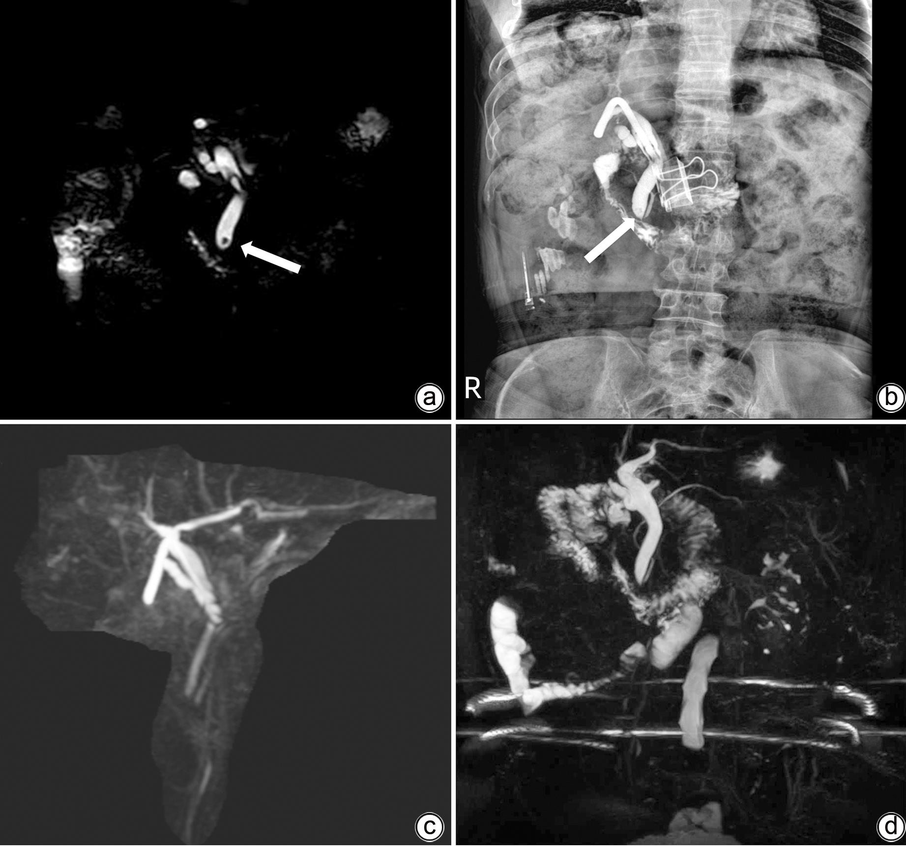
注: a,术前MRCP;b,术前T管造影,箭头处为结石;c,术后MRCP;d,术后随访7个月MRCP,均无结石残留。
图 1 影像学资料
Figure 1. Radiological images
表 1 患者外院血清指标水平变化比较
Table 1. Comparison of the changes of serum indexes of patients in other hospital
指标 术前 术后1 d 术后3 d 术后5 d 术后7 d WBC(×109/L) 14.95 10.21 5.80 5.43 4.96 NEC#(×109/L) 12.17 8.97 3.74 3.37 2.97 CRP(mg/L) 0.28 58.79 42.21 14.39 2.82 ALT(U/L) 26.4 32.6 20.7 33.2 75.5 AST(U/L) 36.3 39.3 20.0 39.7 59.3 TBil(μmol/L) 24.0 19.6 14.7 20.9 18.5 DBil(μmol/L) 13.00 11.33 7.39 11.01 10.64 IBil(μmol/L) 11.00 8.27 7.31 9.89 7.86 注:NEC#,中性粒细胞绝对值; CRP,C反应蛋白。  下载: 导出CSV
-
[1] WU Y, XU CJ, XU SF. Advances in risk factors for recurrence of common bile duct stones[J]. Int J Med Sci, 2021, 18( 4): 1067- 1074. DOI: 10.7150/ijms.52974. [2] WILLIAMS E, BECKINGHAM I, SAYED G EL, et al. Updated guideline on the management of common bile duct stones(CBDS)[J]. Gut, 2017, 66( 5): 765- 782. DOI: 10.1136/gutjnl-2016-312317. [3] HUANG Y, YI JF, ZHOU WC. Research progress on postoperative recurrence factors of choledocholithiasis[J]. Chin J Gen Surg, 2021, 30( 8): 964- 970. DOI: 10.7659/j.issn.1005-6947.2021.08.012.黄瑶, 易剑锋, 周文策. 胆总管结石治疗后复发因素的研究进展[J]. 中国普通外科杂志, 2021, 30( 8): 964- 970. DOI: 10.7659/j.issn.1005-6947.2021.08.012. [4] MO AL, WANG SG, CHEN WQ. Effect analysis of minimally invasive operation in the treatment of cholecystolithiasis complicated with choledocholithiasis[J]. J Laparosc Surg, 2022, 27( 2): 130- 134. DOI: 10.13499/j.cnki.fqjwkzz.2022.02.130.莫阿里, 王三贵, 陈伟强. 微创手术治疗胆囊结石合并胆总管结石的效果分析[J]. 腹腔镜外科杂志, 2022, 27( 2): 130- 134. DOI: 10.13499/j.cnki.fqjwkzz.2022.02.130. [5] TRINGALI A, COSTA D, FUGAZZA A, et al. Endoscopic management of difficult common bile duct stones: Where are we now? A comprehensive review[J]. World J Gastroenterol, 2021, 27( 44): 7597- 7611. DOI: 10.3748/wjg.v27.i44.7597. [6] FENG YP, GAO Y, XIN L, et al. Advances in endoscopic therapy for difficult common bile duct stones[J]. J Clin Hepatol, 2021, 37( 1): 229- 232. DOI: 10.3969/j.issn.1001-5256.2021.01.051.冯拥璞, 高野, 辛磊, 等. 困难胆总管结石的内镜治疗进展[J]. 临床肝胆病杂志, 2021, 37( 1): 229- 232. DOI: 10.3969/j.issn.1001-5256.2021.01.051. [7] KONSTANTAKIS C, TRIANTOS C, THEOPISTOS V, et al. Recurrence of choledocholithiasis following endoscopic bile duct clearance: Long term results and factors associated with recurrent bile duct stones[J]. World J Gastrointest Endosc, 2017, 9( 1): 26- 33. DOI: 10.4253/wjge.v9.i1.26. [8] XIA HT, XIN XL, YANG T, et al. Surgical strategy for recurrent common bile duct stones: A 10-year experience of a single center[J]. Updates Surg, 2021, 73( 4): 1399- 1406. DOI: 10.1007/s13304-020-00882-8. [9] XU W, WANG ZF, WANG HP, et al. Risk factors for common bile duct calculi recurrence and application value of its prediction model after endoscopic retrograde cholangiopancreatography[J]. Chin J Dig Surg, 2021, 20( 8): 890- 897. DOI: 10.3760/cma.j.cn115610-20210720-00356.徐雯, 王正峰, 王海平, 等. 经内镜逆行胰胆管造影术后胆总管结石复发危险因素分析及其预测模型的应用价值[J]. 中华消化外科杂志, 2021, 20( 8): 890- 897. DOI: 10.3760/cma.j.cn115610-20210720-00356. [10] Branch of Biliary Surgery, Chinese Society of Surgery, Chinese Medical Association; Chinese Committee of Biliary Surgeons. Expert consensus on clinical application of choledochoscope(2018 edition)[J]. Chin J Pract Surg, 2018, 38( 1): 21- 24. DOI: 10.19538/j.cjps.issn1005-2208.2018.01.02.中华医学会外科学分会胆道外科学组, 中国医师协会外科医师分会胆道外科医师委员会. 胆道镜临床应用专家共识(2018版)[J]. 中国实用外科杂志, 2018, 38( 1): 21- 24. DOI: 10.19538/j.cjps.issn1005-2208.2018.01.02. [11] CIANCI P, RESTINI E. Management of cholelithiasis with choledocholithiasis: Endoscopic and surgical approaches[J]. World J Gastroenterol, 2021, 27( 28): 4536- 4554. DOI: 10.3748/wjg.v27.i28.4536. [12] ZHANG W, YI HM, ZHANG J, et al. Ultrasound-guided choledochoscopy via T-tube sinus in treatment of complex intrahepatic bile duct stones[J/OL]. Chin J Hepatic Surg Electron Ed, 2021, 10( 4): 394- 397. DOI: 10.3877/cma.j.issn.2095-3232.2021.04.012.张伟, 易惠明, 张剑, 等. 超声引导下经 T 管窦道胆道镜取石治疗复杂型肝内胆管结石[J/OL]. 中华肝脏外科手术学电子杂志, 2021, 10( 4): 394- 397. DOI: 10.3877/cma.j.issn.2095-3232.2021.04.012. [13] ERCP Group, Digestive Endoscopy Branch, Chinese Medical Association, Biliary Pancreatology Group, Digestive Doctor Branch, Chinese Medical Association, National Digestive System Diseases Clinical Medical Research Center. China ERCP guide(2018 edition)[J]. Chin J Dig Endosc, 2018, 35( 11): 777- 813. DOI: 10.3760/cma.j.issn.1007-5232.2018.11.001.中华医学会消化内镜学分会ERCP学组, 中国医师协会消化医师分会胆胰学组, 国家消化系统疾病临床医学研究中心. 中国ERCP指南(2018版)[J]. 中华消化内镜杂志, 2018, 35( 11): 777- 813. DOI: 10.3760/cma.j.issn.1007-5232.2018.11.001. [14] PARK CH. The management of common bile duct stones[J]. Korean J Gastroenterol, 2018, 71( 5): 260- 263. DOI: 10.4166/kjg.2018.71.5.260. [15] Standards of Practice Committee ASGE, CHANDRASEKHARA V, KHASHAB MA, et al. Adverse events associated with ERCP[J]. Gastrointest Endosc, 2017, 85( 1): 32- 47. DOI: 10.1016/j.gie.2016.06.051. [16] STAPFER M, SELBY RR, STAIN SC, et al. Management of duodenal perforation after endoscopic retrograde cholangiopancreatography and sphincterotomy[J]. Ann Surg, 2000, 232( 2): 191- 198. DOI: 10.1097/00000658-200008000-00007. [17] LEE T, TENG TZJ, SHELAT VG. Choledochoscopy: An update[J]. World J Gastrointest Endosc, 2021, 13( 12): 571- 592. DOI: 10.4253/wjge.v13.i12.571. [18] MO ZK, FANG CH, XIANG N, et al. Strategies for preventing the complications of three-dimensional laparoscope combined with rigid choledochoscope lithotripsy guided by three-dimensional visualization technology[J]. Chin J Pract Surg, 2017, 37( 3): 284- 287. DOI: 10.19538/j.cjps.issn1005-2208.2017.03.18.莫志康, 方驰华, 项楠, 等. 三维可视化辅助3D腹腔镜、胆道硬镜肝胆管结石靶向碎石术并发症防治研究[J]. 中国实用外科杂志, 2017, 37( 3): 284- 287. DOI: 10.19538/j.cjps.issn1005-2208.2017.03.18. [19] TÜRK C, PETŘÍK A, NEISIUS A. Ureteral stones: Shockwave lithotripsy or ureteroscopy, which is best?[J]. Eur Urol, 2021, 80( 1): 55- 56. DOI: 10.1016/j.eururo.2021.03.029. [20] VARBAN O, ASSIMOS D, PASSMAN C, et al. Video. Laparoscopic common bile duct exploration and holmium laser lithotripsy: A novel approach to the management of common bile duct stones[J]. Surg Endosc, 2010, 24( 7): 1759- 1764. DOI: 10.1007/s00464-009-0837-0. [21] YAO BH, LI CY. Clinical research progress of ureteroscopy and its treatment of urolithiasis[J]. Prev Med Tribune, 2021, 16( 2): 255- 259. DOI: 10.11864/j.issn.1673.2021.02.27.姚碧海, 黎承杨. 输尿管软镜技术及其治疗尿石症的临床研究进展[J]. 微创医学, 2021, 16( 2): 255- 259. DOI: 10.11864/j.issn.1673.2021.02.27. [22] DOIZI S, TRAXER O. Flexible ureteroscopy: Technique, tips and tricks[J]. Urolithiasis, 2018, 46( 1): 47- 58. DOI: 10.1007/s00240-017-1030-x. [23] MIAO QS, WANG P, WANG L, et al. Application of electronic ureteroscopy in the operation of intrahepatic bile duct stones[J]. J Laparosc Surg, 2014, 19( 3): 171, 181. DOI: 10.13499/j.cnki.fqjwkzz.2014.03.171.苗庆松, 王朋, 王磊, 等. 电子输尿管软镜在肝内胆管结石手术中的应用[J]. 腹腔镜外科杂志, 2014, 19( 3): 171, 181. DOI: 10.13499/j.cnki.fqjwkzz.2014.03.171. [24] HEALY K, CHAMSUDDIN A, SPIVEY J, et al. Percutaneous endoscopic holmium laser lithotripsy for management of complicated biliary calculi[J]. JSLS, 2009, 13( 2): 184- 189. [25] PONSKY LE, GEISINGER MA, PONSKY JL, et al. Contemporary“urologic” intervention in the pancreaticobiliary tree[J]. Urology, 2001, 57( 1): 21- 25. DOI: 10.1016/S0090-4295(00)00888-8. [26] LI XY. Transcystic common bile duct exploration under laparoscope and soft ureteroscope: 35 cases report[J]. Chin J Mod Oper Surg, 2016, 20( 2): 84- 86. DOI: 10.16260/j.cnki.1009-2188.2016.02.002.李学元. 腹腔镜联合输尿管软镜经胆囊管行胆总管探查术(附35例报告)[J]. 中国现代手术学杂志, 2016, 20( 2): 84- 86. DOI: 10.16260/j.cnki.1009-2188.2016.02.002. [27] YAN YK, ZHAO SH, LIU JD, et al. The application in the treatment of intrahepatic lithiasis by laparoscopy with choledochoscope and holmium laser[J/CD]. Chin J Laparosc Surg Electron Ed, 2010, 3( 3): 272- 274. DOI: 10.3969/cma.j.issn.1674-6899.2010.03.017.阎玉矿, 赵守和, 刘继东, 等. 腹腔镜下胆道镜联合钬激光碎石治疗肝内胆管结石[J/CD]. 中华腔镜外科杂志(电子版), 2010, 3( 3): 272- 274. DOI: 10.3969/cma.j.issn.1674-6899.2010.03.017. [28] SARDIWALLA II, KOTO MZ, KUMAR N, et al. Laparoscopic common bile duct exploration use of a rigid ureteroscope: A single institute experience[J]. J Laparoendosc Adv Surg Tech A, 2018, 28( 10): 1169- 1173. DOI: 10.1089/lap.2018.0042. [29] LOFFELD RJLF, PAULUS J, LANGBEIN S, et al. Removal of common bile ducts stones via percutaneous access with a flexible ureteroscope and laser assistance[J]. Case Rep Gastrointest Med, 2019, 2019: 4684631. DOI: 10.1155/2019/4684631. [30] YANG XP, CHEN JC, ZHOU DP, et al. Clinical observatioin of laparoscopic choledochotomy combined with ureteroscopic hard ureterolithotomy T-tube drainage and conventional surgery[J]. Mod Hosp, 2019, 19( 5): 738- 740. DOI: 10.3969/j.issn.1671-332X.2019.05.033.杨小平, 陈剑川, 周冬平, 等. 腹腔镜胆总管切开联合输尿管硬镜取石T管引流术治疗胆总管结石的临床观察[J]. 现代医院, 2019, 19( 5): 738- 740. DOI: 10.3969/j.issn.1671-332X.2019.05.033. [31] QIAN ZF. The clinical effect of the treatment of intrahepatic residual biliary calculi with holmium laser through T-tube sinus under sheath protection[J]. Med Forum, 2022, 26( 1): 4- 6. DOI: 10.19435/j.1672-1721.2022.01.002.钱志峰. 鞘管保护下经T管窦道采用硬镜联合钬激光治疗肝内外胆管残余结石的临床效果观察[J]. 基层医学论坛, 2022, 26( 1): 4- 6. DOI: 10.19435/j.1672-1721.2022.01.002. -

 本文二维码
本文二维码
图(1) / 表(1)
计量
- 文章访问数: 480
- HTML全文浏览量: 135
- PDF下载量: 52
- 被引次数: 0

