中文English
ISSN 1001-5256 (Print)
ISSN 2097-3497 (Online)
CN 22-1108/R
Volume 37 Issue 11
Nov.  2021
Turn off MathJax
Article Contents

Effect of atractylone on the viability and apoptosis of hepatoma HepG2 cells and related mechanism

DOI: 10.3969/j.issn.1001-5256.2021.11.020
Research funding:

The Project of "Chinese Medicine Liver Disease" Clinical TCM Specialty Construction Project of Shanghai Pudong New Area Health Planning Commission (PDZY-2018-0607);

The project of Shanghai Pudong New Area Famous Chinese Medicine Studio Construction (PWRzm2020-14)

  • Received Date: 2021-03-22
  • Accepted Date: 2021-06-08
  • Published Date: 2021-11-20
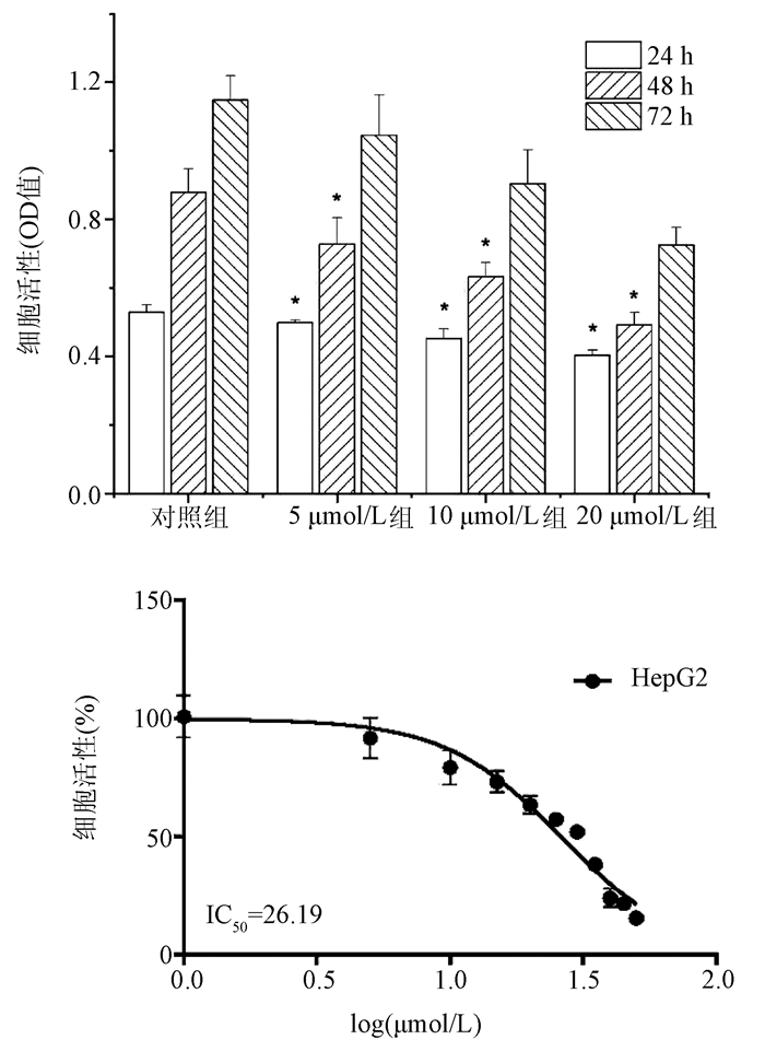
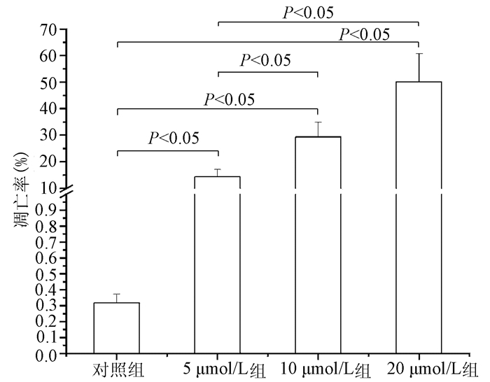
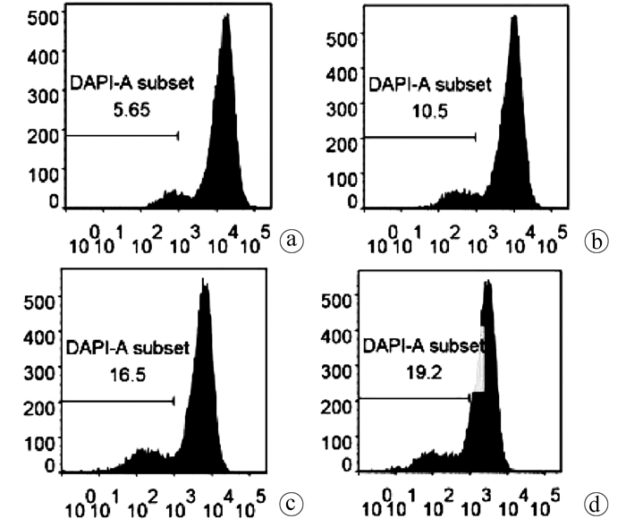
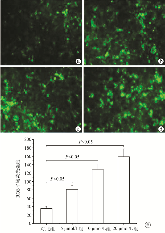
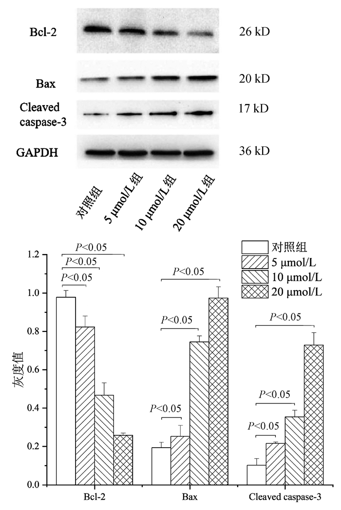
  •   Objective  To investigate the effect of atractylone on the viability and apoptosis of hepatoma HepG2 cells and its mechanism of action.  Methods  Hepatoma HepG2 cells were selected and divided into low-, middle-, and high-dose atractylone groups (5, 10, and 20 μmol/L), and the cells in the control group were added with an equal volume of DMSO. MTT colorimetry was used to measure the viability of HepG2 cells after treatment with different concentrations of atractylone; flow cytometry was used to measure the apoptosis rate and mitochondrial membrane potential of HepG2 cells; the DCFH-DA fluorescent probe labeling method was used to measure the level of reactive oxygen species (ROS) in HepG2 cells; Transwell assay was used to evaluate the effect of atractylone on the migration ability of HepG2 cells; Western blot was used to measure the protein expression levels of Bcl-2, Bax, and cleaved caspase-3. A one-way analysis of variance was used for comparison of continuous data between multiple groups, and the least significant difference t-test was used for comparison between two groups.  Results  After 24 and 48 hours of treatment with atractylone, compared with the control group, the low-, middle-, and high-dose atractylone groups had a tendency of reduction in cell viability (all P < 0.05), with a half inhibitory concentration of 26.19 μmol/L in atractylone treatment of HepG2 cells for 72 hours. The low-, middle-, and high-dose atractylone groups had a significantly higher apoptosis rate than the control group (14.34%/29.32%/50.12% vs 0.32%, all P < 0.05). Compared with the control group, the low-, middle-, and high-dose atractylone groups had a significant increase in the fluorescence intensity of ROS in HepG2 cells (all P < 0.05). After 48 hours of treatment with atractylone, compared with the control group, the low-, middle-, and high-dose atractylone groups had a significant reduction in the number of migrated cells (132.67±18.36/57.00±9.26/31.00±2.45 vs 258.11±38.54, P < 0.05). Compared with the control group, the low-, middle-, and high-dose atractylone groups had a significant reduction in the expression of the anti-apoptotic factor Bcl-2 and significant increases in the expression of the apoptotic factors Bax and cleaved caspase-3 (all P < 0.05).  Conclusion  Atractylone can induce the apoptosis and inhibit the migration of HepG2 cells, which provides an experimental basis for further development and utilization of atractylone.


  • loading
  • [1]
    SUNG H, FERLAY J, SIEGEL RL, et al. Global Cancer Statistics 2020: GLOBOCAN estimates of incidence and mortality worldwide for 36 cancers in 185 countries[J]. CA Cancer J Clin, 2021, 71(3): 209-249. DOI: 10.3322/caac.21660.
    [2]
    WU YX, SHENG QS. Advances in traditional Chinese medicine therapy for primary hepatocarcinoma[J]. Guangxi Med J, 2020, 42(4): 483-485. DOI: 10.1165/j.issn.0253-4304.2020.04.26.

    吴玉潇, 盛庆寿. 中医药治疗原发性肝癌的研究进展[J]. 广西医学, 2020, 42(4): 483-485. DOI: 10.11675/j.issn.0253-4304.2020.04.26.
    [3]
    LI ZW. Analysis of postoperative prognosis and risk factors in patients with primary liver cancer[J]. China's Naturopathy, 2020, 28(13): 73-75. DOI: 10.19621/j.cnki.11-3555/r.2020.1334.

    李柱威. 原发性肝癌患者术后预后及其危险因素分析[J]. 中国民间疗法, 2020, 28(13): 73-75. DOI: 10.19621/j.cnki.11-3555/r.2020.1334.
    [4]
    FORNER A, REIG ME, de LOPE CR, et al. Current strategy for staging and treatment: The BCLC update and future prospects[J]. Semin Liver Dis, 2010, 30(1): 61-74. DOI: 10.1055/s-0030-1247133.
    [5]
    HUANG JH, HUANG ZM, ZHANG TQ, et al. Progress in comprehensive treatment of advanced liver cancer based on interventional therapy[J/CD]. Electronic J Liver Tumor, 2019, 6(4): 27-31. DOI: 10.3969/j.issn.2095-7815.2019.04.007.

    黄金华, 黄职妹, 张天奇, 等. 以介入治疗为基础的中晚期肝癌综合治疗进展[J/CD]. 肝癌电子杂志, 2019, 6(4): 27-31. DOI: 10.3969/j.issn.2095-7815.2019.04.007.
    [6]
    YUAN SX, ZHOU WP. Progress and hot spots of comprehensive treatment for primary liver cancer[J]. Chin J Dig Surg, 2021, 20(2): 163-170. DOI: 10.3760/cma.j.cn115610-20201211-00776.

    袁声贤, 周伟平. 原发性肝癌综合治疗的进展和热点[J]. 中华消化外科杂志, 2021, 20(2): 163-170. DOI: 10.3760/cma.j.cn115610-20201211-00776.
    [7]
    ZHAN YP, ZHAI XF. Study on prescription compatibility law of contemporary famous doctors of TCM in treating primary liver cancer based on data mining[J]. J Liaoning Univ Tradit Chin Med, 2018, 20(3): 159-162. DOI: 10.13194/j.issn.1673-842x.2018.03.046.

    占义平, 翟笑枫. 基于数据挖掘当代名中医治疗原发性肝癌用药配伍规律研究[J]. 辽宁中医药大学学报, 2018, 20(3): 159-162. DOI: 10.13194/j.issn.1673-842x.2018.03.046.
    [8]
    ZHANG Y, CHEN HG, ZHAO C, et al. Research progress on anti-hepatocellular carcinoma mechanism of active ingredients of traditional Chinese medicine[J]. China J Chin Mater Med, 2020, 45(14): 3395-3406. DOI: 10.19540/j.cnki.cjcmm.20200429.601.

    张宇, 陈华国, 赵超, 等. 中药有效成分抗肝癌作用机制研究进展[J]. 中国中药杂志, 2020, 45(14): 3395-3406. DOI: 10.19540/j.cnki.cjcmm.20200429.601.
    [9]
    ZHAO ZJ, XIAO SN, ZHAO YX, et al. Re-evaluation of the literature on the pharmacological effects of Atractylodes macrocephala[J]. Chin J Hosp Pharm, 2011, 31(7): 607-609. https://www.cnki.com.cn/Article/CJFDTOTAL-ZGYZ201107035.htm

    赵子剑, 肖胜男, 赵永新, 等. 苍术药理作用的文献再评价[J]. 中国医院药学杂志, 2011, 31(7): 607-609. https://www.cnki.com.cn/Article/CJFDTOTAL-ZGYZ201107035.htm
    [10]
    LIU C. Progress of medicinal research on Atractylodes macrocephala[J]. Heilongjiang Sci Technol Inform, 2013, 12: 101. DOI: 10.3969/j.issn.1673-1328.2013.12.099.

    刘畅. 苍术药用研究进展[J]. 黑龙江科技信息, 2013, 12: 101. DOI: 10.3969/j.issn.1673-1328.2013.12.099.
    [11]
    CHEN TY, LIU YL, HOU TL, et al. Study on the protective effect of atractylone on acute lung injury in mice[J]. Mod J Integr Tradit Chin West Med, 2018, 27(24): 2623-2626. DOI: 10.3969/j.issn.1008-8849.2018.24.001.

    陈天阳, 刘廷亮, 侯天禄, 等. 苍术酮对急性肺损伤小鼠保护作用的研究[J]. 现代中西医结合杂志, 2018, 27(24): 2623-2626. DOI: 10.3969/j.issn.1008-8849.2018.24.001.
    [12]
    ATHINARAYANAN S, FAN YY, WANG X, et al. Fatty acid desaturase 1 influences hepatic lipid homeostasis by modulating the PPARα-FGF21 axis[J]. Hepatol Commun, 2021, 5(3): 461-477. DOI: 10.1002/hep4.1629.
    [13]
    SUN YP, CHENG Z, WU Z, et al. Research on the inheritance, innovation and high-quality development of Chinese herbal medicine Atractylodes[J]. Hubei Agricult Sci, 2020, 59(11): 203-207. DOI: 10.14088/j.cnki.issn0439-8114.2020.11.040.

    孙元鹏, 程正, 吴喆, 等. 中药材苍术的传承创新与高质量发展研究[J]. 湖北农业科学, 2020, 59(11): 203-207. DOI: 10.14088/j.cnki.issn0439-8114.2020.11.040.
    [14]
    ZHANG MF, SHEN YQ. Advances in studies on anti-inflammation, antitumor, and immunoregulation of Atractylodis Rhizoma[J]. Drug Eval Res, 2016, 39(5): 885-890. DOI: 10.7501/j.issn.1674-6376.2016.05.037.

    张明发, 沈雅琴. 苍术抗炎、抗肿瘤和免疫调节作用的研究进展[J]. 药物评价研究, 2016, 39(5): 885-890. DOI: 10.7501/j.issn.1674-6376.2016.05.037.
    [15]
    ZHOU Y, LU JT, ZHANG XD, et al. Potential target prediction and forward molecular docking verification of atractylon[J]. J Shaoyang Uni(Natural Science Edition), 2019, 16(1): 98-104. DOI: CNKI:SUN:SYXZ.0.2019-01-013.

    周域, 陆建图, 张小丁, 等. 苍术酮潜在靶点预测及正向分子对接验证[J]. 邵阳学院学报(自然科学版), 2019, 16(1): 98-104. DOI: CNKI:SUN:SYXZ.0.2019-01-013.
    [16]
    GENG W, LIANG W, YE ZB, et al. Mechanism of HT29 apoptosis in colorectal cancer cells by atractylone[J]. Chin Tradit Patent Med, 2018, 40(4): 937-940. DOI: 10.3969/j.issn.1001-1528.2018.04.034.

    耿玮, 梁巍, 叶智斌, 等. 苍术酮对结直肠癌细胞HT29凋亡的机制[J]. 中成药, 2018, 40(4): 937-940. DOI: 10.3969/j.issn.1001-1528.2018.04.034.
    [17]
    YAO XY, DOU FF. Role of apoptosis in traditional Chinese medicine treatment of liver cancer[J]. Pract J Cancer, 2020, 35(6): 1040-1044. DOI: 10.3969/j.issn.1001-5930.2020.06.046.

    姚晓云, 钭方芳. 凋亡在中医药治疗肝癌中的作用研究[J]. 实用癌症杂志, 2020, 35(6): 1040-1044. DOI: 10.3969/j.issn.1001-5930.2020.06.046.
    [18]
    SHAHAR N, LARISCH S. Inhibiting the inhibitors: Targeting anti-apoptotic proteins in cancer and therapy resistance[J]. Drug Resist Updat, 2020, 52: 100712. DOI: 10.1016/j.drup.2020.100712.
    [19]
    QIAO Z, CHENG Y, LIU S, et al. Casticin inhibits esophageal cancer cell proliferation and promotes apoptosis by regulating mitochondrial apoptotic and JNK signaling pathways[J]. Naunyn Schmiedebergs Arch Pharmacol, 2019, 392(2): 177-187. DOI: 10.1007/s00210-018-1574-5.
    [20]
    ZHANG Y, WANG Y, ZHAO Y, et al. Novel camphor-based pyrimidine derivatives induced cancer cell death through a ROS-mediated mitochondrial apoptosis pathway[J]. RSC Adv, 2019, 9(51): 29711-29720. DOI: 10.1039/C9RA05900H.
    [21]
    ZHONG FR, CHENG HL, ZHANG H, et al. Effect of kaempferol on the proliferation, migration, invasion, and apoptosis of human hepatoma Bel-7402 cells[J]. J Clin Hepatol, 2020, 36(12): 2725-2729. DOI: 10.3969/j.issn.1001-5256.2020.12.017.

    仲富瑞, 程宦立, 张浩, 等. 山萘酚对人肝癌Bel-7402细胞增殖、迁移、侵袭及凋亡的影响[J]. 临床肝胆病杂志, 2020, 36(12): 2725-2729. DOI: 10.3969/j.issn.1001-5256.2020.12.017.
    [22]
    LI R, ZOU X, ZHU T, et al. Destruction of neutrophil extracellular traps promotesthe apoptosis and inhibits the invasion of gastric cancer cells by regulating the expression of Bcl-2, Bax and NF-κB[J]. Onco Targets Ther, 2020, 13: 5271-5281. DOI: 10.2147/OTT.S227331.
    [23]
    EDLICH F. The great migration of Bax and Bak[J]. Mol Cell Oncol, 2015, 2(3): e995029. DOI: 10.4161/23723556.2014.995029.
    [24]
    GUO DF, WANG RT, HUANG JC, et al. Curcumin derivative C086 induces the apoptosis of hepatoma HepG2 cells via the PI3K-Akt pathway[J]. Chin J Gerontol, 2021, 41(6): 1270-1274. DOI: 10.3969/j.issn.1005-9202.2021.06.042.

    郭登方, 王若涛, 黄建成, 等. 姜黄素衍生物C086通过PI3K-Akt通路诱导肝癌HepG2细胞凋亡[J]. 中国老年学杂志, 2021, 41(6): 1270-1274. DOI: 10.3969/j.issn.1005-9202.2021.06.042.
    [25]
    WU XY, ZHAO YN, WANG XJ, et al. Effect of galectin-3 expression suppression on expressions of Bcl-2 and Bax in gastric cancer MGC-803 cells and its promotion on apoptosis[J]. J Jilin Univ(Med Edit), 2020, 46(2): 335-339. DOI: 10.13481/j.1671-587x.20200221.

    吴雪艳, 赵依纳, 王小杰, 等. 半乳糖凝集素3表达抑制对人胃癌MGC-803细胞中Bcl-2和Bax表达的影响及其促凋亡作用[J]. 吉林大学学报(医学版), 2020, 46(2): 335-339, 前插3. DOI: 10.13481/j.1671-587x.20200221.
    [26]
    FU K, SUI GC, SHI JM. Inhibitory effect of triterpenoid saponins from Dioscorea gracillima on human hepatoma HepG2 cells[J/CD]. Cardiovasc Dis J Integr Tradit Chin Western Med (Electronic), 2020, 8(1): 67-68. DOI: 10.16282/j.cnki.cn11-9336/r.2020.01.052.

    付康, 隋广超, 史金铭. 三萜纤细薯蓣皂苷对人肝癌HepG2细胞的抑制作用研究[J/CD]. 中西医结合心血管病电子杂志, 2020, 8(1): 67-68. DOI: 10.16282/j.cnki.cn11-9336/r.2020.01.052.
    [27]
    NIE SS, LI X, ZHAO YH, et al. Effect of modified Si Junzitang drug serum on expression of apoptosis-related molecules of gastric cancer cell SGC-7901[J]. Chin J Exp Med Formul, 2019, 25(9): 25-30. DOI: 10.13422/j.cnki.syfjx.20190824.

    聂闪闪, 李洵, 赵玉航, 等. 加味四君子汤含药血清对胃癌细胞SGC-7901凋亡相关因子表达的影响[J]. 中国实验方剂学杂志, 2019, 25(9): 25-30. DOI: 10.13422/j.cnki.syfjx.20190824.
    [28]
    MAO M, HUA Y, JIANG X, et al. Expression of tumor necrosis factor alpha and neuronal apoptosis in the developing rat brain after neonatal stroke[J]. Neurosci Lett, 2006, 403(3): 227-232. DOI: 10.1016/j.neulet.2006.03.078.
  • 加载中

Catalog

    通讯作者: 陈斌, bchen63@163.com
    • 1. 

      沈阳化工大学材料科学与工程学院 沈阳 110142

    1. 本站搜索
    2. 百度学术搜索
    3. 万方数据库搜索
    4. CNKI搜索

    Figures(6)  / Tables(1)

    Article Metrics

    Article views (769) PDF downloads(45) Cited by()
    Proportional views
    Related

    /

    DownLoad:  Full-Size Img  PowerPoint
    Return
    Return