中文English
ISSN 1001-5256 (Print)
ISSN 2097-3497 (Online)
CN 22-1108/R
Volume 38 Issue 12
Dec.  2022
Turn off MathJax
Article Contents

Mechanism of carrimycin in regulating the biological function of pancreatic cancer cells

DOI: 10.3969/j.issn.1001-5256.2022.12.020
Research funding:

Science and Technology Research Project of Jilin Province Department of Education (JJKH20220554KJ);

Heatth Science and Technology Capacity Improvement Project of Jilin Province (2021JC063)

More Information
  • Corresponding author: JIN Aihua, drjinah@163.com (ORCID: 0000-0002-3405-8097)
  • Received Date: 2022-05-26
  • Accepted Date: 2022-07-20
  • Published Date: 2022-12-20
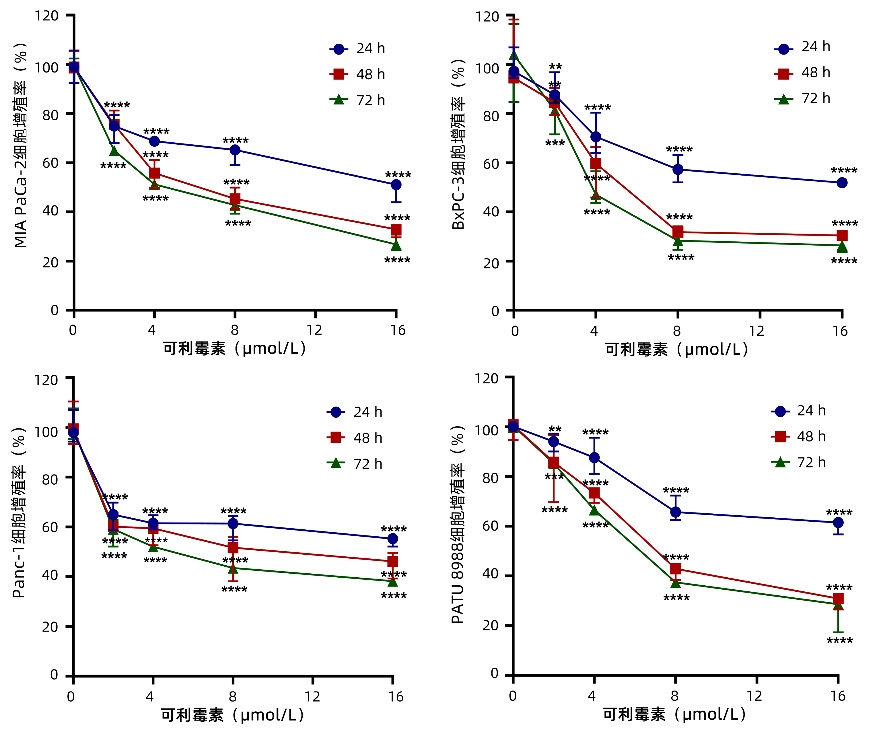
  •   Objective  To investigate the effect of carrimycin on the biological function of pancreatic cancer cells.  Methods  Pancreatic cancer cell lines MIA PaCa-2, BxPC-3, Panc-1, and PATU 8988 were treated with carrimycin at concentrations of 0 (control group), 2, 4, 8, and 16 μmol/L for 24, 48, and 72 hours. MTT assay was used to measure cell viability; EdU cell proliferation assay was used to observe the effect of carrimycin on DNA replication of pancreatic cancer cells; colony formation assay was used to observe the effect of carrimycin on the proliferation of pancreatic cancer cells; flow cytometry was used to analyze the effect of carrimycin on the cell cycle of pancreatic cancer cells; wound healing assay was used to analyze the effect of carrimycin on the migration of pancreatic cancer cells; Western blot was used to measure the expression levels of the markers such as epithelial-mesenchymal transition (EMT) and cell cycle-dependent protein kinase inhibitor 1A (P21); immunofluorescence assay were used to measure the expression levels of EMT-related markers. An analysis of variance was used for comparison between multiple groups, and the least significant difference t-test was used for further comparison between two groups.  Results  Compared with the control group, carrimycin significantly inhibited the proliferative activity of MIA PaCa-2, BxPC-3, Panc-1, and PATU 8988 cells in a concentration- and time-dependent manner (all P < 0.01); carrimycin at concentrations of 4, 8, and 16 μmol/L significantly reduced DNA replication in MIA PaCa-2 cells (t=2.378, 4.984, and 18.970, all P < 0.05) and BxPC-3 cells (t=4.879, 6.089, and 9.521, all P < 0.01); after treatment with carrimycin at concentrations of 4, 8, and 16 μmol/L, colony formation ability significantly decreased with the increase in drug concentration in MIA PaCa-2 cells (t=5.889, 11.240, and 15.840, all P < 0.001) and BxPC-3 cells (t=6.717, 15.800, and 18.850, all P < 0.001). After treatment with carrimycin at concentrations of 4, 8, and 16 μmol/L, there was a significant increase in the proportion of cells in G1 phase in MIA PaCa-2 cells (t=9.071, 12.280, and 19.360, all P < 0.0001) and BxPC-3 cells (t=3.061, 4.962, and 8.868, all P < 0.05), and there was a significant reduction in the proportion of cells in S phase in MIA PaCa-2 cells (t=2.316, 4.165, and 5.562, all P < 0.05) and BxPC-3 cells (t=2.424, 3.264, and 5.744, all P < 0.05). Western blot further demonstrated that compared with the control group, the expression level of the cell cycle-related protein P21 gradually increased with the increase in the concentration of carrimycin in MIA PaCa-2 cells (t=5.437, 6.453, and 8.799, all P < 0.001) and BxPC-3 cells (t=25.130, 44.750, and 52.960, all P < 0.000 1). Wound healing assay showed that after treatment for 12, 24, and 48 hours, carrimycin at concentrations of 0, 4, 8, and 16 μmol/L significantly reduced the lateral migration of MIA PaCa-2 cells (all P < 0.05) and BxPC-3 cells (all P < 0.05). Western blot showed that compared with the control group, carrimycin treatment at concentrations of 4, 8, and 16 μmol/L significantly upregulated the expression of the epithelial marker E-cadherin in MIA PaCa-2 cells (t=2.388, 4.899, and 5.819, all P < 0.05) and BxPC-3 cells (t=2.533, 5.836, and 6.774, all P < 0.05) and significantly downregulated the expression of the interstitial marker Snail in MIA PaCa-2 cells (t=12.440, 14.830, and 16.800, all P < 0.000 1) and BxPC-3 cells (t=5.039, 5.893, and 7.725, all P < 0.01), and it also significantly downregulated the expression of the interstitial marker Vimentin in MIA PaCa-2 cells (t=3.105, 7.752, and 11.200, all P < 0.05) and BxPC-3 cells (t=2.555, 4.883, and 9.153, all P < 0.05).  Conclusion  Carrimycin can effectively inhibit the proliferation, migration, and EMT process of pancreatic cancer cells, thereby exerting an antitumor biological activity.


  • loading
  • [1]
    CHEN W, ZHENG R, BAADE PD, et al. Cancer statistics in China, 2015[J]. CA Cancer J Clin, 2016, 66(2): 115-132. DOI: 10.3322/caac.21338.
    [2]
    General Office of National Health Commission. Standard for diagnosis and treatment of pancreatic cancer (2022 edition)[J]. J Clin Hepatol, 2022, 38(5): 1006-1015. DOI: 10.3969/j.issn.1001-5256.2022.05.007.

    国家卫生健康委办公厅. 胰腺癌诊疗指南(2022年版)[J]. 临床肝胆病杂志, 2022, 38(5): 1006-1015. DOI: 10.3969/j.issn.1001-5256.2022.05.007.
    [3]
    ZHU H, WEI M, XU J, et al. PARP inhibitors in pancreatic cancer: molecular mechanisms and clinical applications[J]. Mol Cancer, 2020, 19(1): 49. DOI: 10.1186/s12943-020-01167-9.
    [4]
    HAN A, XU R, LIU Y, et al. HSDL2 acts as a promoter in pancreatic cancer by regulating cell proliferation and lipid metabolism[J]. Onco Targets Ther, 2021, 14: 435-444. DOI: 10.2147/OTT.S287722.
    [5]
    NEOPTOLEMOS JP, KLEEFF J, MICHL P, et al. Therapeutic developments in pancreatic cancer: current and future perspectives[J]. Nat Rev Gastroenterol Hepatol, 2018, 15(6): 333-348. DOI: 10.1038/s41575-018-0005-x.
    [6]
    HAO TY, HE WQ. Advances in metabolic engineering of macrolide antibiotics[J]. Chin J Biotech, 2021, 37(5): 1737-1747. DOI: 10.13345/j.cjb.200686.

    郝天怡, 赫卫清. 大环内酯类抗生素代谢工程的研究进展[J]. 生物工程学报, 2021, 37(5): 1737-1747. DOI: 10.13345/j.cjb.200686.
    [7]
    JIN Y, ZUO HX, LI MY, et al. Anti-tumor effects of carrimycin and monomeric isovalerylspiramycin I on hepatocellular carcinoma in vitro and in vivo[J]. Front Pharmacol, 2021, 12: 774231. DOI: 10.3389/fphar.2021.774231.
    [8]
    LIANG SY, ZHAO TC, ZHOU ZH, et al. Anti-tumor effect of carrimycin on oral squamous cell carcinoma cells in vitro and in vivo[J]. Transl Oncol, 2021, 14(6): 101074. DOI: 10.1016/j.tranon.2021.101074.
    [9]
    ZHANG XY, HAO RH, LIU F, et al. Progress in Application of Rapamycin in Tumor Treatment[J]. Food Drug, 2018, 20(1): 65-70. DOI: 10.3969/j.issn.1672-979X.2018.01.016.

    张晓元, 郝荣华, 刘飞, 等. 雷帕霉素在肿瘤治疗方面研究进展[J]. 食品与药品, 2018, 20(1): 65-70. DOI: 10.3969/j.issn.1672-979X.2018.01.016.
    [10]
    EDELMAN MJ, SHVARTSBEYN M. Epothilones in development for non—small-cell lung cancer: novel anti-tubulin agents with the potential to overcome taxane resistance[J]. Clin Lung Cancer, 2012, 13(3): 171-180. DOI: 10.1016/j.cllc.2011.02.005.
    [11]
    GIANNAKAKOU P, GUSSIO R, NOGALES E, et al. A common pharmacophore for epothilone and taxanes: molecular basis for drug resistance conferred by tubulin mutations in human cancer cells[J]. Proc Natl Acad Sci U S A, 2000, 97(6): 2904-2909. DOI: 10.1073/pnas.040546297.
    [12]
    LI YL, SUN J, HU X, et al. Epothilone B induces apoptosis and enhances apoptotic effects of ABT-737 on human cancer cells via PI3K/AKT/mTOR pathway[J]. J Cancer Res Clin Oncol, 2016, 142(11): 2281-2289. DOI: 10.1007/s00432-016-2236-y.
    [13]
    CUI J, ZHOU J, HE W, et al. Targeting selenoprotein H in the nucleolus suppresses tumors and metastases by Isovalerylspiramycin I[J]. J Exp Clin Cancer Res, 2022, 41(1): 126. DOI: 10.1186/s13046-022-02350-0.
    [14]
    LIU Y, QUAN Y. Relationship between inflammatory microenvironment and epithelial-mesenchymal transition and its significance in pancreatic carcinoma[J]. Chin J Clin Res, 2018, 31(12): 1619-1623. DOI: 10.13429/j.cnki.cjcr.2018.12.006.

    刘宇, 全颖. 胰腺癌炎性微环境与细胞上皮间质转化的关系及意义[J]. 中国临床研究, 2018, 31(12): 1619-1623. DOI: 10.13429/j.cnki.cjcr.2018.12.006.
    [15]
    BAKIR B, CHIARELLA AM, PITARRESI JR, et al. EMT, MET, plasticity, and tumor metastasis[J]. Trends Cell Biol, 2020, 30(10): 764-776. DOI: 10.1016/j.tcb.2020.07.003.
    [16]
    YEUNG KT, YANG J. Epithelial-mesenchymal transition in tumor metastasis[J]. Mol Oncol, 2017, 11(1): 28-39. DOI: 10.1002/1878-0261.12017.
    [17]
    KONG HR, DAI SJ, CHEN H, et al. α-solanine inhibits epithelial-mesenchymal transition in trichostatin A-resistant human pancreatic cancer cells through the WNT signaling pathway[J]. J Hepatopancreatobiliary Surg, 2020, 32(2): 89-96. DOI: 10.11952/j.issn.1007-1954.2020.02.006.

    孔鸿儒, 戴胜杰, 陈豪, 等. α-龙葵碱通过WNT信号通路抑制曲古抑菌素A-耐药人胰腺癌细胞的上皮-间质转化进程[J]. 肝胆胰外科杂志, 2020, 32(2): 89-96. DOI: 10.11952/j.issn.1007-1954.2020.02.006.
    [18]
    LIU JB, QI M, LI QQ, et al. The mechanism of cordycepin in inhibition of pancreatic cancer stem cells proliferation and metastasis[J]. Acta Pharm Sin, 2017, 52(9): 1404-1409. DOI: 10.16438/j.0513-4870.2017-0353.

    刘建兵, 戚梦, 李巧琪, 等. 虫草素抑制胰腺癌干细胞增殖及转移的机制研究[J]. 药学学报, 2017, 52(9): 1404-1409. DOI: 10.16438/j.0513-4870.2017-0353.
  • 加载中

Catalog

    通讯作者: 陈斌, bchen63@163.com
    • 1. 

      沈阳化工大学材料科学与工程学院 沈阳 110142

    1. 本站搜索
    2. 百度学术搜索
    3. 万方数据库搜索
    4. CNKI搜索

    Figures(4)  / Tables(1)

    Article Metrics

    Article views (1414) PDF downloads(49) Cited by()
    Proportional views
    Related

    /

    DownLoad:  Full-Size Img  PowerPoint
    Return
    Return