中文English
ISSN 1001-5256 (Print)
ISSN 2097-3497 (Online)
CN 22-1108/R
Volume 37 Issue 9
Sep.  2021
Turn off MathJax
Article Contents

Application of liver three-dimensional visualized reconstruction technique in hepatectomy for children with complicated hepatoblastoma

DOI: 10.3969/j.issn.1001-5256.2021.09.025
Research funding:

National Key R & D Program of China (2017YFC0110401)

  • Received Date: 2021-01-18
  • Accepted Date: 2021-02-18
  • Published Date: 2021-09-20
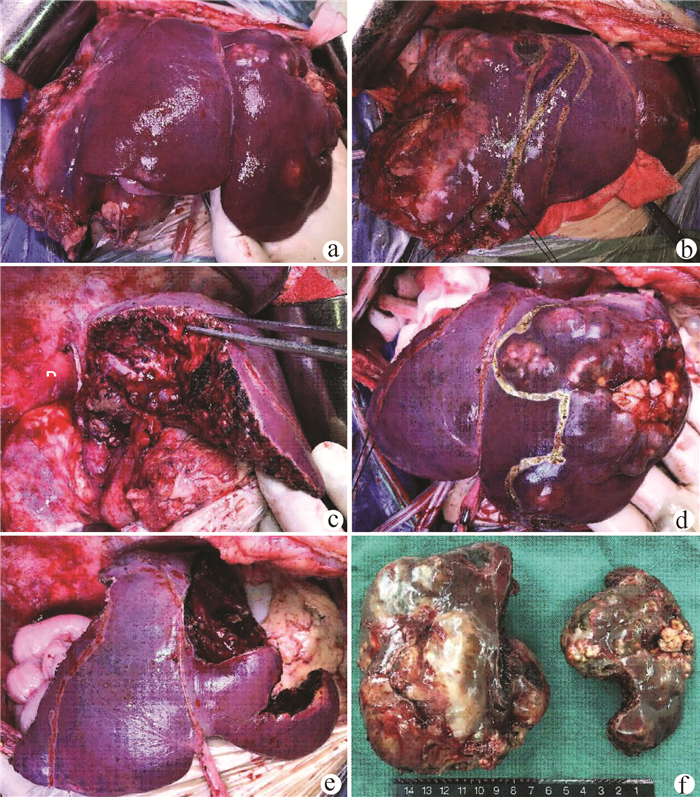
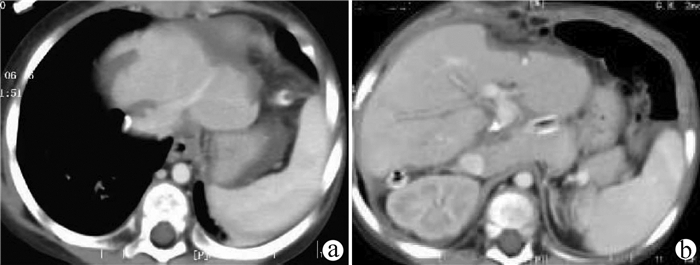
  •   Objective  To investigate the application of liver three-dimensional (3D) visualized reconstruction technique in hepatectomy for children with complicated hepatoblastoma.  Methods  A retrospective analysis was performed for the clinical data of 30 children with hepatoblastoma who underwent hepatectomy for radical resection in PLA Rocket Force Characteristic Medical Center from January 2018 to October 2020, and according to whether liver 3D visualization with IQQA-Liver system was performed before surgery, the children were divided into 3D reconstruction group with 15 children and control group with 15 children. The two groups were compared in terms of perioperative parameters, short-term prognosis, and follow-up conditions. The independent samples t-test was used for comparison of normally distributed continuous data between two groups, and the Mann-Whitney U test was used for comparison of non-normally distributed continuous data between two groups; the Fisher's exact test was used for comparison of categorical data between two groups.  Results  Compared with the control group, the 3D reconstruction group had a significantly higher mean age (55.7±10.2 years vs 28.2±2.7 years, P < 0.05) and a significantly higher number of patients with POSTTEXT stage III/VI hepatoblastoma (12 vs 5, P < 0.05) or involvement of the hepatic vein or the inferior vena cava (11 vs 3, P < 0.05). All children completed the surgery successfully, and there were no significant differences between the two groups in blood loss, time of operation, number of times and duration of hepatic portal occlusion, and number of children receiving segmental hepatectomy or partial hepatectomy (all P > 0.05). The median follow-up after surgery was 9.5 months. In the 3D reconstruction group, 2 children experienced recurrence and were diagnosed at 10 and 12 months, respectively, after surgery, and they were treated with chemotherapy at the moment; in the control group, 4 children experienced recurrence, which was higher than that in the 3D reconstruction group (P=0.651), and among these 4 children, 2 had recurrence at 7 months after surgery, received liver transplantation, and survived up to now, and the other 2 children died shortly after recurrence.  Conclusion  3D visualized reconstruction technique helps to perform hepatectomy for children with complicated hepatoblastoma more safely and accurately, especially extended hepatectomy for patients with stage POST TEXT III/IV hepatoblastoma, thereby avoiding liver transplantation.


  • loading
  • [1]
    YANG T, WHITLOCK RS, VASUDEVAN SA. Surgical management of hepatoblastoma and recent advances[J]. Cancers (Basel), 2019, 11(12): 1944. DOI: 10.3390/cancers11121944.
    [2]
    HAFBERG E, BORINSTEIN SC, ALEXOPOULOS SP. Contemporary management of hepatoblastoma[J]. Curr Opin Organ Transplant, 2019, 24(2): 113-117. DOI: 10.1097/MOT.0000000000000618.
    [3]
    WANG XS, YANG W, HUANG C, et al. Clinical features and outcome evaluation for children with hepatoblastoma[J]. J Capit Med Univ, 2019, 40(2): 169-173. DOI: 10.3969/j.issn.1006-7795.2019.02.003.

    王希思, 杨维, 黄程, 等. 儿童肝母细胞瘤的临床特征和治疗效果评价[J]. 首都医科大学学报, 2019, 40(2): 169-173. DOI: 10.3969/j.issn.1006-7795.2019.02.003.
    [4]
    LAKE CM, TIAO GM, BONDOC AJ. Surgical management of locally-advanced and metastatic hepatoblastoma[J]. Semin Pediatr Surg, 2019, 28(6): 150856. DOI: 10.1016/j.sempedsurg.2019.150856.
    [5]
    Compilation and Examination Expert Group for Guidelines for the diagnosis and treatment of hepatoblastoma (2019). Guidelines for the diagnosis and treatment of hepatoblastoma (2019)[J]. J Clin Hepatol, 2019, 35(11): 2431-2434. DOI: 10.3969/j.issn.1001-5256.2019.11.008.

    儿童肝母细胞瘤诊疗规范(2019年版)编写审定专家组. 儿童肝母细胞瘤诊疗规范(2019年版)[J]. 临床肝胆病杂志, 2019, 35(11): 2431-2434. DOI: 10.3969/j.issn.1001-5256.2019.11.008.
    [6]
    EL-GENDI A, FADEL S, EL-SHAFEI M, et al. Avoiding liver transplantation in post-treatment extent of disease III and IV hepatoblastoma[J]. Pediatr Int, 2018, 60(9): 862-868. DOI: 10.1111/ped.13634.
    [7]
    ZHU W, HE SS, ZENG SL, et al. Three-dimensional visual assessment and virtual reality study of centrally located hepatocellular carcinoma on the axis of blood vessels[J]. Chin J Surg, 2019, 57(5): 358-365. DOI: 10.3760/cma.j.issn.0529-5815.2019.05.008.

    祝文, 何松盛, 曾思略, 等. 以血管为轴心的中央型肝癌三维可视化评估及虚拟现实的研究[J]. 中华外科杂志, 2019, 57(5): 358-365. DOI: 10.3760/cma.j.issn.0529-5815.2019.05.008.
    [8]
    Chinese Society of Digital Medicine, Liver Cancer Committee of Chinese Medical Doctor Association, Clinical Precision Medicine Committee of Chinese Medical Doctor Association, et al. Specification for technical operation and clinical application of three-dimensional visualization technology for primary liver cancer (2020 edition)[J]. Chin J Dig Surg, 2020, 19(9): 897-918. DOI: 10.3760/cma.j.cn115610-20200720-00499.

    中华医学会数字医学分会, 中国医师协会肝癌专业委员会, 中国医师协会精准医学专业委员会, 等. 原发性肝癌三维可视化技术操作及诊疗规范(2020版)[J]. 中华消化外科杂志, 2020, 19(9): 897-918. DOI: 10.3760/cma.j.cn115610-20200720-00499.
    [9]
    FANG C, AN J, BRUNO A, et al. Consensus recommendations of three-dimensional visualization for diagnosis and management of liver diseases[J]. Hepatol Int, 2020, 14(4): 437-453. DOI: 10.1007/s12072-020-10052-y.
    [10]
    ZHANG G, ZHOU XJ, ZHU CZ, et al. Usefulness of three-dimensional(3D) simulation software in hepatectomy for pediatric hepatoblastoma[J]. Surg Oncol, 2016, 25(3): 236-243. DOI: 10.1016/j.suronc.2016.05.023.
    [11]
    SU L, DONG Q, ZHANG H, et al. Clinical application of a three-dimensional imaging technique in infants and young children with complex liver tumors[J]. Pediatr Surg Int, 2016, 32(4): 387-395. DOI: 10.1007/s00383-016-3864-7.
    [12]
    SU L, DONG Q, ZHANG H, et al. 3D Application of 3D visualization technology in precise hepatectomy for complex liver tumors in infants[J/CD]. Chin J Hepat Surg(Electronic Edition), 2015, 4(5): 274-278. DOI: 10.3877/cma.j.issn.2095-3232.2015.05.005.

    苏琳, 董蒨, 张虹, 等. 3D可视化技术在婴幼儿复杂肝肿瘤精准肝切除中的应用[J/CD]. 中华肝脏外科手术学电子杂志, 2015, 4(5): 274-278. DOI: 10.3877/cma.j.issn.2095-3232.2015.05.005.
    [13]
    ZHAO J, ZHOU XJ, ZHU CZ, et al. 3D simulation assisted resection of giant hepatic mesenchymal hamartoma in children[J]. Comput Assist Surg (Abingdon), 2017, 22(1): 54-59. DOI: 10.1080/24699322.2017.1358401.
    [14]
    FUCHS J, CAVDAR S, BLUMENSTOCK G, et al. POST-TEXT III and IV hepatoblastoma: Extended hepatic resection avoids liver transplantation in selected cases[J]. Ann Surg, 2017, 266(2): 318-323. DOI: 10.1097/SLA.0000000000001936.
    [15]
    ARONSON DC, WEEDA VB, MAIBACH R, et al. Microscopically positive resection margin after hepatoblastoma resection: What is the impact on prognosis? A Childhood Liver Tumours Strategy Group (SIOPEL) report[J]. Eur J Cancer, 2019, 106: 126-132. DOI: 10.1016/j.ejca.2018.10.013.
    [16]
    SONG D, SUN D, JIANG DS. Comparative study of three-dimensional visualization technology and two-dimensional imaging technology in liver resection of liver cancer patients[J]. J Clin Exp Med, 2020, 19(6): 656-660. DOI: 10.3969/j.issn.1671-4695.2020.06.026.

    宋铎, 孙铎, 姜德帅. 三维可视化技术与二维影像技术在肝癌患者肝切除术中的疗效比较研究[J]. 临床和实验医学杂志, 2020, 19(6): 656-660. DOI: 10.3969/j.issn.1671-4695.2020.06.026.
  • 加载中

Catalog

    通讯作者: 陈斌, bchen63@163.com
    • 1. 

      沈阳化工大学材料科学与工程学院 沈阳 110142

    1. 本站搜索
    2. 百度学术搜索
    3. 万方数据库搜索
    4. CNKI搜索

    Figures(3)  / Tables(4)

    Article Metrics

    Article views (471) PDF downloads(36) Cited by()
    Proportional views
    Related

    /

    DownLoad:  Full-Size Img  PowerPoint
    Return
    Return