PDF下载 ( 678 KB)

超声新技术在肝细胞癌诊疗中的应用现状与展望
DOI: 10.12449/JCH260704
利益冲突声明:本文不存在任何利益冲突。
作者贡献声明:林思嘉和李玲玲负责构思及撰写文章;周建华负责全面指导、终审及定稿。
Current application and prospects of new ultrasound technologies in the diagnosis and treatment of hepatocellular carcinoma
-
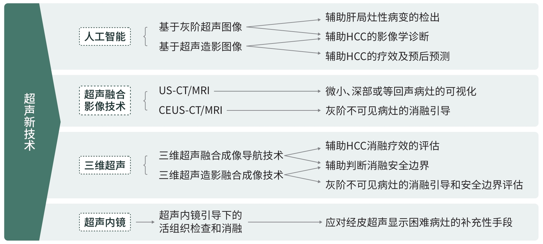
摘要: 作为原发性肝癌的常见类型,肝细胞癌是严重威胁我国民众生命健康的恶性疾病之一。超声作为肝细胞癌临床诊疗过程中的重要影像学手段,其新技术的发展与应用具有较高的临床价值。目前,超声及超声造影联合人工智能在肝局灶性病变的检出、影像学诊断、疗效和预后预测方面已展现出应用潜力;超声融合成像技术在经皮热消融术的引导及疗效评估中展现出临床价值;三维超声在辅助消融与消融灶评估中也有一定优势;而超声内镜可作为经皮超声扫查或引导困难肝细胞癌病灶的一种补充影像学手段。Abstract: As a common type of primary liver cancer, hepatocellular carcinoma (HCC) is a major health threat in China. Ultrasound is a crucial imaging modality in the clinical diagnosis and treatment of HCC, and the development and application of new ultrasound technologies have high clinical value. Currently, ultrasound and contrast-enhanced ultrasound combined with artificial intelligence have shown promising potential in the detection of focal liver lesions, image-based HCC diagnosis, outcome assessment, and prognostic prediction; furthermore, ultrasound fusion imaging technology has shown certain clinical value in guidance and efficacy evaluation of percutaneous thermal ablation; three-dimensional ultrasound has certain advantages in assisting ablation and assessing ablation margins; endoscopic ultrasonography can be used as a supplementary method for HCC cases that are difficult to scan or guide via percutaneous ultrasound.
-
[1] Vogel A, Meyer T, Sapisochin G, et al. Hepatocellular carcinoma[J]. Lancet, 2022, 400( 10360): 1345- 1362. DOI: 10.1016/s0140-6736(22)01200-4. [2] Bray F, Laversanne M, Sung H, et al. Global cancer statistics 2022: GLOBOCAN estimates of incidence and mortality worldwide for 36 cancers in 185 countries[J]. CA Cancer J Clin, 2024, 74( 3): 229- 263. DOI: 10.3322/caac.21834. [3] Wu C H, Sheu J C, Chou P L, et al. Automatic real-time detection and diagnosis of liver tumor with ultrasound[J]. J Hepatocell Carcinoma, 2025, 12: 1599- 1611. DOI: 10.2147/JHC.S524311. [4] Rhyou S Y, Yoo J C. Automated ultrasonography of hepatocellular carcinoma using discrete wavelet transform based deep-learning neural network[J]. Med Image Anal, 2025, 101: 103453. DOI: 10.1016/j.media.2025.103453. [5] Tiyarattanachai T, Apiparakoon T, Marukatat S, et al. The feasibility to use artificial intelligence to aid detecting focal liver lesions in real-time ultrasound: A preliminary study based on videos[J]. Sci Rep, 2022, 12: 7749. DOI: 10.1038/s41598-022-11506-z. [6] Tiyarattanachai T, Apiparakoon T, Chaichuen O, et al. Artificial intelligence assists operators in real-time detection of focal liver lesions during ultrasound: A randomized controlled study[J]. Eur J Radiol, 2023, 165: 110932. DOI: 10.1016/j.ejrad.2023.110932. [7] Xi I L, Wu J, Guan J, et al. Deep learning for differentiation of benign and malignant solid liver lesions on ultrasonography[J]. Abdom Radiol, 2021, 46( 2): 534- 543. DOI: 10.1007/s00261-020-02564-w. [8] Schmauch B, Herent P, Jehanno P, et al. Diagnosis of focal liver lesions from ultrasound using deep learning[J]. Diagn Interv Imaging, 2019, 100( 4): 227- 233. DOI: 10.1016/j.diii.2019.02.009. [9] Lu R F, She C Y, He D N, et al. AI enhanced diagnostic accuracy and workload reduction in hepatocellular carcinoma screening[J]. npj Digit Med, 2025, 8: 500. DOI: 10.1038/s41746-025-01892-9. [10] Dadoun H, Rousseau A L, de Kerviler E, et al. Deep learning for the detection, localization, and characterization of focal liver lesions on abdominal US images[J]. Radiol Artif Intell, 2022, 4( 3): e210110. DOI: 10.1148/ryai.210110. [11] Hu H T, Wang W, Chen L D, et al. Artificial intelligence assists identifying malignant versus benign liver lesions using contrast-enhanced ultrasound[J]. J Gastroenterol Hepatol, 2021, 36( 10): 2875- 2883. DOI: 10.1111/jgh.15522. [12] Urhut M C, Săndulescu L D, Streba C T, et al. Diagnostic performance of an artificial intelligence model based on contrast-enhanced ultrasound in patients with liver lesions: A comparative study with clinicians[J]. Diagnostics(Basel), 2023, 13( 21): 3387. DOI: 10.3390/diagnostics13213387. [13] Căleanu C D, Sîrbu C L, Simion G. Deep neural architectures for contrast enhanced ultrasound(CEUS) focal liver lesions automated diagnosis[J]. Sensors(Basel), 2021, 21( 12): 4126. DOI: 10.3390/s21124126. [14] Ding W Z, Meng Y Q, Ma J, et al. Contrast-enhanced ultrasound-based AI model for multi-classification of focal liver lesions[J]. J Hepatol, 2025, 83( 2): 426- 439. DOI: 10.1016/j.jhep.2025.01.011. [15] National Health Commission of the People’s Republic of China. Guideline for diagnosis and treatment of primary liver cancer(2026edition)[J]. J Clin Hepatol, 2026, 42( 4): 794- 827. DOI: 10.12449/JCH260409.中华人民共和国国家卫生健康委员会. 原发性肝癌诊疗指南(2026年版)[J]. 临床肝胆病杂志, 2026, 42( 4): 794- 827. DOI: 10.12449/JCH260409. [16] American College of Radiology. CEUS LI-RADS v2017 core[R/OL]. https://edge.sitecorecloud.io/americancoldf5f-acrorgf92a-productioncb02-3650/media/ACR/Files/RADS/LI-RADS/CEUS-LI-RADS-2017-Core.pdf. https://edge.sitecorecloud.io/americancoldf5f-acrorgf92a-productioncb02-3650/media/ACR/Files/RADS/LI-RADS/CEUS-LI-RADS-2017-Core.pdf [17] Hoopes J R, Lyshchik A, Xiao T S, et al. Using machine learning to improve the contrast-enhanced ultrasound liver imaging reporting and data system diagnosis of hepatocellular carcinoma in indeterminate liver nodules[J]. Ultrasound Med Biol, 2025, 51( 11): 1953- 1960. DOI: 10.1016/j.ultrasmedbio.2025.06.029. [18] Sheng R F, Huang J, Zhang W G, et al. A semi-automatic step-by-step expert-guided LI-RADS grading system based on gadoxetic acid-enhanced MRI[J]. J Hepatocell Carcinoma, 2021, 8: 671- 683. DOI: 10.2147/JHC.S316385. [19] Tran Q H, Sam T B, Tang V H, et al. Feature-driven LI-RADS classification in liver cancer: A CNN-based approach for CT imaging under data scarcity[C]// 2025 IEEE 22nd International Symposium on Biomedical Imaging(ISBI). April 14-17, 2025, Houston, TX, USA. IEEE, 2025: 1- 5. DOI: 10.1109/ISBI60581.2025.10980650. [20] Wang K, Liu Y H, Chen H X, et al. Fully automating LI-RADS on MRI with deep learning-guided lesion segmentation, feature characterization, and score inference[J]. Front Oncol, 2023, 13: 1153241. DOI: 10.3389/fonc.2023.1153241. [21] Hai Y, Savsani E, Chong W, et al. Meta-analysis and systematic review of contrast-enhanced ultrasound in evaluating the treatment response after locoregional therapy of hepatocellular carcinoma[J]. Abdom Radiol(NY), 2021, 46( 11): 5162- 5179. DOI: 10.1007/s00261-021-03248-9. [22] Oezdemir I, Wessner C E, Shaw C, et al. Tumor vascular networks depicted in contrast-enhanced ultrasound images as a predictor for transarterial chemoembolization treatment response[J]. Ultrasound Med Biol, 2020, 46( 9): 2276- 2286. DOI: 10.1016/j.ultrasmedbio.2020.05.010. [23] Liu D, Liu F, Xie X Y, et al. Accurate prediction of responses to transarterial chemoembolization for patients with hepatocellular carcinoma by using artificial intelligence in contrast-enhanced ultrasound[J]. Eur Radiol, 2020, 30( 4): 2365- 2376. DOI: 10.1007/s00330-019-06553-6. [24] Zhang W B, Guo Q H, Zhu Y L, et al. Cross-institutional evaluation of deep learning and radiomics models in predicting microvascular invasion in hepatocellular carcinoma: Validity, robustness, and ultrasound modality efficacy comparison[J]. Cancer Imaging, 2024, 24( 1): 142. DOI: 10.1186/s40644-024-00790-9. [25] Hu H T, Wang Z, Huang X W, et al. Ultrasound-based radiomics score: A potential biomarker for the prediction of microvascular invasion in hepatocellular carcinoma[J]. Eur Radiol, 2019, 29( 6): 2890- 2901. DOI: 10.1007/s00330-018-5797-0. [26] Zhang D, Wei Q, Wu G G, et al. Preoperative prediction of microvascular invasion in patients with hepatocellular carcinoma based on radiomics nomogram using contrast-enhanced ultrasound[J]. Front Oncol, 2021, 11: 709339. DOI: 10.3389/fonc.2021.709339. [27] Liu F, Liu D, Wang K, et al. Deep learning radiomics based on contrast-enhanced ultrasound might optimize curative treatments for very-early or early-stage hepatocellular carcinoma patients[J]. Liver Cancer, 2020, 9( 4): 397- 413. DOI: 10.1159/000505694. [28] Ma Q P, He X L, Li K, et al. Dynamic contrast-enhanced ultrasound radiomics for hepatocellular carcinoma recurrence prediction after thermal ablation[J]. Mol Imaging Biol, 2021, 23( 4): 572- 585. DOI: 10.1007/s11307-021-01578-0. [29] Calandri M, Mauri G, Yevich S, et al. Fusion imaging and virtual navigation to guide percutaneous thermal ablation of hepatocellular carcinoma: A review of the literature[J]. Cardiovasc Intervent Radiol, 2019, 42( 5): 639- 647. DOI: 10.1007/s00270-019-02167-z. [30] Ahn S J, Lee J M, Lee D H, et al. Real-time US-CT/MR fusion imaging for percutaneous radiofrequency ablation of hepatocellular carcinoma[J]. J Hepatol, 2017, 66( 2): 347- 354. DOI: 10.1016/j.jhep.2016.09.003. [31] Song K D, Lee M W, Rhim H, et al. Percutaneous US/MRI fusion-guided radiofrequency ablation for recurrent subcentimeter hepatocellular carcinoma: Technical feasibility and therapeutic outcomes[J]. Radiology, 2018, 288( 3): 878- 886. DOI: 10.1148/radiol.2018172743. [32] Xu E J, Li K, Long Y L, et al. Intra-procedural CT/MR-ultrasound fusion imaging helps to improve outcomes of thermal ablation for hepatocellular carcinoma: Results in 502 nodules[J]. Ultraschall Med, 2021, 42( 2): e9- e19. DOI: 10.1055/a-1021-1616. [33] You Y J, Long Y L, Yan R H, et al. Improving ablation safety for hepatocellular carcinoma proximal to the hilar bile ducts by ultrasound-MR fusion imaging: A preliminary comparative study[J]. Front Oncol, 2021, 11: 570312. DOI: 10.3389/fonc.2021.570312. [34] Lu Y B, Huang Y N, Weng Y C, et al. Contrast-enhanced ultrasonography guidance avoids US-CT/MR fusion error for percutaneous radiofrequency ablation of hepatocellular carcinoma[J]. BMC Med Imaging, 2024, 24( 1): 323. DOI: 10.1186/s12880-024-01508-w. [35] Huang Q N, Zeng Q J, Long Y L, et al. Fusion imaging techniques and contrast-enhanced ultrasound for thermal ablation of hepatocellular carcinoma-A prospective randomized controlled trial[J]. Int J Hyperth, 2019, 36( 1): 1206- 1214. DOI: 10.1080/02656736.2019.1687945. [36] Ma Q P, Xu E J, Zeng Q J, et al. Intraprocedural computed tomography/magnetic resonance-contrast-enhanced ultrasound fusion imaging improved thermal ablation effect of hepatocellular carcinoma: Comparison with conventional ultrasound[J]. Hepatol Res, 2019, 49( 7): 799- 809. DOI: 10.1111/hepr.13336. [37] Minami Y, Minami T, Takita M, et al. Radiofrequency ablation for hepatocellular carcinoma: Clinical value of ultrasound-ultrasound overlay fusion for optimal ablation and local controllability[J]. Hepatol Res, 2020, 50( 1): 67- 74. DOI: 10.1111/hepr.13407. [38] Bo X W, Xu H X, Guo L H, et al. Ablative safety margin depicted by fusion imaging with post-treatment contrast-enhanced ultrasound and pre-treatment CECT/CEMRI after radiofrequency ablation for liver cancers[J]. Br J Radiol, 2017, 90( 1078): 20170063. DOI: 10.1259/bjr.20170063. [39] Xu E J, Lv S M, Li K, et al. Immediate evaluation and guidance of liver cancer thermal ablation by three-dimensional ultrasound/contrast-enhanced ultrasound fusion imaging[J]. Int J Hyperth, 2018, 34( 6): 870- 876. DOI: 10.1080/02656736.2017.1373306. [40] Minami Y, Kudo M. Image guidance in ablation for hepatocellular carcinoma: Contrast-enhanced ultrasound and fusion imaging[J]. Front Oncol, 2021, 11: 593636. DOI: 10.3389/fonc.2021.593636. [41] Carriero S, Della Pepa G, Monfardini L, et al. Role of fusion imaging in image-guided thermal ablations[J]. Diagnostics(Basel), 2021, 11( 3): 549. DOI: 10.3390/diagnostics11030549. [42] Sun Yueting, Guo Yuqing, Liu Jiaming, et al. Preliminary application of three-dimensional ultrasound fusion imaging visualization technology guiding precise needle placement for thermal ablation of hepatocellular carcinoma[J]. Chin J Ultrasonography, 2024, 33( 2): 158- 164. DOI: 10.3760/cma.j.cn131148-20230926-00142.孙月婷, 郭语清, 刘家铭, 等. 三维超声融合成像可视化技术在引导肝细胞癌热消融精准布针中的初步应用[J]. 中华超声影像学杂志, 2024, 33( 2): 158- 164. DOI: 10.3760/cma.j.cn131148-20230926-00142. [43] Lan Sirong, Xu Jiwei, Zhang Yaoming, et al. Guidance of real-time contrast-enhanced ultrasonography and 3D ultrasound fusion imaging for ablation area in patients with primary liver cancer during microwave ablation therapy[J]. J Pract Hepatol, 2022, 25( 6): 889- 892. DOI: 10.3969/j.issn.1672-5069.2022.06.033.蓝思荣, 徐继威, 张耀明, 等. 实时超声造影与3D超声融合成像导航评估微波消融治疗原发性肝癌患者价值研究[J]. 实用肝脏病杂志, 2022, 25( 6): 889- 892. DOI: 10.3969/j.issn.1672-5069.2022.06.033. [44] You Y J, Zhang M, Li K, et al. Feasibility of 3D US/CEUS-US/CEUS fusion imaging-based ablation planning in liver tumors: A retrospective study[J]. Abdom Radiol(NY), 2021, 46( 6): 2865- 2874. DOI: 10.1007/s00261-020-02909-5. [45] Long H Y, Zhou X Y, Zhang X E, et al. 3D fusion is superior to 2D point-to-point contrast-enhanced US to evaluate the ablative margin after RFA for hepatocellular carcinoma[J]. Eur Radiol, 2024, 34( 2): 1247- 1257. DOI: 10.1007/s00330-023-10023-5. [46] Hotta N, Maeno T, Ayada M, et al. Four-dimensional ultrasonography for therapeutic radiofrequency ablation for hepatocellular carcinoma[J]. Hepatogastroenterology, 2006, 53( 70): 521- 525. [47] Hotta N, Yamada S, Murase K, et al. Usefulness of real-time 4D ultrasonography during radiofrequency ablation in a case of hepatocellular carcinoma[J]. Case Rep Gastroenterol, 2011, 5( 1): 82- 87. DOI: 10.1159/000321721. [48] Bogstad J, Vilmann P, Burcharth F. Early detection of recurrent hepatocellular carcinoma by endosonographically guided fine-needle aspiration biopsy[J]. Endoscopy, 1997, 29( 4): 322- 324. DOI: 10.1055/s-2007-1004198. [49] Chen F, Bao H W, Deng Z, et al. Endoscopic ultrasound-guided sampling using core biopsy needle for diagnosis of left-lobe hepatocellular carcinoma in patients with underlying cirrhosis[J]. J Cancer Res Ther, 2020, 16( 5): 1100- 1105. DOI: 10.4103/jcrt.JCRT_723_19. [50] Tantau A, Zaharie T. EUS-guided fine needle biopsy for hepatocarcinoma of the right liver lobe as a rescue diagnostic technique after a negative percutaneousguided liver biopsy[J]. Med Ultrason, 2023, 25( 1): 115- 116. DOI: 10.11152/mu-3993. [51] Takano Y, Yamawaki M, Noda J, et al. Endoscopic ultrasound-guided tissue acquisition for focal liver lesions in the caudate lobe: A report of seven cases[J]. Clin J Gastroenterol, 2024, 17( 2): 334- 337. DOI: 10.1007/s12328-023-01906-7. [52] Xu M, Xu D X, Deng Z, et al. Long-term outcomes of endoscopic ultrasound-guided laser ablation for liver tumors in the caudate lobe: 5 years of experience[J]. Scand J Gastroenterol, 2023, 58( 5): 558- 564. DOI: 10.1080/00365521.2022.2148833. -

 下载:
下载:
 本文二维码
本文二维码
图(1)
计量
- 文章访问数: 510
- HTML全文浏览量: 93
- PDF下载量: 64
- 被引次数: 0