PDF下载 ( 18386 KB)

2,5-二羟基苯甲酸对代谢相关脂肪性肝病体外细胞模型的影响及其作用机制
DOI: 10.12449/JCH260715
Effect and mechanism of action of 2,5-dihydroxybenzoic acid on an in vitro cell model of metabolic dysfunction-associated fatty liver disease
-
摘要:
目的 探究2,5-二羟基苯甲酸(2,5-DHBA)治疗代谢相关脂肪性肝病(MAFLD)潜在靶点和调控磷脂酰肌醇3-激酶(PI3K)/蛋白激酶B(Akt)信号通路的分子机制。 方法 首先进行网络药理学研究,通过数据库筛选2,5-DHBA与MAFLD的共同作用靶点,进行分子对接与通路富集分析预测其潜在调控的生物学过程与信号通路。其次,进一步通过细胞(小鼠正常肝细胞AML-12)实验验证,通过细胞计数试剂盒8筛选2,5-DHBA 3个最佳干预浓度。将细胞分为5组:对照组、模型组(2 mmol/L的游离脂肪酸构建的MAFLD细胞模型)以及2,5-DHBA低、中、高3个干预剂量组(2 mmol/L的游离脂肪酸+3个不同浓度的2,5-DHBA)。采用油红O染色观察细胞内脂质沉积情况,荧光探针DCFH-DA检测细胞内活性氧的水平,生化法检测甘油三酯(TG)和总胆固醇(TC)含量;采用实时聚合酶链式反应与Western Blot检测基因及蛋白表达水平。多组间比较采用单因素方差分析,进一步两两比较采用Tukey’s检验。 结果 共筛选出32个2,5-DHBA与MAFLD的共同作用靶点。在1.25~20 μmol/L浓度范围内,2,5-DHBA处理组自2.5 μmol/L浓度起,呈剂量依赖性提高AML-12细胞活力。选取最低起效浓度 1.25 μmol/L、中间梯度浓度5 μmol/L及有效区间内最高安全浓度20 μmol/L作为低、中、高3个干预剂量。与模型组相比,1.25、5和20 μmol/L浓度的2,5-DHBA干预均能降低细胞内脂滴积累(P值均<0.05);与模型组比较,3个剂量组TG和TC水平差异均有统计学意义(P值均<0.05)。与对照组比较,模型组内ROS荧光强度显著升高(P<0.05);与模型组相比,经5 μmol/L和20 μmol/L浓度的2,5-DHBA处理后,细胞内ROS水平呈剂量依赖性下降(P值均<0.05);与模型组相比,5 μmol/L和20 μmol/L浓度的2,5-DHBA治疗显著下调了促脂生成基因固醇调节元件转录因子1、脂肪酸合成酶的mRNA表达水平,并上调了抗氧化核心基因核转录因子红系2相关因子2及其下游靶基因血红素加氧酶1的mRNA表达(P值均<0.05)。与对照组相比,模型组p-PI3K/PI3K、p-Akt/Akt表达均显著升高(P值均<0.05);与模型组相比,经5 μmol/L的2,5-DHBA治疗后,p-PI3K/PI3K、p-Akt/Akt的蛋白表达水平均显著降低(P<0.05)。与对照组相比,模型组Nrf2、HO-1蛋白表达水平显著降低(P值均<0.05);经1.25 μmol/L的2,5-DHBA治疗后Nrf2和HO-1蛋白水平均显著升高(P<0.05)。 结论 网络药理学联合细胞实验证实,2,5-DHBA能通过调控PI3K/Akt信号通路,减轻脂质沉积与氧化应激,进而发挥治疗MAFLD的作用。 -
关键词:
- 代谢相关脂肪性肝病 /
- 2,5-二羟基苯甲酸 /
- 磷酸肌醇3-激酶类
Abstract:Objective To investigate the potential targets of 2,5-dihydroxybenzoic acid (2,5-DHBA) in the treatment of metabolic dysfunction-associated fatty liver disease (MAFLD) and the molecular mechanism by which it regulates the phosphatidylinositol 3-kinase (PI3K)/protein kinase B (Akt) signaling pathway. Methods A network pharmacology analysis was performed at first, and related databases were used to obtain the common action targets of 2,5-DHBA and MAFLD, followed by molecular docking and pathway enrichment analysis to predict the potential biological processes and signaling pathways regulated by these targets. Then mouse normal hepatocytes AML-12 were used for cell experiments, and CCK-8 assay was used to determine the three optimal intervention concentrations of 2,5-DHBA. AML-12 cells were divided into control group, model group (cells cultured in a medium containing 2 mmol/L free fatty acid [FFA] to establish a cell model of MAFLD), and three 2,5-DHBA intervention groups (cells cultured with FFA and 2,5-DHBA at the three different concentrations of 1.25, 5, and 20 μmol/L, respectively). Oil red O staining was used to observe intracellular lipid accumulation; the fluorescent probe DCFH-DA was used to measure the level of intracellular reactive oxygen species (ROS); biochemical assays were used to measure the content of triglyceride (TG) and total cholesterol (TC); quantitative real-time PCR and Western Blot were used to measure the expression levels of related genes and proteins. A one-way analysis of variance was used for comparison between multiple groups, and the Tukey’s test was used for further comparison between two groups. Results A total of 32 common targets were obtained for 2,5-DHBA and MAFLD. Within a concentration range of 1.25 — 20 μmol/L, 2,5-DHBA treatment, starting from the concentration of 2.5 μmol/L, increased the viability of AML-12 cells in a dose-dependent manner. The minimum effective concentration of 1.25 μmol/L, the intermediate gradient concentration of 5 μmol/L, and the maximum safe concentration within the effective range of 20 μmol/L were selected for low-, middle-, and high-dose intervention, respectively. Compared with the model group, 2,5-DHBA intervention at concentrations of 1.25, 5, and 20 μmol/L could significantly reduce lipid droplet accumulation in cells (all P<0.05), and there were significant differences in TG and TC between the model group and the three intervention groups (all P<0.05). Compared with the control group, the model group had a significant increase in ROS fluorescence intensity (P<0.05), and compared with the model group, there was a dose-dependent reduction in intracellular ROS level after treatment with 5 μmol/L or 20 μmol/L 2,5-DHBA (P<0.05). Compared with the model group, 2,5-DHBA treatment at concentrations of 5 and 20 μmol/L significantly downregulated the mRNA expression levels of the lipogenic genes SREBf1 and FASN and upregulated the mRNA expression levels of the key antioxidant gene Nrf2 and its downstream target gene HO-1 (all P<0.05). Compared with the control group, the expression of p-PI3K/PI3K and p-Akt/Akt in the model group was significantly increased (all P<0.05). Compared with the model group, the protein expression levels of p-PI3K/PI3K and p-Akt/Akt were significantly decreased after 5 μmol/L 2,5-DHBA treatment (P<0.05). Compared with the control group, the expression levels of Nrf2 and HO-1 protein in the model group were significantly decreased (all P<0.05). The protein levels of Nrf2 and HO-1 were significantly increased after treatment with 1.25 μmol/L 2,5-DHBA (P<0.05). Conclusion Network pharmacology and cellular experiments confirm that 2,5-DHBA can alleviate lipid deposition and oxidative stress by regulating the PI3K/Akt signaling pathway, thereby exerting a therapeutic effect on MAFLD. -
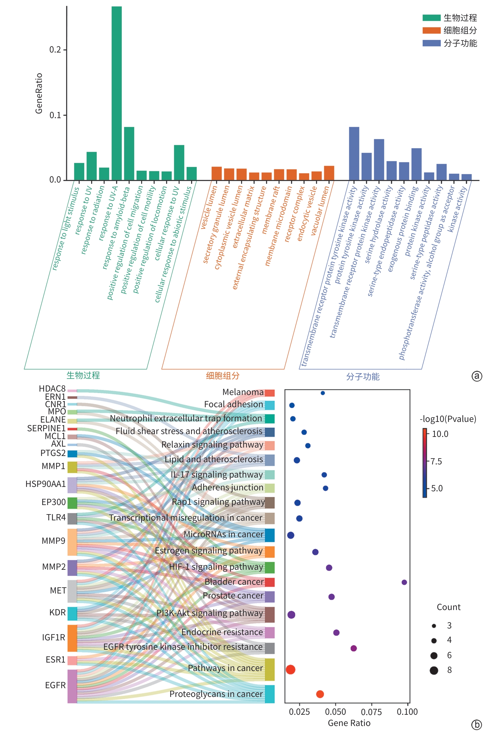
注: a,GO功能富集分析结果,展示了BP、CC和MF中富集显著性前10的条目;b,KEGG通路富集分析前20的信号通路。MAFLD,代谢相关脂肪性肝病;GO,基因本体;KEGG,京都基因与基因组百科全书;HDAC8,组蛋白去乙酰化酶8;ERN1,内质网到细胞核信号转导1;CNR1,大麻素受体1;MPO,髓过氧化物酶;ELANE,中性粒细胞弹性蛋白酶;SERPINE1,丝氨酸蛋白酶抑制剂家族E成员1;MCL1,髓细胞白血病1;AXL,AXL受体酪氨酸激酶;PTGS2,前列腺素内过氧化物合酶2;MMP1,基质金属蛋白酶1;HSP90AA1,热休克蛋白90α家族A类成员1;EP300,蛋白乙酰转移酶P300;TLR4,Toll样受体4;MMP9;基质金属蛋白酶9;MMP2,基质金属蛋白酶2;MET,间质-上皮转化因子;KDR,激酶插入域受体;IGF1R,胰岛素样生长因子1受体;ESR1,雌激素受体1;EGFR,表皮生长因子受体;IL-17,白细胞介素17;Rap1,Ras相关蛋白1;HIF-1,低氧诱导因子1;PI3K-Akt,磷脂酰肌酶3-激酶-蛋白激酶B。
图 1 2,5-DHBA治疗MAFLD靶点的GO与KEGG富集分析
Figure 1. GO and KEGG enrichment analysis of 2,5-DHBA for MAFLD targets
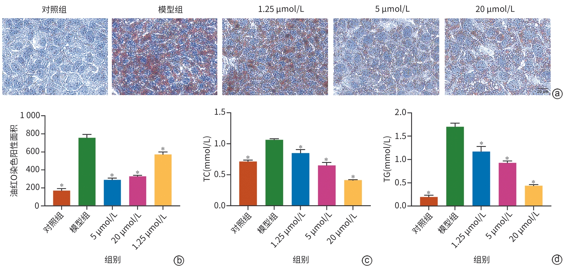
注: a,油红O染色结果(×400);b,油红O染色定量分析;c,TC水平测定;d,TG水平测定。与模型组比较,*P<0.05。FFA,游离脂肪酸;TC,总胆固醇;TG,甘油三酯;2,5-DHBA,2,5-二羟基苯甲酸。
图 2 2,5-DHBA减轻FFA诱导的AML-12细胞脂质堆积
Figure 2. 2,5-DHBA alleviates FFA-induced lipid accumulation in AML-12 cells
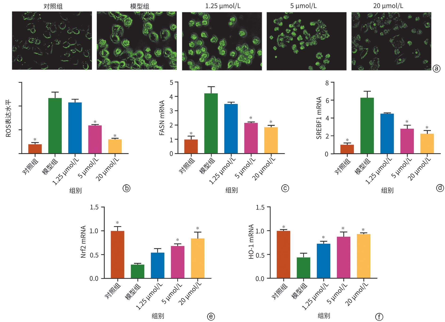
注: a,ROS荧光探针代表性图像(×400);b,ROS定量分析;c~f,FASN、SREBF1、Nrf2和HO-1的mRNA表达水平。与模型组比较,*P<0.05。ROS,活性氧;FFA,游离脂肪酸;FASN,脂肪酸合成酶;SREBF1,固醇调控元件结合转录因子1;Nrf2,核转录因子红系2相关因子2;HO-1,血红素加氧酶1。
图 3 2,5-DHBA减轻FFA诱导的AML-12细胞脂质沉积和氧化应激
Figure 3. 2,5-DHBA alleviates FFA-induced lipid deposition and oxidative stress in AML-12 cells
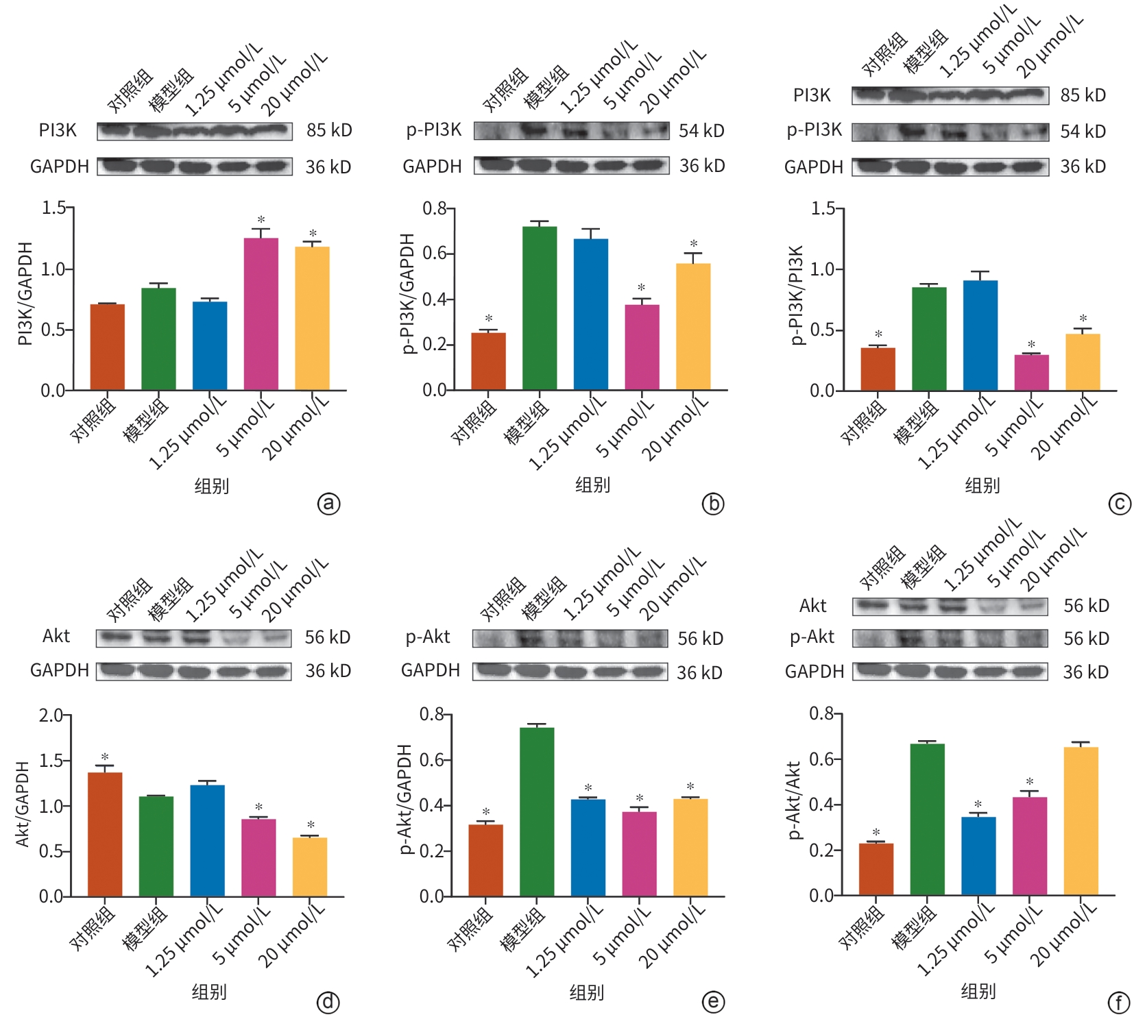
注: a、b,PI3K、p-PI3K的蛋白表达及相对表达量统计图;c,p-PI3K/PI3K的蛋白表达及相对表达量统计图;d、e,Akt、p-Akt的蛋白表达及相对表达量统计图;f,p-AKT/AKT的蛋白表达及相对表达量统计图。与模型组比较,*P<0.05。MAFLD,代谢相关脂肪性肝病;FFA,游离脂肪酸;PI3K,磷脂酰肌醇3-激酶;GAPDH,甘油醛-3-磷酸脱氢酶;p-PI3K,磷酸化磷脂酰肌醇3-激酶;Akt,蛋白激酶B;p-Akt,磷酸化蛋白激酶B。
图 4 2,5-DHBA调节PI3K/AKT信号通路改善MAFLD
Figure 4. 2,5-DHBA ameliorates MAFLD by modulating the PI3K/AKT signaling pathway
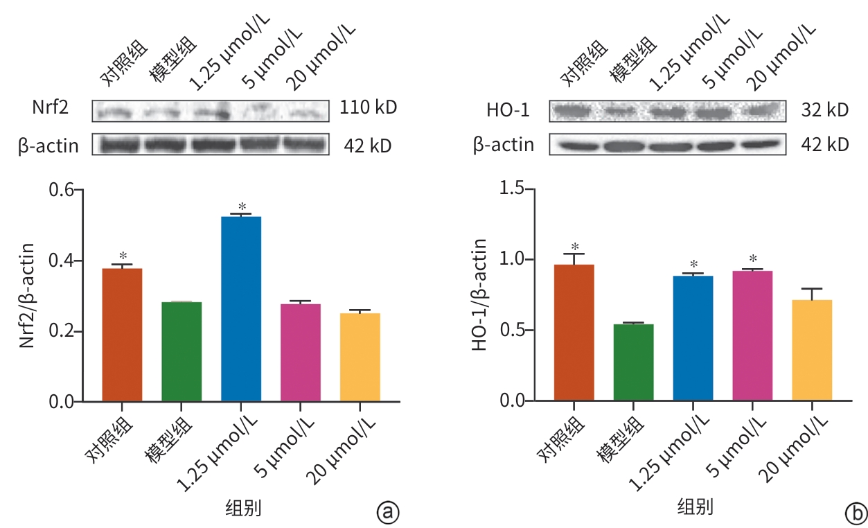
注: a,Nrf2的蛋白表达及相对表达量统计图;b,HO-1的蛋白表达及相对表达量统计图。与模型组比较,*P<0.05。Nrf2,核转录因子红系2相关因子2;HO-1,血红素加氧酶1;GAPDH,甘油醛-3-磷酸脱氢酶;2,5-DHBA,2,5-二羟基苯甲酸;MAFLD,代谢相关脂肪性肝病。
图 5 2,5-DHBA抑制氧化应激改善MAFLD
Figure 5. 2,5-DHBA inhibits oxidative stress to ameliorate MAFLD
表 1 引物序列
Table 1. Primer sequences
引物 方向 序列(5'-3') Nrf2 F AAGCAGACGGAGGAGGAG R GGAGGTGTTCAGGCAAGG SREBF1 F CATCCACATGGCTCTGAGTG R CTCATCCACAAAGAAGCGGT FASN F GAGAAAGCTTGCCAAACAGG R GAGGGTCTTGCAGGAGACAG HO-1 F GCTCAACATCCAGCTCTTTGAGG R GACAAAGTTCATGGCCCTGGGA GAPDH F GTTCCCACCCTAGAAAGTCCA R ACTTGGAGGAGGTTTGCTGG 注:Nrf2,核转录因子红系2相关因子2;SREBF1,固醇调控元件结合转录因子1;FASN,脂肪酸合成酶;HO-1,血红素加氧酶1;GAPDH,甘油醛-3-磷酸脱氢酶。
 下载: 导出CSV
-
[1] Machado M V, Diehl A M. Pathogenesis of nonalcoholic steatohepatitis[J]. Gastroenterology, 2016, 150( 8): 1769- 1777. DOI: 10.1053/j.gastro.2016.02.066. [2] Younossi Z M, Golabi P, Paik J M, et al. The global epidemiology of nonalcoholic fatty liver disease(NAFLD) and nonalcoholic steatohepatitis(NASH): A systematic review[J]. Hepatology, 2023, 77( 4): 1335- 1347. DOI: 10.1097/HEP.0000000000000004. [3] Younossi Z M, Kalligeros M, Henry L. Epidemiology of metabolic dysfunction-associated steatotic liver disease[J]. Clin Mol Hepatol, 2025, 31( Suppl): S32- S50. DOI: 10.3350/cmh.2024.0431. [4] Semmler G, Datz C, Trauner M. Eating, diet, and nutrition for the treatment of non-alcoholic fatty liver disease[J]. Clin Mol Hepatol, 2023, 29( Suppl): S244- S260. DOI: 10.3350/cmh.2022.0364. [5] Devasia A G, Ramasamy A, Leo C H. Current therapeutic landscape for metabolic dysfunction-associated steatohepatitis[J]. Int J Mol Sci, 2025, 26( 4): 1778. DOI: 10.3390/ijms26041778. [6] Buzzetti E, Pinzani M, Tsochatzis E A. The multiple-hit pathogenesis of non-alcoholic fatty liver disease(NAFLD)[J]. Metabolism, 2016, 65( 8): 1038- 1048. DOI: 10.1016/j.metabol.2015.12.012. [7] Heeren J, Scheja L. Metabolic-associated fatty liver disease and lipoprotein metabolism[J]. Mol Metab, 2021, 50: 101238. DOI: 10.1016/j.molmet.2021.101238. [8] Li Y X, Yang P P, Ye J L, et al. Updated mechanisms of MASLD pathogenesis[J]. Lipids Health Dis, 2024, 23( 1): 117. DOI: 10.1186/s12944-024-02108-x. [9] Chen Z, Tian R F, She Z G, et al. Role of oxidative stress in the pathogenesis of nonalcoholic fatty liver disease[J]. Free Radic Biol Med, 2020, 152: 116- 141. DOI: 10.1016/j.freeradbiomed.2020.02.025. [10] Miura Y, Voican C, Sakai Y, et al. A computational model of the crosstalk between hepatocyte fatty acid metabolism and oxidative stress highlights the key enzymes, metabolites, and detoxification pathways in the context of MASLD[J]. Toxicol Appl Pharmacol, 2025, 495: 117185. DOI: 10.1016/j.taap.2024.117185. [11] Yang K L, Chen J P, Zhang T Q, et al. Efficacy and safety of dietary polyphenol supplementation in the treatment of non-alcoholic fatty liver disease: A systematic review and meta-analysis[J]. Front Immunol, 2022, 13: 949746. DOI: 10.3389/fimmu.2022.949746. [12] Abedi F, Razavi B M, Hosseinzadeh H. A review on gentisic acid as a plant derived phenolic acid and metabolite of aspirin: Comprehensive pharmacology, toxicology, and some pharmaceutical aspects[J]. Phytother Res, 2020, 34( 4): 729- 741. DOI: 10.1002/ptr.6573. [13] Han X, Guo J L, Gao Y X, et al. Gentisic acid prevents diet-induced obesity in mice by accelerating the thermogenesis of brown adipose tissue[J]. Food Funct, 2021, 12( 3): 1262- 1270. DOI: 10.1039/d0fo02474k. [14] Hsiang C Y, Hsu K T, Lo H Y, et al. 2, 5-dihydroxybenzoic acid ameliorates metabolic dysfunction-associated steatotic liver disease by targeting the CCL2-CCR2 axis to reduce lipid accumulation[J]. Nutrients, 2025, 17( 11): 1835. DOI: 10.3390/nu17111835. [15] Bajpai A K, Davuluri S, Tiwary K, et al. Systematic comparison of the protein-protein interaction databases from a user's perspective[J]. J Biomed Inform, 2020, 103: 103380. DOI: 10.1016/j.jbi.2020.103380. [16] Zhou Y Y, Zhou B, Pache L, et al. Metascape provides a biologist-oriented resource for the analysis of systems-level datasets[J]. Nat Commun, 2019, 10( 1): 1523. DOI: 10.1038/s41467-019-09234-6. [17] Aniceto N, Martinho N, Rufino I, et al. LigExtract: Large-scale automated identification of ligands from protein structures in the protein data bank[J]. Genomics Proteomics Bioinformatics, 2025, 23( 4): qzaf018. DOI: 10.1093/gpbjnl/qzaf018. [18] Li Z Q, Guo C Q, Wang T J, et al. Exploring the mechanism of PFTrDA on human hepatic metabolic diseases based on network toxicology and molecular docking[J]. Sci Rep, 2025, 15( 1): 38872. DOI: 10.1038/s41598-025-22665-0. [19] Snigireva A V, Morenkov O S, Skarga Y Y, et al. A 2, 5-dihydroxybenzoic acid-gelatin conjugate inhibits the basal and Hsp90-stimulated migration and invasion of tumor cells[J]. J Funct Biomater, 2020, 11( 2): 39. DOI: 10.3390/jfb11020039. [20] Baidya R, Gautheron J, Crawford D H G, et al. Inhibition of MLKL attenuates necroptotic cell death in a murine cell model of ischaemia injury[J]. J Clin Med, 2021, 10( 2): 212. DOI: 10.3390/jcm10020212. [21] Geh D, Anstee Q M, Reeves H L. NAFLD-associated HCC: Progress and opportunities[J]. J Hepatocell Carcinoma, 2021, 8: 223- 239. DOI: 10.2147/JHC.S272213. [22] Abdelmalek M F. Nonalcoholic fatty liver disease: Another leap forward[J]. Nat Rev Gastroenterol Hepatol, 2021, 18( 2): 85- 86. DOI: 10.1038/s41575-020-00406-0. [23] Jurek J M, Zablocka-sowinska K, Clavero mestres H, et al. The impact of dietary interventions on metabolic outcomes in metabolic dysfunction-associated steatotic liver disease(MASLD) and comorbid conditions, including obesity and type 2 diabetes[J]. Nutrients, 2025, 17( 7): 1257. DOI: 10.3390/nu17071257. [24] Yaskolka meir A, Rinott E, Tsaban G, et al. Effect of green-Mediterranean diet on intrahepatic fat: The DIRECT PLUS randomised controlled trial[J]. Gut, 2021, 70( 11): 2085- 2095. DOI: 10.1136/gutjnl-2020-323106. [25] Wang Z W, Jin Z, Xiong Z F. Research progress of epidermal growth factor receptor in metabolic dysfunction-associated steatotic liver disease and related diseases[J]. Diabetes Obes Metab, 2025, 27( 10): 5418- 5431. DOI: 10.1111/dom.16653. [26] Lv M F, Zhai Y X, Yu H, et al. Linarin alleviates high-fat diet-induced NAFLD via modulating the PI3K/Akt/mTOR pathway, autophagy, and gut microbiota[J]. Biochim Biophys Acta Mol Cell Biol Lipids, 2025, 1870( 7): 159666. DOI: 10.1016/j.bbalip.2025.159666. [27] Hai S P, Li X T, Xie E L, et al. Intestinal IL-33 promotes microbiota-derived trimethylamine N-oxide synthesis and drives metabolic dysfunction-associated steatotic liver disease progression by exerting dual regulation on HIF-1α[J]. Hepatology, 2025, 82( 1): 184- 198. DOI: 10.1097/HEP.0000000000000985. [28] Ogawa N, Seki A, Nasti A, et al. Therapeutic effects of adipose tissue-derived mesenchymal stem cells on ER stress in a murine model of metabolic dysfunction-associated steatohepatitis: An in vivo and in vitro study[J]. Stem Cell Res Ther, 2025, 16( 1): 349. DOI: 10.1186/s13287-025-04482-4. [29] Dömer D, Walther T, Möller S, et al. Neutrophil extracellular traps activate proinflammatory functions of human neutrophils[J]. Front Immunol, 2021, 12: 636954. DOI: 10.3389/fimmu.2021.636954. [30] van der Windt D J, Sud V, Zhang H J, et al. Neutrophil extracellular traps promote inflammation and development of hepatocellular carcinoma in nonalcoholic steatohepatitis[J]. Hepatology, 2018, 68( 4): 1347- 1360. DOI: 10.1002/hep.29914. [31] Shahzad M H, Rayes R F, Cools-lartigue J, et al. Neutrophil extracellular traps in cancer[J]. Nat Rev Cancer, 2026, 26( 2): 104- 117. DOI: 10.1038/s41568-025-00888-7. [32] Feješ A, Belvončíková P, Bečka E, et al. Myeloperoxidase, extracellular DNA and neutrophil extracellular trap formation in the animal models of metabolic dysfunction-associated steatotic liver disease[J]. World J Gastroenterol, 2025, 31( 27): 106166. DOI: 10.3748/wjg.v31.i27.106166. [33] Xiao H M, Sun X H, Lin Z Y, et al. Gentiopicroside targets PAQR3 to activate the PI3K/AKT signaling pathway and ameliorate disordered glucose and lipid metabolism[J]. Acta Pharm Sin B, 2022, 12( 6): 2887- 2904. DOI: 10.1016/j.apsb.2021.12.023. [34] Lien E C, Dibble C C, Toker A. PI3K signaling in cancer: Beyond AKT[J]. Curr Opin Cell Biol, 2017, 45: 62- 71. DOI: 10.1016/j.ceb.2017.02.007. [35] Wang Z, Wang L Y, Hou Y C, et al. Dietary carbohydrate-to-protein caloric ratio availability affects hepatic fat deposition via the IGF-1/PI3K/Akt signaling pathway[J]. Food Res Int, 2025, 219: 117035. DOI: 10.1016/j.foodres.2025.117035. [36] Yuan Y W, Gong M X, Pan R L, et al. Artificially fermented dark loose tea ameliorates metabolic-associated fatty liver disease by activating the PI3K/AKT signalling pathway and regulating gut microbiota dysbiosis[J]. Food Chem X, 2025, 31: 103099. DOI: 10.1016/j.fochx.2025.103099. [37] Li J D, Wang T Q, Liu P P, et al. Hesperetin ameliorates hepatic oxidative stress and inflammation via the PI3K/AKT-Nrf2-ARE pathway in oleic acid-induced HepG2 cells and a rat model of high-fat diet-induced NAFLD[J]. Food Funct, 2021, 12( 9): 3898- 3918. DOI: 10.1039/d0fo02736G. [38] Li L P, Huang L Y, Lei R C, et al. DEHP and DBP, common phthalates, induce glucose metabolism disorders in rats via oxidative damage of PI3K/Akt/GLUT4 signaling[J]. Environ Pollut, 2024, 341: 122948. DOI: 10.1016/j.envpol.2023.122948. [39] Liu S N, Pi J B, Zhang Q. Signal amplification in the KEAP1-NRF2-ARE antioxidant response pathway[J]. Redox Biol, 2022, 54: 102389. DOI: 10.1016/j.redox.2022.102389. [40] Zhang Y M, Zhang Z G. Modulating nuclear factor erythroid 2-related factor 2 and heme oxygenase-1 in liver-brain axis disorders[J]. World J Psychiatry, 2025, 15( 9): 108382. DOI: 10.5498/wjp.v15.i9.108382. [41] Cheng Y, Gao Y, Li J, et al. TrkB agonist N-acetyl serotonin promotes functional recovery after traumatic brain injury by suppressing ferroptosis via the PI3K/Akt/Nrf2/Ferritin H pathway[J]. Free Radic Biol Med, 2023, 194: 184- 198. DOI: 10.1016/j.freeradbiomed.2022.12.002. [42] Schieber M, Chandel N S. ROS function in redox signaling and oxidative stress[J]. Curr Biol, 2014, 24( 10): R453- R462. DOI: 10.1016/j.cub.2014.03.034. [43] Li Y, Xu S Q, Mihaylova M M, et al. AMPK phosphorylates and inhibits SREBP activity to attenuate hepatic steatosis and atherosclerosis in diet-induced insulin-resistant mice[J]. Cell Metab, 2011, 13( 4): 376- 388. DOI: 10.1016/j.cmet.2011.03.009. [44] Salazar M, Rojo A I, Velasco D, et al. Glycogen synthase kinase-3beta inhibits the xenobiotic and antioxidant cell response by direct phosphorylation and nuclear exclusion of the transcription factor Nrf2[J]. J Biol Chem, 2006, 281( 21): 14841- 14851. DOI: 10.1074/jbc.M513737200. -

 本文二维码
本文二维码
计量
- 文章访问数: 351
- HTML全文浏览量: 87
- PDF下载量: 55
- 被引次数: 0