PDF下载 ( 16002 KB)

4例肝内胆管内乳头状肿瘤的临床及病理学特征分析
DOI: 10.12449/JCH260723
Clinical and pathological features of intrahepatic intraductal papillary neoplasm of the bile duct: An analysis of four cases
-
摘要:
目的 探讨肝内胆管内乳头状肿瘤的临床及病理学特征,提高对该疾病的认识和诊疗水平。 方法 回顾性分析2010年1月—2025年12月于首都医科大学附属北京朝阳医院接受手术切除且病理确诊为肝内胆管内乳头状肿瘤的4例病例的临床资料,总结其临床表现、影像学及病理学特征,并结合国内外文献进行分析。 结果 4例患者中女性3例、男性1例,中位年龄65岁,临床表现包括黄疸、腹痛或腹泻,1例为无症状体检发现。影像学主要表现为胆管扩张或囊实性占位性病变。病理学分型包括胃型、肠型、胰胆管型和嗜酸细胞型各1例,其中伴低级别上皮内瘤变1例、高级别上皮内瘤变3例(其中1例伴浸润性癌)。 结论 肝内胆管内乳头状肿瘤是一种少见的肝脏癌前病变,其临床、影像及病理表现多样,根治性手术切除是治疗的关键。 Abstract:Objective To investigate the clinical and pathological features of intrahepatic intraductal papillary neoplasm of the bile duct (IPNB), and to improve the understanding, diagnosis, and treatment of this disease. Methods A retrospective analysis was performed for four patients who underwent surgical resection in Beijing Chaoyang Hospital, Capital Medical University, from January 2010 to December 2025 and were diagnosed with intrahepatic IPNB based on pathological examination. The clinical, imaging, and pathological features of these patients were summarized, and a literature review was performed for related articles in China and globally. Results Among the four patients, there were three female patients and one male patient, with a median age of 65 years, and clinical manifestations included jaundice, abdominal pain or diarrhea, and asymptomatic findings on physical examination. The main imaging manifestation was bile duct dilatation or solid-cystic space-occupying lesion. Pathological typing revealed one case each of the gastric, intestinal, pancreatobiliary, and oncocytic types, and there was one patient with low-grade intraepithelial neoplasia and three patients with high-grade intraepithelial neoplasia, among whom one patient also had invasive carcinoma. Conclusion Intrahepatic IPNB is a rare precancerous lesion of the liver with diverse clinical, imaging, and pathological manifestations, and radical surgical resection remains the cornerstone of treatment. -
Key words:
- Intrahepatic Biliary Papillomatosis /
- Disease Attributes /
- Diagnosis
-
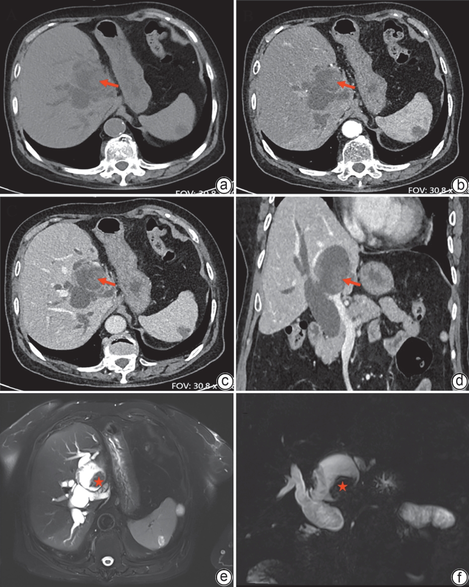
注: a,平扫CT可见肝内胆管扩张,左肝管乳头状肿物(红色箭头);b、c,动脉期及门静脉期增强CT可见肿物强化(红色箭头);d,冠状位增强计算机体层成像可见肝内外胆管扩张,左肝管内见强化乳头状肿物(红色箭头);e、f,轴位及冠状位磁共振胰胆管成像显示左肝内胆管乳头状实性肿物(红色星号)。
图 1 肝内胆管内乳头状肿瘤的增强计算机体层成像及磁共振胰胆管成像表现
Figure 1. CT scan and MRCP appearance of intraductal papillary neoplasms of the intrahepatic bile duct
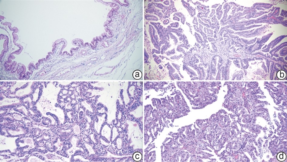
注: a,胃型:乳头大部分为单纯乳头,胞浆富含黏液,类似胃小凹上皮(HE染色,×100);b,肠型:乳头结构大部分为弥漫增生的单纯乳头,类似结肠绒毛管状腺瘤,表面被覆假复层上皮细胞(HE染色,×100);c,胰胆管型:复杂或分支状乳头,细胞呈立方或低柱状,细胞核居中、圆形,(HE染色,×200);d,嗜酸细胞型:复杂或分支乳头状生长,增生密集,细胞呈柱状,胞浆丰富、嗜酸性,细胞核居中、圆形(HE染色,×200)。
图 2 肝内胆管内乳头状肿瘤的显微镜下表现
Figure 2. Microscopic appearance of intraductal papillary neoplasms of the intrahepatic bile duct
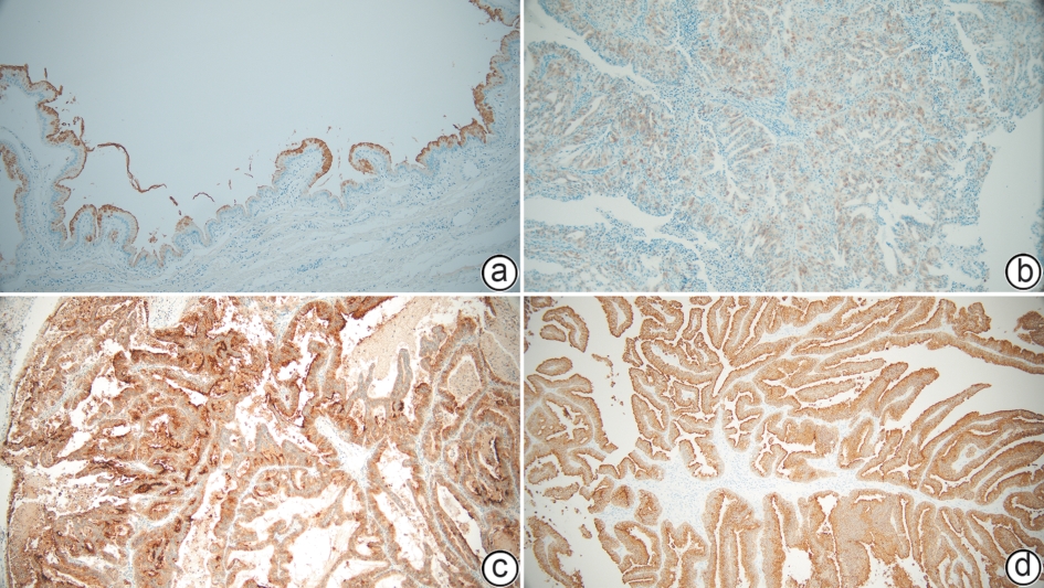
注: a,黏蛋白5AC阳性,细胞质内着色;b,黏蛋白6阳性,细胞质内着色;c,上皮膜抗原(黏蛋白1)阳性,细胞膜质内着色;d,黏蛋白2阳性,细胞质内着色。
图 3 免疫组织化学结果(EnVision法,×100)
Figure 3. Immunohistochemical results using EnVision method (×100)
表 1 4例肝内胆管内乳头状肿瘤患者的临床资料及病理学特征
Table 1. Clinical data and pathological features of 4 patients with intraductal papillary neoplasms of the intrahepatic bile duct
项目 病例1 病例2 病例3 病例4 性别 女 女 男 女 年龄(岁) 56 73 62 68 临床症状 黄疸 消化不良 腹泻及黄疸 无 天冬氨酸氨基转移酶1) 122.0 1201.0 185.0 30.0 总胆红素2) 485.3 36.5 329.4 6.2 部位/大小(cm) 肝左叶/1.5×1.5 肝左叶/2.0×1.5 肝右叶/2.5×2.0 肝右叶/8.8×7.1 手术方式 肝移植 左半肝切除术+肝外胆管
切除术+胆肠吻合术右半肝切除术+肝外胆管切除
术+胆肠吻合术+淋巴结清扫肝部分切除术 随访时间/结果 40个月/死亡 5个月/存活 39个月/存活 115个月/死亡 组织学分级 低级别 高级别 高级别伴浸润癌 高级别 亚型 胃型 肠型 胰胆管型 嗜酸细胞型 改良分级 1级 1级 2级 2级 免疫表型 EMA部分+,MUC2-,
MUC5AC+,MUC6部
分+,CDX2-EMA灶+,MUC2+,MUC5
AC部分+,MUC6部分+,
CDX2+EMA+,MUC2-,MUC5AC+,
MUC6部分+,CDX2-EMA部分+,MUC2-,
MUC5AC+,MUC6部
分+,CDX2-注:1)正常范围:<40 U/L;2)正常范围:<20 μmol/L。EMA,上皮膜抗原;MUC,黏蛋白;CDX2:尾型同源盒转录因子2。
 下载: 导出CSV
-
[1] Fléjou J F. Classification OMS 2010 des tumeurs digestives: La quatrième édition[J]. Ann De Pathol, 2011, 31( 5): S27- S31. DOI: 10.1016/j.annpat.2011.08.001. [2] Nagtegaal I D, Odze R D, Klimstra D, et al. The 2019 WHO classification of tumours of the digestive system[J]. Histopathology, 2020, 76( 2): 182- 188. DOI: 10.1111/his.13975. [3] Rocha F G, Lee H, Katabi N, et al. Intraductal papillary neoplasm of the bile duct: A biliary equivalent to intraductal papillary mucinous neoplasm of the pancreas?[J]. Hepatology, 2012, 56( 4): 1352- 1360. DOI: 10.1002/hep.25786. [4] Youn J M, Hwang S, Ahn C S, et al. Clinicopathological features and long-term outcomes of intraductal papillary neoplasms of the bile duct of the liver: Single-institution experience with 146 patients[J]. J Gastrointest Surg, 2022, 26( 7): 1394- 1405. DOI: 10.1007/s11605-022-05268-2. [5] Aoki Y, Mizuma M, Hata T, et al. Intraductal papillary neoplasms of the bile duct consist of two distinct types specifically associated with clinicopathological features and molecular phenotypes[J]. J Pathol, 2020, 251( 1): 38- 48. DOI: 10.1002/path.5398. [6] Nakanuma Y, Uesaka K, Kakuda Y, et al. Intraductal papillary neoplasm of bile duct: Updated clinicopathological characteristics and molecular and genetic alterations[J]. J Clin Med, 2020, 9( 12): 3991. DOI: 10.3390/jcm9123991. [7] Nakanuma Y, Jang K T, Fukushima N, et al. A statement by the Japan-Korea expert pathologists for future clinicopathological and molecular analyses toward consensus building of intraductal papillary neoplasm of the bile duct through several opinions at the present stage[J]. J Hepato Biliary Pancreat Sci, 2018, 25( 3): 181- 187. DOI: 10.1002/jhbp.532. [8] Kubota K, Jang J Y, Nakanuma Y, et al. Clinicopathological characteristics of intraductal papillary neoplasm of the bile duct: A Japan-Korea collaborative study[J]. J Hepato Biliary Pancreat Sci, 2020, 27( 9): 581- 597. DOI: 10.1002/jhbp.785. [9] Lluís N, Onoe S, Serradilla-Martín M, et al. Intraductal papillary neoplasms of the bile ducts: A comparative study of a rare disease in Europe and Nagoya, Japan[J]. HPB, 2024, 26( 4): 565- 575. DOI: 10.1016/j.hpb.2024.01.009. [10] Huang X R, Zhang Z, Zhu D S, et al. Clinicopathological and prognosis analysis of intraductal papillary neoplasm of the bile duct: A dual-center retrospective study[J]. Eur J Surg Oncol, 2025, 51( 9): 110212. DOI: 10.1016/j.ejso.2025.110212. [11] Chang Xiaoyan, Li Ji, Ren Xinyu, et al. Intraductal papillary neoplasms of intrahepatic bile duct: A clinicopathologic study of 25 cases[J]. Chin J Pathol, 2016, 45( 4): 243- 247. DOI: 10.3760/cma.j.issn.0529-5807.2016.04.006.常晓燕, 李霁, 任新瑜, 等. 肝内胆管内乳头状肿瘤临床及病理学分析[J]. 中华病理学杂志, 2016, 45( 4): 243- 247. DOI: 10.3760/cma.j.issn.0529-5807.2016.04.006. [12] Han S Y, Kim D U, Nam H S, et al. Comparison of the malignant predictors in intrahepatic and extrahepatic intraductal papillary neoplasm of the bile duct[J]. J Clin Med, 2022, 11( 7): 1985. DOI: 10.3390/jcm11071985. [13] Kim J R, Jang K T, Jang J Y, et al. Clinicopathologic analysis of intraductal papillary neoplasm of bile duct: Korean multicenter cohort study[J]. HPB, 2020, 22( 8): 1139- 1148. DOI: 10.1016/j.hpb.2019.11.007. [14] Chun J, Sung Y N, An S, et al. Oncocytic type has distinct immunohistochemical and recurrence-free survival than other histologic types of the intraductal papillary neoplasm of the bile duct[J]. Hum Pathol, 2024, 148: 72- 80. DOI: 10.1016/j.humpath.2024.05.006. [15] Nakanuma Y, Kakuda Y, Fukumura Y, et al. The pathologic and genetic characteristics of the intestinal subtype of intraductal papillary neoplasms of the bile duct[J]. Am J Surg Pathol, 2019, 43( 9): 1212- 1220. DOI: 10.1097/PAS.0000000000001295. [16] Nakanuma Y, Kakuda Y, Sugino T, et al. Pathologies of precursor lesions of biliary tract carcinoma[J]. Cancers, 2022, 14( 21): 5358. DOI: 10.3390/cancers14215358. [17] Chung T, Oh S, Won J, et al. Genomic and transcriptomic signatures of sequential carcinogenesis from papillary neoplasm to biliary tract cancer[J]. J Hepatol, 2025, 83( 1): 119- 130. DOI: 10.1016/j.jhep.2025.01.007. [18] Huang Deng, Gan Lang. Clinical status and research progress of intraductal papillary neoplasm of the bile duct[J]. Chin J Dig Surg, 2024, 23( 7): 919- 925. DOI: 10.3760/cma.j.cn115610-20240523-00259.黄登, 甘浪. 胆管内乳头状肿瘤的临床现状和研究进展[J]. 中华消化外科杂志, 2024, 23( 7): 919- 925. DOI: 10.3760/cma.j.cn115610-20240523-00259. [19] Wang Haonian, Sun Bei, Chen Hua. Diagnosis and treatment strategies for intraductal papillary neoplasm of the bile duct[J/OL]. Chin J Liver Dis(Electronic Version), 2024, 13( 2): 140- 144. DOI: 10.3877/cma.j.issn.2095-3232.2024.02.004王浩年, 孙备, 陈华. 胆管内乳头状肿瘤的诊治策略[J/OL]. 中华肝脏外科手术学电子杂志, 2024, 13( 2): 140- 144. DOI: 10.3877/cma.j.issn.2095-3232.2024.02.004. [20] Noguchi D, Kuriyama N, Kozuka Y, et al. A recent update on the morphological classification of intraductal papillary neoplasm of the bile duct: Correlation with postoperative prognosis and pathological features[J]. PLoS One, 2025, 20( 5): e0325081. DOI: 10.1371/journal.pone.0325081. [21] Zhang Y Q, Liang Y, Liu Y, et al. Photodynamic therapy for hepatic hilar intraductal papillary neoplasm of the bile duct: A case report[J]. VideoGIE, 2022, 7( 5): 178- 181. DOI: 10.1016/j.vgie.2022.01.016. -

 本文二维码
本文二维码
计量
- 文章访问数: 710
- HTML全文浏览量: 261
- PDF下载量: 49
- 被引次数: 0