PDF下载 ( 947 KB)

肠道通透性改变在肝脏疾病中的作用机制与临床意义
DOI: 10.12449/JCH260733
利益冲突声明:本文不存在任何利益冲突。
作者贡献声明:杜家升负责查阅文献,论文撰写与修改;何欢负责归纳文献与分析;尹兴隆、杨小番参与修改论文;陈刚负责指导论文撰写并定稿。
Mechanism of action and clinical significance of altered intestinal permeability in liver diseases
-
摘要: 肠道通透性在肠-肝轴中扮演核心角色,已成为肝脏疾病防治的重要靶点。本文基于近年来国内外相关研究,综述了肠道屏障功能与肠道通透性的定义及相互关系、肠道通透性的评估方法,并重点探讨了肠道通透性在多种肝脏疾病发生发展中的作用机制与临床意义。研究表明, 肠道通透性的增加是多种肝脏疾病的重要特征和驱动因素,该状态导致细菌及有害物质易位,引发全身性炎症反应,从而加剧肝损伤、纤维化及代谢紊乱,与代谢相关脂肪性肝病、酒精性肝病、自身免疫性肝病及其终末事件(包括肝硬化、肝衰竭以及肝细胞癌)的发生发展密切相关。未来研究应致力于评估方法的标准化、深入探索作用机制及相关成果的临床转化。Abstract: Intestinal permeability plays a pivotal role in the gut-liver axis and has emerged as a critical target for the prevention and treatment of liver diseases. Based on recent studies in China and globally, this article reviews the definitions and interrelationship of intestinal barrier function and intestinal permeability, the methods for assessing intestinal permeability, and the mechanism of action and clinical significance of intestinal permeability in the development and progression of various liver diseases. Studies have shown that increased intestinal permeability is a significant hallmark and an important driving factor for multiple liver diseases, and this state leads to the translocation of bacteria and harmful substances, trigger a systemic inflammatory response, and thus exacerbates liver injury, fibrosis, and metabolic dysregulation. Therefore, it is closely associated with the development and progression of metabolic associated fatty liver disease, alcoholic liver disease, autoimmune liver diseases, and their end-stage events, including liver cirrhosis, liver failure, and hepatocellular carcinoma. Future research should focus on the standardization of assessment methods, the exploration of underlying mechanisms, and the clinical translation of these findings.
-
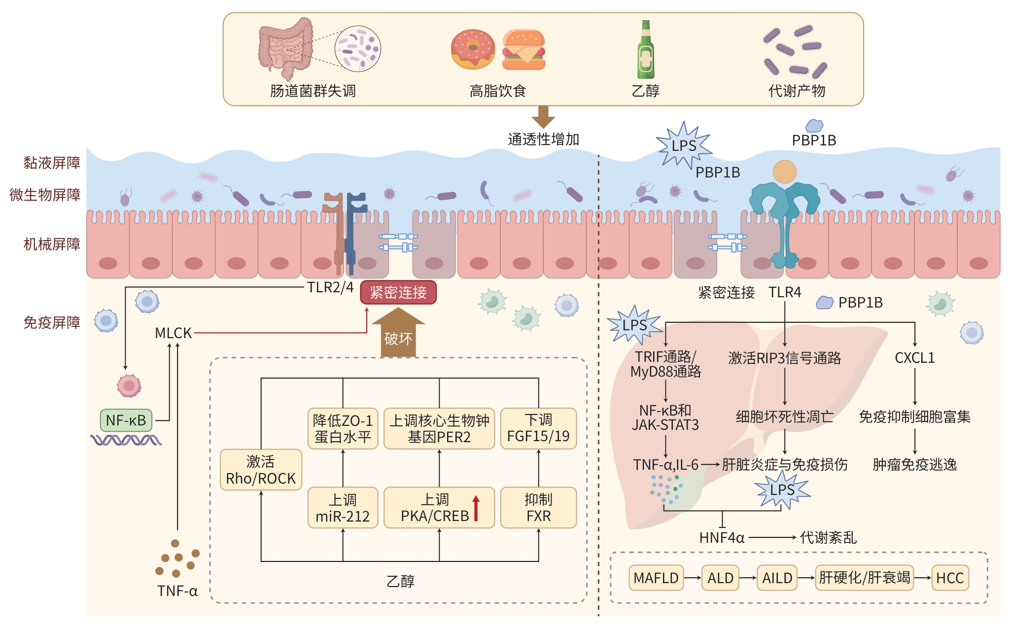
注: TLR,Toll样受体;MLCK,肌球蛋白轻链激酶;NF-κB,核因子κB;TNF-α,肿瘤坏死因子α;Rho/ROCK,Rho/Rho关联卷曲螺旋形成蛋白激酶通路;ZO-1,紧密连接蛋白1;miR-212,微RNA-212;PER2,昼夜节律蛋白2;PKA/CREB,蛋白激酶A/环磷酸腺苷应答元件结合蛋白质;FGF,成纤维细胞生长因子;FXR,法尼醇X受体;LPS,脂多糖;PBP1B,青霉素结合蛋白1B;TRIF,β干扰素TIR结构域衔接蛋白;RIP3,受体相互作用蛋白激酶3;MyD88,髓样分化因子88;JAK-STAT3,Janus激酶-信号转导及转录激活蛋白3;IL-6,白细胞介素6;HNF-4α,肝细胞核因子4α,CXCL1,CXC基序趋化因子配体1;MAFLD,代谢相关脂肪性肝病;ALD,酒精性肝病;AILD,自身免疫性肝病;HCC,肝细胞癌。
图 1 肠道通透性增加及促进肝脏疾病进展的机制
Figure 1. Mechanistic diagram of increased intestinal permeability and its role in promoting liver disease progression
-
[1] Song C Y, Chai Z L, Chen S, et al. Intestinal mucus components and secretion mechanisms: What we do and do not know[J]. Exp Mol Med, 2023, 55( 4): 681- 691. DOI: 10.1038/s12276-023-00960-y. [2] Perez-diaz-del-campo N, Castelnuovo G, Ribaldone D G, et al. Fecal and circulating biomarkers for the non-invasive assessment of intestinal permeability[J]. Diagnostics, 2023, 13( 11): 1976. DOI: 10.3390/diagnostics13111976. [3] France M M, Turner J R. The mucosal barrier at a glance[J]. J Cell Sci, 2017, 130( 2): 307- 314. DOI: 10.1242/jcs.193482. [4] Horowitz A, Chanez-paredes S D, Haest X, et al. Paracellular permeability and tight junction regulation in gut health and disease[J]. Nat Rev Gastroenterol Hepatol, 2023, 20( 7): 417- 432. DOI: 10.1038/s41575-023-00766-3. [5] Paone P, Cani P D. Mucus barrier, mucins and gut microbiota: The expected slimy partners?[J]. Gut, 2020, 69( 12): 2232- 2243. DOI: 10.1136/gutjnl-2020-322260. [6] Yang M Z, Jiang Z P, Zhou L T, et al. 3’-Sialyllactose and B. infantis synergistically alleviate gut inflammation and barrier dysfunction by enriching cross-feeding bacteria for short-chain fatty acid biosynthesis[J]. Gut Microbes, 2025, 17( 1): 2486512. DOI: 10.1080/19490976.2025.2486512. [7] Oami T, Abtahi S, Shimazui T, et al. Claudin-2 upregulation enhances intestinal permeability, immune activation, dysbiosis, and mortality in sepsis[J]. Proc Natl Acad Sci USA, 2024, 121( 10): e2217877121. DOI: 10.1073/pnas.2217877121. [8] Chanez-paredes S D, Abtahi S, Zha J M, et al. Mechanisms underlying distinct subcellular localization and regulation of epithelial long myosin light-chain kinase splice variants[J]. J Biol Chem, 2024, 300( 2): 105643. DOI: 10.1016/j.jbc.2024.105643. [9] Garg A, Zhao A, Erickson S L, et al. Pregnane X receptor activation attenuates inflammation-associated intestinal epithelial barrier dysfunction by inhibiting cytokine-induced myosin light-chain kinase expression and c-Jun N-terminal kinase 1/2 activation[J]. J Pharmacol Exp Ther, 2016, 359( 1): 91- 101. DOI: 10.1124/jpet.116.234096. [10] Schoultz I, Keita Å V. The intestinal barrier and current techniques for the assessment of gut permeability[J]. Cells, 2020, 9( 8): 1909. DOI: 10.3390/cells9081909. [11] Seethaler B, Basrai M, Neyrinck AM, et al. Biomarkers for assessment of intestinal permeability in clinical practice[J]. Am J Physiol Gastrointest Liver Physiol, 2021, 321( 1): G11- G17. DOI: 10.1152/ajpgi.00113.2021. [12] Alizadeh A, Akbari P, Garssen J, et al. Epithelial integrity, junctional complexes, and biomarkers associated with intestinal functions[J]. Tissue Barriers, 2022, 10( 3): 1996830. DOI: 10.1080/21688370.2021.1996830. [13] Remund B, Yilmaz B, Sokollik C. D-lactate: Implications for gastrointestinal diseases[J]. Children, 2023, 10( 6): 945. DOI: 10.3390/children10060945. [14] Stephens M, von der Weid P Y. Lipopolysaccharides modulate intestinal epithelial permeability and inflammation in a species-specific manner[J]. Gut Microbes, 2020, 11( 3): 421- 432. DOI: 10.1080/19490976.2019.1629235. [15] Sturgeon C, Fasano A. Zonulin, a regulator of epithelial and endothelial barrier functions, and its involvement in chronic inflammatory diseases[J]. Tissue Barriers, 2016, 4( 4): e1251384. DOI: 10.1080/21688370.2016.1251384. [16] Huang X, Zhou Y C, Sun Y W, et al. Intestinal fatty acid binding protein: A rising therapeutic target in lipid metabolism[J]. Prog Lipid Res, 2022, 87: 101178. DOI: 10.1016/j.plipres.2022.101178. [17] Narum M, Seljeflot I, Bratseth V, et al. Intestinal fatty acid binding protein is associated with coronary artery disease in long-term type 1 diabetes-the Dialong study[J]. Cardiovasc Diabetol, 2024, 23( 1): 419. DOI: 10.1186/s12933-024-02509-3. [18] Martínez Velasco S, González García A, Irastorza Terradillos I X, et al. Chapter 4-Intestinal permeability assessment using lactulose and mannitol in celiac disease[M]//Castellanos-rubio A, Galluzzi L. Methods in Cell Biology. Academic Press, 2023: 39- 50. [19] Streekstra E J, Keuper-navis M, van den Heuvel J J M W, et al. Human enteroid monolayers as a potential alternative for Ussing chamber and Caco-2 monolayers to study passive permeability and drug efflux[J]. Eur J Pharm Sci, 2024, 201: 106877. DOI: 10.1016/j.ejps.2024.106877. [20] Kourula S, Derksen M, Jardi F, et al. Intestinal organoids as an in vitro platform to characterize disposition, metabolism, and safety profile of small molecules[J]. Eur J Pharm Sci, 2023, 188: 106481. DOI: 10.1016/j.ejps.2023.106481. [21] Rosso C, Caviglia G P, Younes R, et al. Circulating zonulin is related to hepatic necroinflammation in patients with non alcoholic fatty liver disease[J]. Clin Lab, 2020, 66( 4). DOI: 10.7754/Clin.Lab.2019.190922. [22] de Munck T J I, Verhaegh P, Spooren C, et al. Colonic permeability is increased in non-cirrhotic patients with nonalcoholic fatty liver disease[J]. Dig Liver Dis, 2023, 55( 5): 614- 621. DOI: 10.1016/j.dld.2022.12.022. [23] Rosso C, Caviglia G P, Armandi A, et al. Association between gut permeability and insulin resistance: Any role for zonulin in patients with non-alcoholic fatty liver disease?[J]. Clin Res Hepatol Gastroenterol, 2021, 45( 2): 101611. DOI: 10.1016/j.clinre.2020.101611. [24] Zhuang Y P, Zhang Y T, Zhang R X, et al. The gut-liver axis in nonalcoholic fatty liver disease: Association of intestinal permeability with disease severity and treatment outcomes[J]. Int J Clin Pract, 2022, 2022: 4797453. DOI: 10.1155/2022/4797453. [25] Kaushal K, Agarwal S, Sharma S, et al. Demonstration of gut-barrier dysfunction in early stages of non-alcoholic fatty liver disease: A proof-of-concept study[J]. J Clin Exp Hepatol, 2022, 12( 4): 1102- 1113. DOI: 10.1016/j.jceh.2022.01.006. [26] Carpino G, Del Ben M, Pastori D, et al. Increased liver localization of lipopolysaccharides in human and experimental NAFLD[J]. Hepatology, 2020, 72( 2): 470- 485. DOI: 10.1002/hep.31056. [27] Li P, Chen X, Dong M L, et al. Gut inflammation exacerbates high-fat diet induced steatosis by suppressing VLDL-TG secretion through HNF4α pathway[J]. Free Radic Biol Med, 2021, 172: 459- 469. DOI: 10.1016/j.freeradbiomed.2021.06.027. [28] Longo L, Tonin Ferrari J, Rampelotto P H, et al. Gut dysbiosis and increased intestinal permeability drive microRNAs, NLRP-3 inflammasome and liver fibrosis in a nutritional model of non-alcoholic steatohepatitis in adult male sprague dawley rats[J]. Clin Exp Gastroenterol, 2020, 13: 351- 368. DOI: 10.2147/CEG.S262879. [29] Wang Yang, Li Jiaxi, Liu Changrui, et al. Hexavitamin soya lecithin mitigates alcoholic liver disease via intestinal barrier restoration[J/CD]. Chin J Liver Dis(Electronic Version), 2025, 17( 4): 11- 20. DOI: 10.3969/j.issn.1674-7380.2025.04.003.王阳, 李家玺, 刘常睿, 等. 六维磷脂通过恢复肠道屏障改善酒精性肝炎[J/CD]. 中国肝脏病杂志(电子版), 2025, 17( 4): 11- 20. DOI: 10.3969/j.issn.1674-7380.2025.04.003. [30] Lebrun E S, Nighot M, Dharmaprakash V, et al. The gut microbiome and alcoholic liver disease: Ethanol consumption drives consistent and reproducible alteration in gut microbiota in mice[J]. Life, 2020, 11( 1): 7. DOI: 10.3390/life11010007. [31] Chen P, Stärkel P, Turner J R, et al. Dysbiosis-induced intestinal inflammation activates tumor necrosis factor receptor I and mediates alcoholic liver disease in mice[J]. Hepatology, 2015, 61( 3): 883- 894. DOI: 10.1002/hep.27489. [32] Wang H Y, Chi C, Xu Y Q, et al. Occludin endocytosis is involved in the disruption of the intestinal epithelial barrier in a mouse model of alcoholic steatohepatitis[J]. J Dig Dis, 2019, 20( 9): 476- 485. DOI: 10.1111/1751-2980.12800. [33] Tang Y M, Banan A, Forsyth C B, et al. Effect of alcohol on miR-212 expression in intestinal epithelial cells and its potential role in alcoholic liver disease[J]. Alcohol Clin Exp Res, 2008, 32( 2): 355- 364. DOI: 10.1111/j.1530-0277.2007.00584.x. [34] Elamin E, Masclee A, Dekker J, et al. Ethanol disrupts intestinal epithelial tight junction integrity through intracellular calcium-mediated Rho/ROCK activation[J]. Am J Physiol Gastrointest Liver Physiol, 2014, 306( 8): G677- G685. DOI: 10.1152/ajpgi.00236.2013. [35] B T 4th Davis, Voigt R M, Shaikh M, et al. CREB protein mediates alcohol-induced circadian disruption and intestinal permeability[J]. Alcohol Clin Exp Res, 2017, 41( 12): 2007- 2014. DOI: 10.1111/acer.13513. [36] Zhang Yuxiu, He Bo, Zhang Lijun, et al. Research progress on pathogenesis and drug intervention of alcoholic liver disease[J]. Chin J Clin Pharmacol Ther, 2026, 31( 3): 409- 419. DOI: 10.12092/j.issn.1009-2501.2026.03.013.张玉秀, 贺博, 张立君, 等. 酒精性肝病的发病机制和药物干预研究新进展[J]. 中国临床药理学与治疗学, 2026, 31( 3): 409- 419. DOI: 10.12092/j.issn.1009-2501.2026.03.013. [37] Hartmann P, Chen W C, Schnabl B. The intestinal microbiome and the leaky gut as therapeutic targets in alcoholic liver disease[J]. Front Physiol, 2012, 3: 402. DOI: 10.3389/fphys.2012.00402. [38] Kitahata S, Yamamoto Y, Yoshida O, et al. Ileal mucosa-associated microbiota overgrowth associated with pathogenesis of primary biliary cholangitis[J]. Sci Rep, 2021, 11( 1): 19705. DOI: 10.1038/s41598-021-99314-9. [39] Liu M, Ji Y L, Hu Y J, et al. Lactococcus garvieae aggravates cholestatic liver disease by increasing intestinal permeability and enhancing bile acid reabsorption[J]. World J Gastroenterol, 2025, 31( 10): 101014. DOI: 10.3748/wjg.v31.i10.101014. [40] Zhang H X, Liu M, Zhong W L, et al. Leaky gut driven by dysbiosis augments activation and accumulation of liver macrophages via RIP3 signaling pathway in autoimmune hepatitis[J]. Front Immunol, 2021, 12: 624360. DOI: 10.3389/fimmu.2021.624360. [41] Hlavaty M, Brezina J, Osadcha T, et al. Serological markers of intestinal barrier function and inflammation as potential predictors of recurrent primary sclerosing cholangitis[J]. Clin Exp Gastroenterol, 2025, 18: 171- 178. DOI: 10.2147/CEG.S508794. [42] Bloom P P, Rao K, Bassis C, et al. Regional changes in intestinal permeability in cirrhosis are associated with mucosal bacteria[J]. Hepatol Commun, 2023, 7( 10): e0221. DOI: 10.1097/HC9.0000000000000221. [43] Midori Y, Nosaka T, Hiramatsu K, et al. Isolation of mucosa-associated microbiota dysbiosis in the ascending colon in hepatitis C virus post-sustained virologic response cirrhotic patients[J]. Front Cell Infect Microbiol, 2024, 14: 1371429. DOI: 10.3389/fcimb.2024.1371429. [44] Simbrunner B, Hofer B S, Schwabl P, et al. FXR-FGF19 signaling in the gut-liver axis is dysregulated in patients with cirrhosis and correlates with impaired intestinal defence[J]. Hepatol Int, 2024, 18( 3): 929- 942. DOI: 10.1007/s12072-023-10636-4. [45] Jothimani D, Paramasivam R, Manoharan M, et al. Fecal calprotectin in patients with liver cirrhosis[J]. Indian J Gastroenterol, 2023, 42( 6): 818- 823. DOI: 10.1007/s12664-023-01450-9. [46] Lin Y H, Kuo N R, Shen H C, et al. Prediction models combining zonulin, LPS, and LBP predict acute kidney injury and hepatorenal syndrome-acute kidney injury in cirrhotic patients[J]. Sci Rep, 2023, 13( 1): 13048. DOI: 10.1038/s41598-023-40088-7. [47] Haedge F, Reuken P A, Reißing J, et al. Surrogate markers of intestinal permeability, bacterial translocation and gut-vascular barrier damage across stages of cirrhosis[J]. Liver Int, 2025, 45( 6): e70119. DOI: 10.1111/liv.70119. [48] Huang Deng, Zhai Fude, Li Jianwei. Mechanisms and therapeutic strategies of gut microbiota in the immune‑suppressive micro-environment of hepatocellular carcinoma[J]. Chin J Dig Surg, 2025, 24( 11): 1451- 1458. DOI: 10.3760/cma.j.cn115610-20251008-00616.黄登, 翟富德, 李建伟. 肠道菌群在肝细胞癌免疫抑制微环境中的作用机制与治疗策略[J]. 中华消化外科杂志, 2025, 24( 11): 1451- 1458. DOI: 10.3760/cma.j.cn115610-20251008-00616. [49] Bian C F, Wang Y, Yu A, et al. Gut microbiota changes and biological mechanism in hepatocellular carcinoma after transarterial chemoembolization treatment[J]. Front Oncol, 2022, 12: 1002589. DOI: 10.3389/fonc.2022.1002589. [50] Wang X L, Fang Y, Liang W, et al. Gut-liver translocation of pathogen Klebsiella pneumoniae promotes hepatocellular carcinoma in mice[J]. Nat Microbiol, 2025, 10( 1): 169- 184. DOI: 10.1038/s41564-024-01890-9. [51] Zhang Q F, Ma C, Duan Y, et al. Gut microbiome directs hepatocytes to recruit MDSCs and promote cholangiocarcinoma[J]. Cancer Discov, 2021, 11( 5): 1248- 1267. DOI: 10.1158/2159-8290.CD-20-0304. [52] Schneider K M, Mohs A, Gui W F, et al. Imbalanced gut microbiota fuels hepatocellular carcinoma development by shaping the hepatic inflammatory microenvironment[J]. Nat Commun, 2022, 13( 1): 3964. DOI: 10.1038/s41467-022-31312-5. -

 下载:
下载:
 本文二维码
本文二维码
图(1)
计量
- 文章访问数: 488
- HTML全文浏览量: 100
- PDF下载量: 45
- 被引次数: 0