PDF下载 ( 2690 KB)

不同血脂成分与代谢相关脂肪性肝病肝脂肪变性严重程度的关联性分析
DOI: 10.12449/JCH260910
Association of different blood lipid components with the severity of hepatic steatosis in metabolic dysfunction-associated fatty liver disease
-
摘要:
目的 阐明关键血脂异常在代谢相关脂肪性肝病(MAFLD)肝脂肪变性进展中的独立及协同作用。 方法 选取2022年1月—2024年12月于首都医科大学附属北京友谊医院接受健康体检的7 697例参与者,最终纳入2 177例MAFLD患者为研究对象。采用瞬时弹性成像技术量化肝脂肪变性程度(S1~S3级)。应用多变量有序Logistic回归分析血清甘油三酯(TG)、高密度脂蛋白胆固醇(HDL-C)和低密度脂蛋白胆固醇(LDL-C)与肝脂肪变性严重程度的独立及协同关联,并检验其与代谢因素的交互作用及非线性关系。计量资料多组间比较采用单因素方差分析或Kruskal-Wallis H检验,计数资料多组间比较采用χ2检验。 结果 TG升高[比值比(OR)=1.77,95%置信区间(CI):1.48~2.10,P<0.001]与HDL-C水平降低(OR=1.35,95%CI:1.11~1.65,P=0.003)均与更严重的肝脂肪变性独立相关,且二者并存时关联最强(OR=2.40,95%CI:1.87~3.07,P<0.001)。剂量-反应关系分析提示,TG水平对肝脂肪变性存在非线性阈值效应,阈值为1.51 mmol/L(非线性P=0.001),低于此水平时肝脂肪变性程度随TG升高加剧更显著(P=0.001)。亚组分析提示,HDL-C降低与肝脂肪变性严重程度的关联仅限于男性、年轻成人(<45岁)及代谢更健康的亚组(包括非糖尿病、非肥胖、非高尿酸血症及非高血压者)。是否肥胖与异常的TG水平和HDL-C水平之间存在显著的交互作用(交互作用P值均<0.05)。LDL-C升高与肝脂肪变性的关联则局限于特定亚组人群(非肥胖、无高尿酸血症)。 结论 TG水平升高与HDL-C水平降低是与MAFLD肝脂肪变性严重程度相关的独立因素。脂质的协同效应及其与代谢因素的交互作用可进一步细化风险分层,为MAFLD的精准管理提供依据。 Abstract:Objective To clarify the independent and synergistic effects of dyslipidemia in the progression of hepatic steatosis in metabolic dysfunction-associated fatty liver disease (MAFLD). Methods A total of 7 697 participants who underwent physical examination in Beijing Friendship Hospital, Capital Medical University, from January 2022 to December 2024 were enrolled, and finally 2 177 patients with MAFLD were enrolled as subjects. Transient elastography was used to quantify the severity of hepatic steatosis (grades S1-S3). The multivariable ordinal Logistic regression analysis was used to investigate the independent and synergistic associations of serum triglyceride (TG), high-density lipoprotein cholesterol (HDL-C), and low-density lipoprotein cholesterol (LDL-C) with the severity of hepatic steatosis, and their interactions and potential nonlinear relationships with metabolic factors were also examined. One-way analysis of variance or Kruskal-Wallis H test was used for comparison of continuous data between groups, and chi-square test was used for comparison of categorical data between groups. Results An increase in TG (odds ratio [OR]=1.77, 95% confidence interval [CI]: 1.48 — 2.10, P<0.001) and a reduction in HDL-C (OR=1.35, 95%CI: 1.11 — 1.65, P=0.003) were independently associated with a greater severity of hepatic steatosis, with the strongest association when both abnormalities coexisted (OR=2.40, 95%CI: 1.87 — 3.07, P<0.001). The dose-response relationship analysis further revealed a nonlinear threshold effect of TG on hepatic steatosis at the threshold of 1.51 mmol/L (Pnonlinearity=0.001), below which the severity of steatosis increased more rapidly with the increase in TG concentration (P=0.001). The subgroup analysis showed that the association between the reduction in HDL-C and the severity of steatosis was observed only in males, young adults (aged <45 years), and metabolically healthier subgroups (without diabetes, obesity, hyperuricemia or hypertension). Significant interactions were observed between the presence or absence of obesity and the abnormal levels of TG and HDL‑C (both Pinteraction<0.05). The association of the increase in LDL-C with steatosis was confined to specific subgroups (without obesity or hyperuricemia). Conclusion The increase in TG and the reduction in HDL-C are independently associated with the severity of hepatic steatosis in MAFLD. The synergistic effect of blood lipids and their interactions with metabolic factors may further refine risk stratification, so as to provide a basis for precise management of MAFLD. -
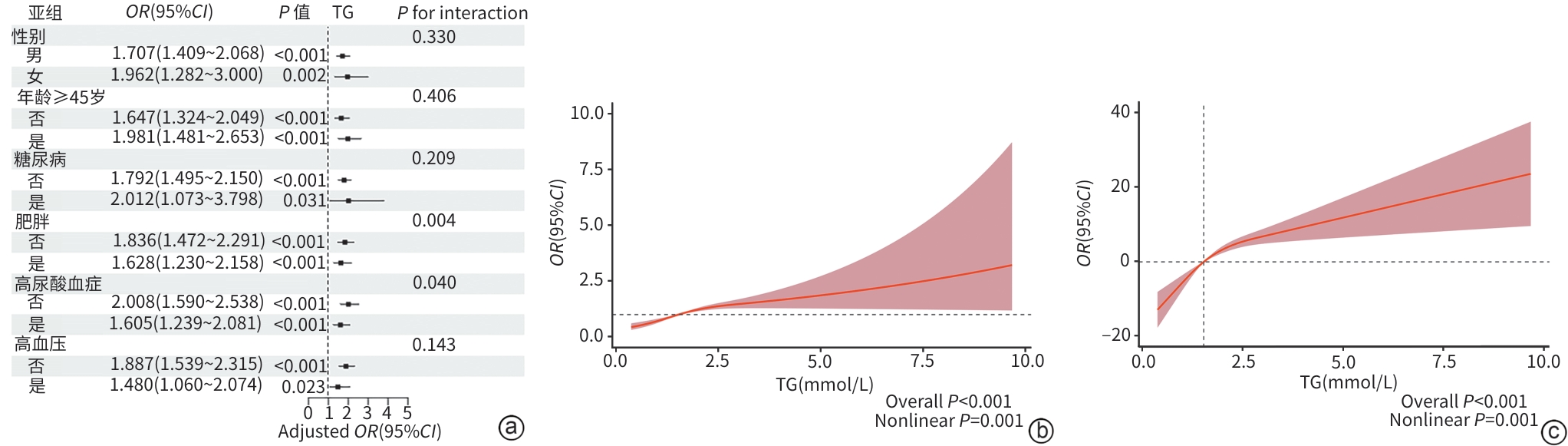
注: a,不同亚组中TG升高与肝脂肪变性严重程度的关联;b,TG水平与肝脂肪变性严重程度的剂量-反应关系,Y轴为更高肝脏脂肪含量等级;c,TG水平与CAP的剂量-反应关系,Y轴为校正后CAP的变化值,参考基准为CAP中位数。TG,甘油三酯;CAP,受控衰减参数;MAFLD,代谢相关脂肪性肝病;OR,比值比;CI,置信区间。
图 2 血清TG对MAFLD患者肝脂肪变性严重程度的影响
Figure 2. Influence of serum TG on liver steatosis severity in MAFLD patients
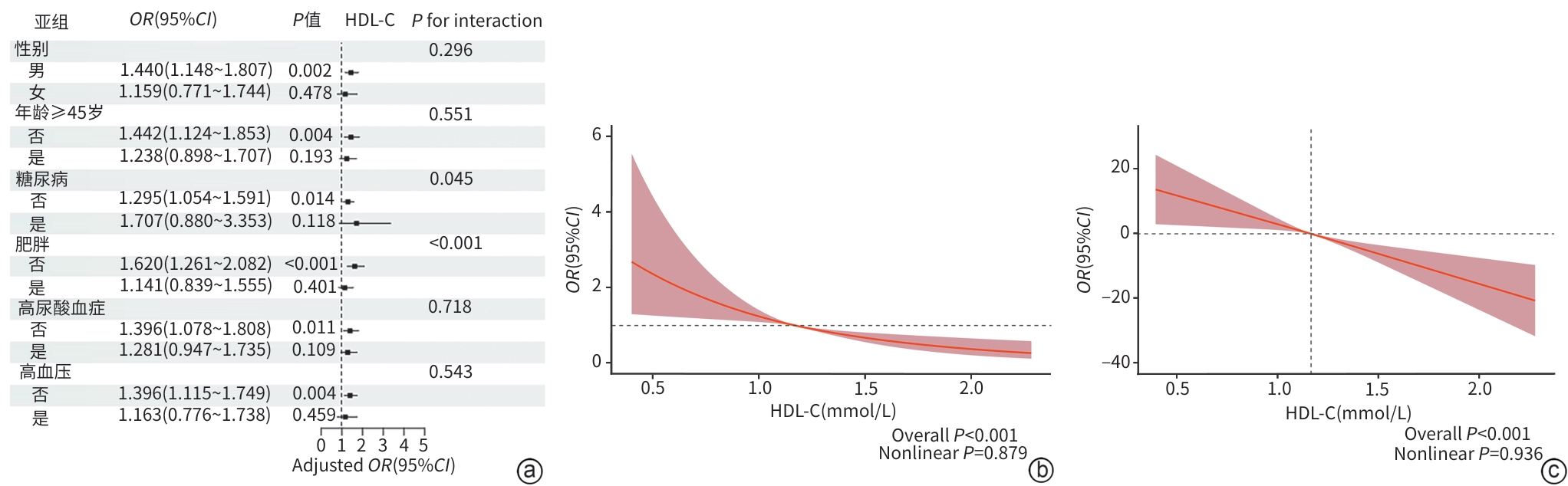
注: a,不同亚组中HDL-C降低与肝脂肪变性严重程度的关联;b,HDL-C水平与肝脂肪变性严重程度的剂量-反应关系,Y轴为更高肝脏脂肪含量等级;c,HDL-C水平与CAP的剂量-反应关系,Y轴为校正后CAP的变化值,参考基准为CAP中位数。HDL-C,高密度脂蛋白胆固醇;CAP,受控衰减参数;MAFLD,代谢相关脂肪性肝病;OR,比值比;CI,置信区间。
图 3 血清HDL-C对MAFLD患者肝脂肪变性严重程度的影响
Figure 3. Influence of serum HDL-C on liver steatosis severity in MAFLD patients
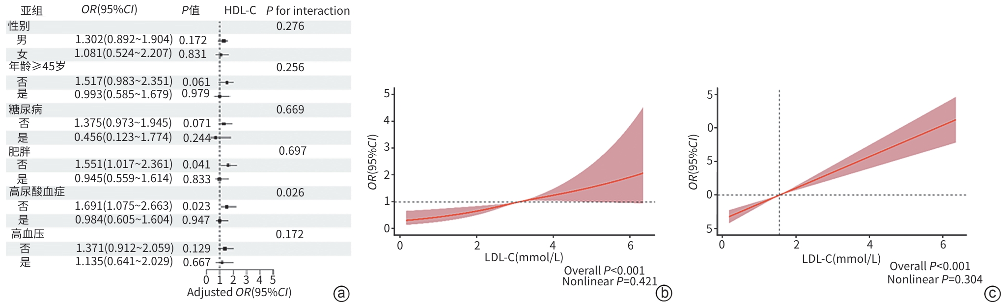
注: a,不同亚组中LDL-C升高与肝脂肪变性严重程度的关联;b,LDL-C水平与肝脂肪变性严重程度的剂量-反应关系,Y轴为更高肝脏脂肪含量等级;c,LDL-C水平与CAP的剂量-反应关系,Y轴为校正后CAP的变化值,参考基准为CAP中位数。LDL-C,低密度脂蛋白胆固醇;CAP,受控衰减参数;MAFLD,代谢相关脂肪性肝病;OR,比值比;CI,置信区间。
图 4 血清LDL-C对MAFLD患者肝脂肪变性严重程度的影响
Figure 4. Influence of serum LDL-C on liver steatosis severity in MAFLD patients
表 1 不同肝脂肪变性程度MAFLD患者的基线特征
Table 1. Baseline characteristics of MAFLD patients in different liver steatosis grades
指标 S1级(n=870) S2级(n=615) S3级(n=692) 统计值 P值 性别[例(%)] χ2=29.231 <0.001 女 232(26.7) 126(20.5) 120(17.3) 男 638(73.3) 489(79.5) 572(82.7) 年龄(岁) 42.8±8.8 43.2±8.2 42.1±8.0 F=3.202 0.041 吸烟[例(%)]1) 50(5.8) 40(6.5) 49(7.1) χ2=1.214 0.548 高血压[例(%)] 43(4.9) 33(5.4) 64(9.2) χ2=14.772 0.001 糖尿病[例(%)] 18(2.1) 10(1.6) 24(3.5) χ2=7.404 0.025 BMI(kg/m2) 26.1±2.6 27.4±3.0 28.7±3.2 F=173.471 <0.001 SBP(mmHg) 123.9±13.9 127.0±13.9 130.0±14.2 F=45.160 <0.001 DBP(mmHg) 78.0±10.6 80.4±10.8 82.8±11.1 F=44.920 <0.001 脉搏(次/min) 73.5±13.2 73.8±9.9 75.4±10.1 F=2.982 0.051 TG(mmol/L) 1.3(1.0~1.8) 1.6(1.2~2.2) 1.9(1.4~2.6) H=260.791 <0.001 HDL-C(mmol/L) 1.2±0.2 1.2±0.2 1.1±0.2 F=23.158 <0.001 LDL-C(mmol/L) 3.0±0.7 3.1±0.7 3.2±0.7 F=15.660 <0.001 ALT(U/L) 20.0(16.0~29.0) 25.0(18.0~37.0) 34.0(24.0~52.0) H=308.542 <0.001 UA(μmol/L) 383.1±89.2 404.3±84.5 429.8±91.4 F=69.691 <0.001 HbA1c(%) 5.7±0.6 5.7±0.6 5.9±0.9 F=17.203 <0.001 FBG(mmol/L) 5.3±1.0 5.5±0.9 5.7±1.3 F=28.337 <0.001 Cr(μmol/L) 75.0±14.6 76.1±13.5 75.7±13.0 F=1.224 0.299 WBC(×109/L) 5.8±1.3 6.0±1.4 6.3±1.6 F=32.001 <0.001 HGB(g/L) 150.2±14.2 153.6±12.9 154.9±12.2 F=31.411 <0.001 PLT(×109/L) 239.8±54.3 243.6±51.2 248.9±57.2 F=5.462 0.004 LSM(kPa) 5.3±1.1 5.5±1.3 6.0±1.5 F=90.217 <0.001 注:1)吸烟定义为现在吸烟,不包括既往吸烟者。BMI,体重指数;SBP,收缩压;DBP,舒张压;TG,甘油三酯;HDL-C,高密度脂蛋白胆固醇;LDL-C,低密度脂蛋白胆固醇;ALT,丙氨酸氨基转移酶;UA,尿酸;HbA1c,糖化血红蛋白;FBG,空腹血糖;Cr,肌酐;WBC,白细胞;HGB,血红蛋白;PLT,血小板;LSM,肝硬度值;MAFLD,代谢相关脂肪性肝病。
 下载: 导出CSV
表 2 血脂异常类型与肝脂肪变性严重程度的关联性分析
Table 2. The associations between different dyslipidemias and severity of hepatic steatosis
变量 模型1 模型2 模型3 OR(95%CI) P值 OR(95%CI) P值 OR(95%CI) P值 单一血脂异常 TG升高 2.23(1.90~2.62) <0.001 2.11(1.80~2.49) <0.001 1.77(1.48~2.10) <0.001 HDL-C降低 1.32(1.11~1.57) 0.002 1.52(1.26~1.83) <0.001 1.35(1.11~1.65) 0.003 LDL-C升高 1.40(1.02~1.92) 0.035 1.48(1.08~2.03) 0.014 1.28(0.92~1.79) 0.145 联合血脂异常:TG与HDL-C TG正常HDL-C正常 Reference Reference Reference TG升高HDL-C正常 2.25(1.85~2.73) <0.001 2.13(1.76~2.60) <0.001 1.74(1.42~2.14) <0.001 TG正常HDL-C降低 1.27(0.99~1.61) 0.056 1.44(1.12~1.84) 0.005 1.27(0.98~1.66) 0.074 TG升高HDL-C降低 2.92(2.32~3.67) <0.001 3.18(2.52~4.02) <0.001 2.40(1.87~3.07) <0.001 联合血脂异常:TG与LDL-C TG正常LDL-C正常 Reference Reference Reference TG升高LDL-C正常 2.37(2.01~2.80) <0.001 2.32(1.96~2.74) <0.001 1.89(1.59~2.26) <0.001 TG正常LDL-C升高 1.55(0.97~2.46) 0.063 1.58(0.99~2.50) 0.053 1.38(0.84~2.24) 0.198 TG升高LDL-C升高 2.68(1.79~4.06) <0.001 2.65(1.76~4.01) <0.001 1.98(1.27~3.07) 0.002 注:模型1未校正;模型2校正年龄和性别;模型3校正年龄、性别、体重指数、收缩压、糖化血红蛋白、尿酸及血小板。TG,甘油三酯;HDL-C,高密度脂蛋白胆固醇;LDL-C,低密度脂蛋白胆固醇;OR,比值比;CI,置信区间。
下载: 导出CSV
-
[1] Younossi Z M, Golabi P, Paik J M, et al. The global epidemiology of nonalcoholic fatty liver disease(NAFLD) and nonalcoholic steatohepatitis(NASH): A systematic review[J]. Hepatology, 2023, 77( 4): 1335- 1347. DOI: 10.1097/HEP.0000000000000004. [2] Huang D Q, Wong V W S, Rinella M E, et al. Metabolic dysfunction-associated steatotic liver disease in adults[J]. Nat Rev Dis Primers, 2025, 11: 14. DOI: 10.1038/s41572-025-00599-1. [3] Vilar-Gomez E, Vuppalanchi R, Gawrieh S, et al. CAP and LSM as determined by VCTE are independent predictors of all-cause mortality in the US adult population[J]. Hepatology, 2023, 77( 4): 1241- 1252. DOI: 10.1097/HEP.0000000000000023. [4] Semova I, Biddinger S B. Triglycerides in nonalcoholic fatty liver disease: Guilty until proven innocent[J]. Trends Pharmacol Sci, 2021, 42( 3): 183- 190. DOI: 10.1016/j.tips.2020.12.001. [5] García-Ruiz I, Solís-Muñoz P, Fernández-Moreira D, et al. NADPH oxidase is implicated in the pathogenesis of oxidative phosphorylation dysfunction in mice fed a high-fat diet[J]. Sci Rep, 2016, 6: 23664. DOI: 10.1038/srep23664. [6] Du T T, Sun X X, Yuan G, et al. Lipid phenotypes in patients with nonalcoholic fatty liver disease[J]. Metabolism, 2016, 65( 9): 1391- 1398. DOI: 10.1016/j.metabol.2016.06.006. [7] Tsutsumi T, Kawaguchi T, Fujii H, et al. Low HDL cholesterol levels in women and hypertriglyceridemia in men: Predictors of MASLD onset in individuals without steatosis[J]. J Gastroenterol, 2025, 60( 7): 891- 904. DOI: 10.1007/s00535-025-02242-y. [8] Yuan J, He X Q, Lu Y, et al. Triglycerides/high-density lipoprotein-cholesterol ratio outperforms traditional lipid indicators in predicting metabolic dysfunction-associated steatotic liver disease among U.S. adults[J]. Front Endocrinol, 2025, 16: 1591241. DOI: 10.3389/fendo.2025.1591241. [9] Li B Y, Liu Y W, Ma X R, et al. The association between non-high-density lipoprotein cholesterol to high-density lipoprotein cholesterol ratio and hepatic steatosis and liver fibrosis among US adults based on NHANES[J]. Sci Rep, 2025, 15( 1): 6527. DOI: 10.1038/s41598-025-90773-y. [10] Fan J G, Xu X Y, Yang R X, et al. Guideline for the prevention and treatment of metabolic dysfunction-associated fatty liver disease(version 2024)[J]. J Clin Transl Hepatol, 2024, 12( 11): 955- 974. DOI: 10.14218/JCTH.2024.00311. [11] Liu W Y, Huang S S, Ji H S, et al. From“burnt-out” to“burning-out”: Capturing liver fat loss in patients with advanced metabolic dysfunction–associated steatotic liver disease from a dynamic perspective[J]. Gastroenterology, 2025, 169( 2): 326- 336. DOI: 10.1053/j.gastro.2025.02.034. [12] Karlas T, Petroff D, Sasso M, et al. Individual patient data meta-analysis of controlled attenuation parameter(CAP) technology for assessing steatosis[J]. J Hepatol, 2017, 66( 5): 1022- 1030. DOI: 10.1016/j.jhep.2016.12.022. [13] Joint Committee on the Chinese Guidelines for Lipid Management. Chinese guidelines for lipid management(2023)[J]. Chin Circulation J, 2023, 38( 3): 237- 271. DOI: 10.3969/j.issn.1000-3614.2023.03.001.中国血脂管理指南修订联合专家委员会. 中国血脂管理指南(2023年)[J]. 中国循环杂志, 2023, 38( 3): 237- 271. DOI: 10.3969/j.issn.1000-3614.2023.03.001. [14] Donnelly K L, Smith C I, Schwarzenberg S J, et al. Sources of fatty acids stored in liver and secreted via lipoproteins in patients with nonalcoholic fatty liver disease[J]. J Clin Invest, 2005, 115( 5): 1343- 1351. DOI: 10.1172/JCI23621. [15] Lee E, Korf H, Vidal-Puig A. An adipocentric perspective on the development and progression of non-alcoholic fatty liver disease[J]. J Hepatol, 2023, 78( 5): 1048- 1062. DOI: 10.1016/j.jhep.2023.01.024. [16] Adiels M, Westerbacka J, Soro-Paavonen A, et al. Acute suppression of VLDL1 secretion rate by insulin is associated with hepatic fat content and insulin resistance[J]. Diabetologia, 2007, 50( 11): 2356- 2365. DOI: 10.1007/s00125-007-0790-1. [17] Scorletti E, Carr R M. A new perspective on NAFLD: Focusing on lipid droplets[J]. J Hepatol, 2022, 76( 4): 934- 945. DOI: 10.1016/j.jhep.2021.11.009. [18] Adiels M, Taskinen M R, Packard C, et al. Overproduction of large VLDL particles is driven by increased liver fat content in man[J]. Diabetologia, 2006, 49( 4): 755- 765. DOI: 10.1007/s00125-005-0125-z. [19] Fabbrini E, Mohammed B S, Magkos F, et al. Alterations in adipose tissue and hepatic lipid kinetics in obese men and women with nonalcoholic fatty liver disease[J]. Gastroenterology, 2008, 134( 2): 424- 431. DOI: 10.1053/j.gastro.2007.11.038. [20] Jakubowski H. The molecular bases of anti-oxidative and anti-inflammatory properties of paraoxonase 1[J]. Antioxidants, 2024, 13( 11): 1292. DOI: 10.3390/antiox13111292. [21] Petersen M C, Shulman G I. Roles of diacylglycerols and ceramides in hepatic insulin resistance[J]. Trends Pharmacol Sci, 2017, 38( 7): 649- 665. DOI: 10.1016/j.tips.2017.04.004. [22] Dongiovanni P, Stender S, Pietrelli A, et al. Causal relationship of hepatic fat with liver damage and insulin resistance in nonalcoholic fatty liver[J]. J Intern Med, 2018, 283( 4): 356- 370. DOI: 10.1111/joim.12719. [23] Vilar-Gomez E, Nephew L D, Vuppalanchi R, et al. High-quality diet, physical activity, and college education are associated with low risk of NAFLD among the US population[J]. Hepatology, 2022, 75( 6): 1491- 1506. DOI: 10.1002/hep.32207. [24] Vávrová L, Kodydková J, Zeman M, et al. Altered activities of antioxidant enzymes in patients with metabolic syndrome[J]. Obes Facts, 2013, 6( 1): 39- 47. DOI: 10.1159/000348569. [25] Barrera J, Chambliss K L, Ahmed M, et al. Bazedoxifene and conjugated estrogen prevent diet-induced obesity, hepatic steatosis, and type 2 diabetes in mice without impacting the reproductive tract[J]. Am J Physiol Endocrinol Metab, 2014, 307( 3): E345- E354. DOI: 10.1152/ajpendo.00653.2013. [26] Zhang Y J, Klein K, Sugathan A, et al. Transcriptional profiling of human liver identifies sex-biased genes associated with polygenic dyslipidemia and coronary artery disease[J]. PLoS One, 2011, 6( 8): e23506. DOI: 10.1371/journal.pone.0023506. [27] Palmisano B T, Zhu L, Stafford J M. Role of estrogens in the regulation of liver lipid metabolism[M]// Sex and Gender Factors Affecting Metabolic Homeostasis, Diabetes and Obesity. Cham: Springer International Publishing, 2017: 227- 256. DOI: 10.1007/978-3-319-70178-3_12. [28] Bhathena J, Kulamarva A, Martoni C, et al. Diet-induced metabolic Hamster model of nonalcoholic fatty liver disease[J]. Diabetes Metab Syndr Obes, 2011, 4: 195- 203. DOI: 10.2147/DMSO.S18435. [29] Côté I, Ngo Sock E T, Lévy É, et al. An atherogenic diet decreases liver FXR gene expression and causes severe hepatic steatosis and hepatic cholesterol accumulation: Effect of endurance training[J]. Eur J Nutr, 2013, 52( 5): 1523- 1532. DOI: 10.1007/s00394-012-0459-5. -

 本文二维码
计量
- 文章访问数: 16
- HTML全文浏览量: 9
- PDF下载量: 0
- 被引次数: 0