PDF下载 ( 37776 KB)

巨噬细胞脂解刺激脂蛋白受体缺失对代谢相关脂肪性肝炎小鼠模型的影响
DOI: 10.12449/JCH260914
Effect of macrophage lipolysis-stimulated lipoprotein receptor deficiency on a mouse model of metabolic dysfunction-associated steatohepatitis
-
摘要:
目的 探究巨噬细胞脂解刺激脂蛋白受体(LSR)缺失是否通过激活Yes相关蛋白(YAP)信号通路影响代谢相关脂肪性肝炎(MASH)的疾病进展。 方法 选取8周龄、体重相近的雄性野生型(WT)和巨噬细胞特异性Lsr基因敲除(LsrMφKO,KO)小鼠,分别给予普通饮食、高脂饮食(HFD)以及高脂高胆固醇饮食(HFHC)喂养30周。采用苏木精-伊红及油红O染色评估肝脏病理改变;采用免疫荧光染色检测F4/80及平滑肌肌动蛋白α表达;采用实时荧光定量逆转录聚合酶链式反应和蛋白质印迹法检测脂质代谢、炎症及纤维化相关指标。通过骨髓移植实验(WT→KO组 vs KO→KO组)验证巨噬细胞LSR的作用。在细胞水平上,分离WT和KO小鼠的肝巨噬细胞,检测YAP信号通路相关蛋白及下游靶基因表达;并采用YAP抑制剂维替泊芬体外处理上述细胞,验证该通路对炎症因子表达的调控作用。符合正态分布的计量资料两组间比较采用独立样本t检验,非正态分布的计量资料两组间比较采用Mann-Whitney U检验。 结果 普通饮食条件下,WT小鼠与KO小鼠肝脏各项指标比较,差异均无统计学意义。HFD及HFHC喂养后KO小鼠肝脏脂滴沉积显著增加,脂代谢紊乱,炎症及纤维化相关指标均显著升高(P值均<0.05)。骨髓移植结果显示,与KO→KO组相比,WT→KO组小鼠的肝损伤程度显著减轻(P值均<0.05)。机制研究表明,KO小鼠肝巨噬细胞中磷酸化大肿瘤抑制激酶1(p-LATS1)及磷酸化YAP水平显著降低,总YAP蛋白及其下游靶基因表达上调(P值均<0.05)。体外维替泊芬干预可显著抑制KO小鼠肝巨噬细胞中炎症因子的表达(P值均<0.05)。 结论 巨噬细胞LSR缺失通过抑制LATS1/2激酶活性激活YAP信号通路,促进炎症因子表达,从而加速MASH小鼠模型的肝损伤进程。 Abstract:Objective To investigate whether macrophage lipolysis-stimulated lipoprotein receptor (LSR) deficiency affects the progression of metabolic dysfunction-associated steatohepatitis (MASH) by activating the Yes-associated protein (YAP) signaling pathway. Methods Male wild-type (WT) mice and macrophage-specific Lsr knockout (LsrMφKO, KO) mice, with an age of 8 weeks and similar body weights, were selected and fed with a normal diet, a high-fat diet (HFD), or a high-fat high-cholesterol (HFHC) diet for 30 weeks. HE staining and oil red O staining were used to evaluate liver pathological changes; immunofluorescent staining was used to measure the expression levels of F4/80 and α-smooth muscle actin; RT-qPCR and Western blot were used to measure the levels of markers associated with lipid metabolism, inflammation, and fibrosis. Bone marrow transplantation rescue experiments (WT→KO vs KO→KO groups) were conducted to explore the functional role of LSR in macrophages. At the cellular level, hepatic macrophages were isolated from WT and KO mice to measure the expression levels of YAP signaling pathway-related proteins and their downstream target genes, and in addition, these cells were treated in vitro with verteporfin (VP), a specific inhibitor of YAP, to verify the regulatory effect of this pathway on the expression of inflammatory factors. The independent samples t-test was used for comparison of normally distributed measurement data between two groups, and the Mann-Whitney U test was used for comparison of non-normally distributed measurement data. Results Under the condition of normal diet, there were no significant differences in hepatic indicators between WT and KO mice. KOmice fed an HFD or HFHC diet showed a significant increase in hepatic lipid droplet accumulation, accompanied by lipid metabolism disorder and significant increases in the levels of indicators associated with inflammation and fibrosis (P<0.05). Bone marrow transplantation experiments confirmed that compared with the KO→KO group, the WT→KO group had significant alleviation of liver injury (P<0.05). Mechanistic studies showed that there were significant reductions in the levels of phosphorylated large tumor suppressor kinase 1 (LATS1) and phosphorylated YAP and significant increases in the expression levels of total YAP protein and its downstream target genes in hepatic macrophages of KO mice (P<0.05). In vitro VP intervention significantly inhibited the expression levels of inflammatory factors in hepatic macrophages isolated from KO mice (P<0.05). Conclusion Macrophage LSR deficiency activates the YAP signaling pathway by inhibiting LATS1/2 kinase activity and promotes the expression of inflammatory factors, thereby accelerating the progression of liver injury in a mouse model of MASH. -
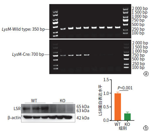
注: a,小鼠基因型鉴定;b,蛋白质印迹法验证KO小鼠基因敲除效率。LSR,脂解刺激脂蛋白受体。
图 1 KO 小鼠模型构建与验证
Figure 1. Construction and validation of KO mouse model
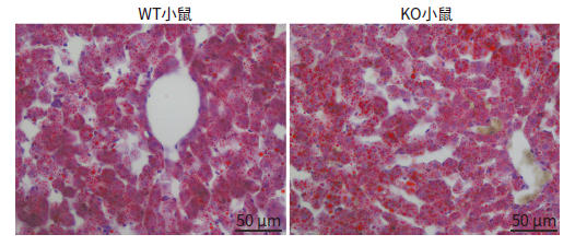
图 2 普通饮食小鼠38周肝组织油红O染色结果比较(×400)
Figure 2. Comparison of oil red O staining results in liver tissues under normal diet (×400)
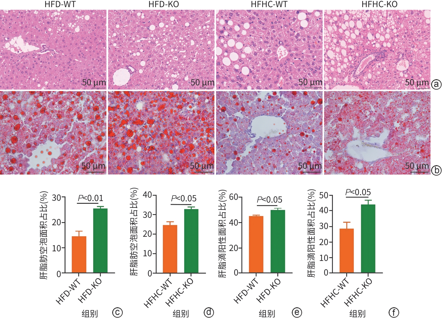
注: a,肝组织HE染色结果(×400);b,肝组织油红O染色结果(×400);c,HFD喂养小鼠HE染色结果比较;d,HFHC喂养小鼠HE染色结果比较;e,HFD喂养小鼠油红O染色结果比较;f,HFHC喂养小鼠油红O染色结果比较。HFD-WT,高脂饮食野生型;HFD-KO,高脂饮食巨噬细胞Lsr基因敲除型;HFHC-WT,高脂高胆固醇饮食野生型;HFHC-KO,高脂高胆固醇饮食巨噬细胞Lsr基因敲除型;LSR,脂解刺激脂蛋白受体;HE,苏木精-伊红。
图 3 HFD和HFHC喂养小鼠肝组织HE染色和油红O染色结果
Figure 3. HFD and HFHC diet-induced mouse liver HE staining and oil red O staining

注: a,HFD模型蛋白质印迹法检测肝脏中LY6G、F4/80、MCP-1蛋白表达;b,HFHC模型蛋白质印迹法检测肝脏中LY6G、F4/80、MCP-1蛋白表达水平;c,免疫荧光检测HFD及HFHC喂养小鼠肝脏中F4/80阳性细胞表达(×200)。HFD-WT,高脂饮食野生型;HFD-KO,高脂饮食巨噬细胞Lsr基因敲除型;HFHC-WT,高脂高胆固醇饮食野生型;HFHC-KO,高脂高胆固醇饮食巨噬细胞Lsr基因敲除型;LY6G,淋巴细胞抗原6G;F4/80,EGF样模块黏蛋白样激素受体样序列1;MCP-1,单核细胞趋化蛋白1。
图 4 HFD及HFHC喂养小鼠肝脏炎症相关蛋白表达
Figure 4. Hepatic expression of inflammation-related proteins in mice fed HFD or HFHC
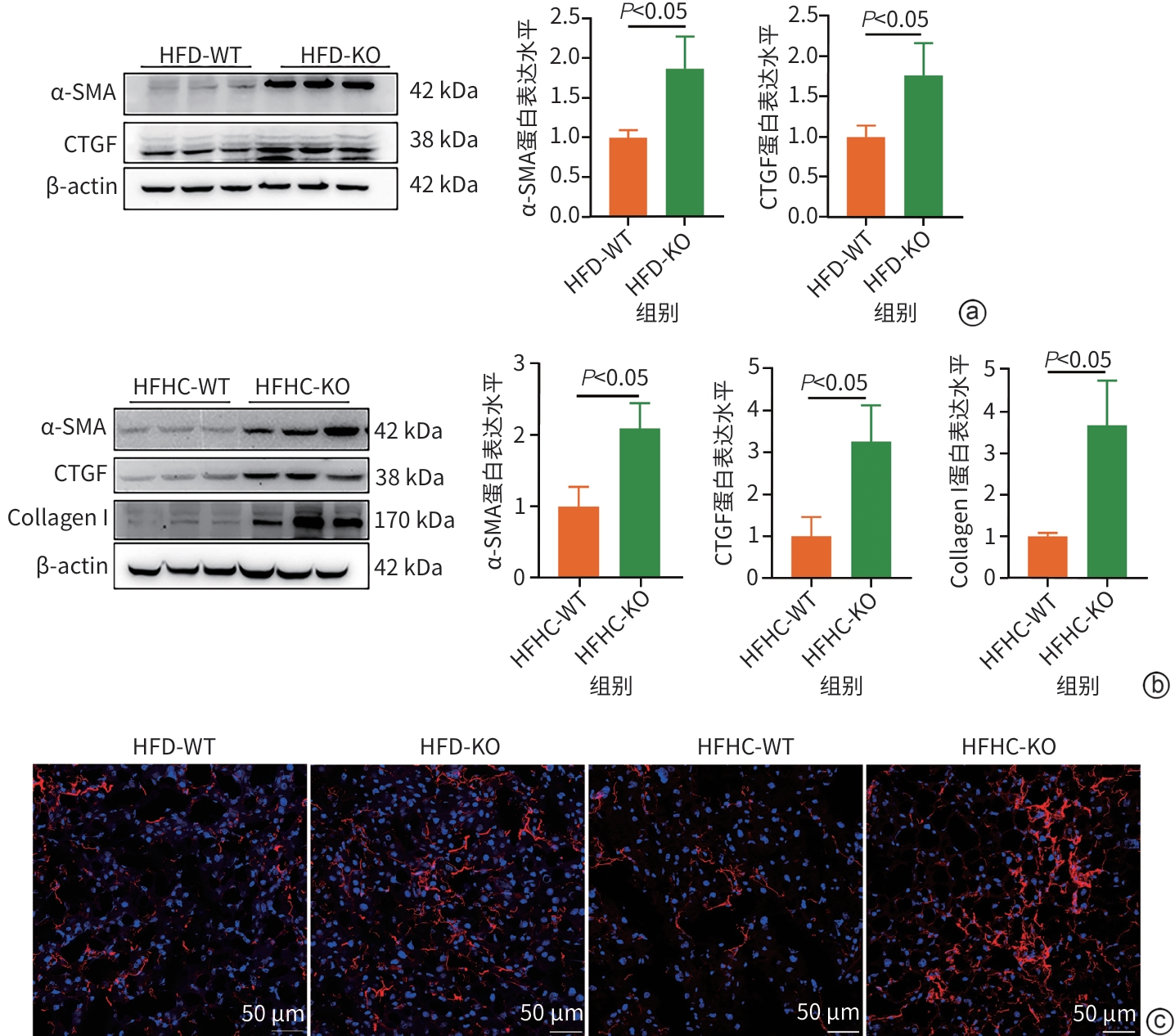
注: a,HFD模型蛋白质印迹法检测肝脏中α-SMA、CTGF蛋白表达;b,HFHC模型蛋白质印迹法检测肝脏中α-SMA、CTGF、Col Ⅰ(Collagen Ⅰ)蛋白表达;c,免疫荧光检测HFD与HFHC饮食小鼠肝脏中Collagen Ⅰ阳性细胞表达(×200)。HFD-WT,高脂饮食野生型;HFD-KO,高脂饮食巨噬细胞Lsr基因敲除型;HFHC-WT,高脂高胆固醇饮食野生型;HFHC-KO,高脂高胆固醇饮食巨噬细胞Lsr基因敲除型;α-SMA,平滑肌肌动蛋白α;CTGF,结缔组织生长因子;Collagen Ⅰ,Ⅰ型胶原蛋白;LSR,脂解刺激脂蛋白受体。
图 5 HFD与HFHC喂养小鼠肝脏纤维化相关蛋白表达
Figure 5. Hepatic expression of fibrosis-related proteins in mice fed HFD or HFHC
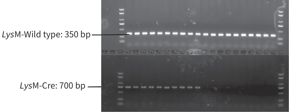
图 6 骨髓移植后基因型鉴定
Figure 6. Identification of genotypes after bone marrow transplantation
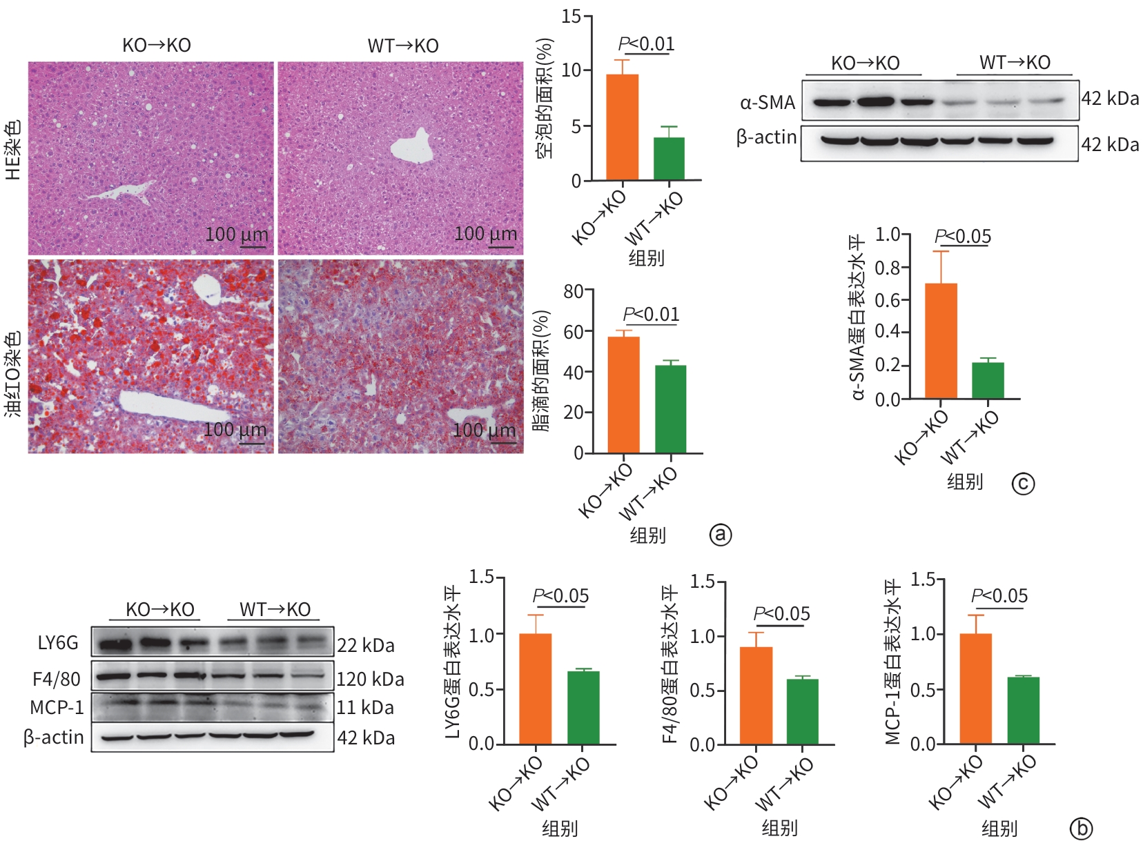
注: a,骨髓移植小鼠肝组织HE与油红O染色(×200);b,骨髓移植小鼠蛋白质印迹法检测肝脏中LY6G、F4/80、MCP-1蛋白表达水平;c,骨髓移植小鼠蛋白质印迹法检测肝脏中α-SMA蛋白表达水平。KO→KO,巨噬细胞Lsr敲除供体→敲除受体;WT→KO,野生型供体→巨噬细胞Lsr敲除受体;LY6G,淋巴细胞抗原6G;F4/80,EGF样模块黏蛋白样激素受体样序列1;MCP-1,单核细胞趋化蛋白1;α-SMA,平滑肌肌动蛋白α;LSR,脂解刺激脂蛋白受体;MASH,代谢相关脂肪性肝炎;HE,苏木精-伊红。
图 7 巨噬细胞LSR功能回补可缓解KO小鼠HFHC诱导的MASH进程
Figure 7. Functional rescue of macrophage LSR alleviates HFHC-induced MASH progression in KO mice
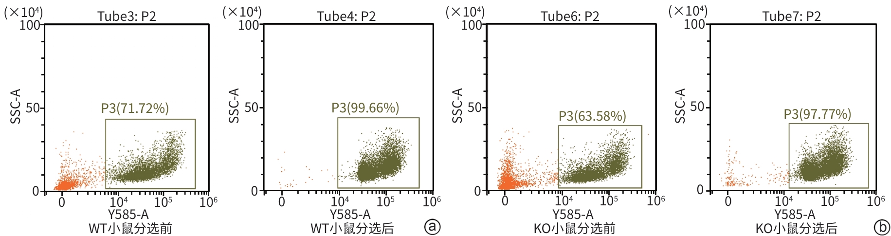
注: a,WT小鼠肝巨噬细胞经分选前后F4/80⁺细胞群所占比例;b,KO小鼠肝巨噬细胞经分选前后F4/80⁺细胞群所占比例。P3门控区域为F4/80⁺细胞群,门控内数值为F4/80⁺细胞占总细胞百分比。F4/80,EGF样模块黏蛋白样激素受体样序列1。
图 8 流式细胞术鉴定肝巨噬细胞分选前后F4/80⁺细胞纯度
Figure 8. Flow cytometric validation of F4/80⁺ hepatic macrophages purity before and after FACS
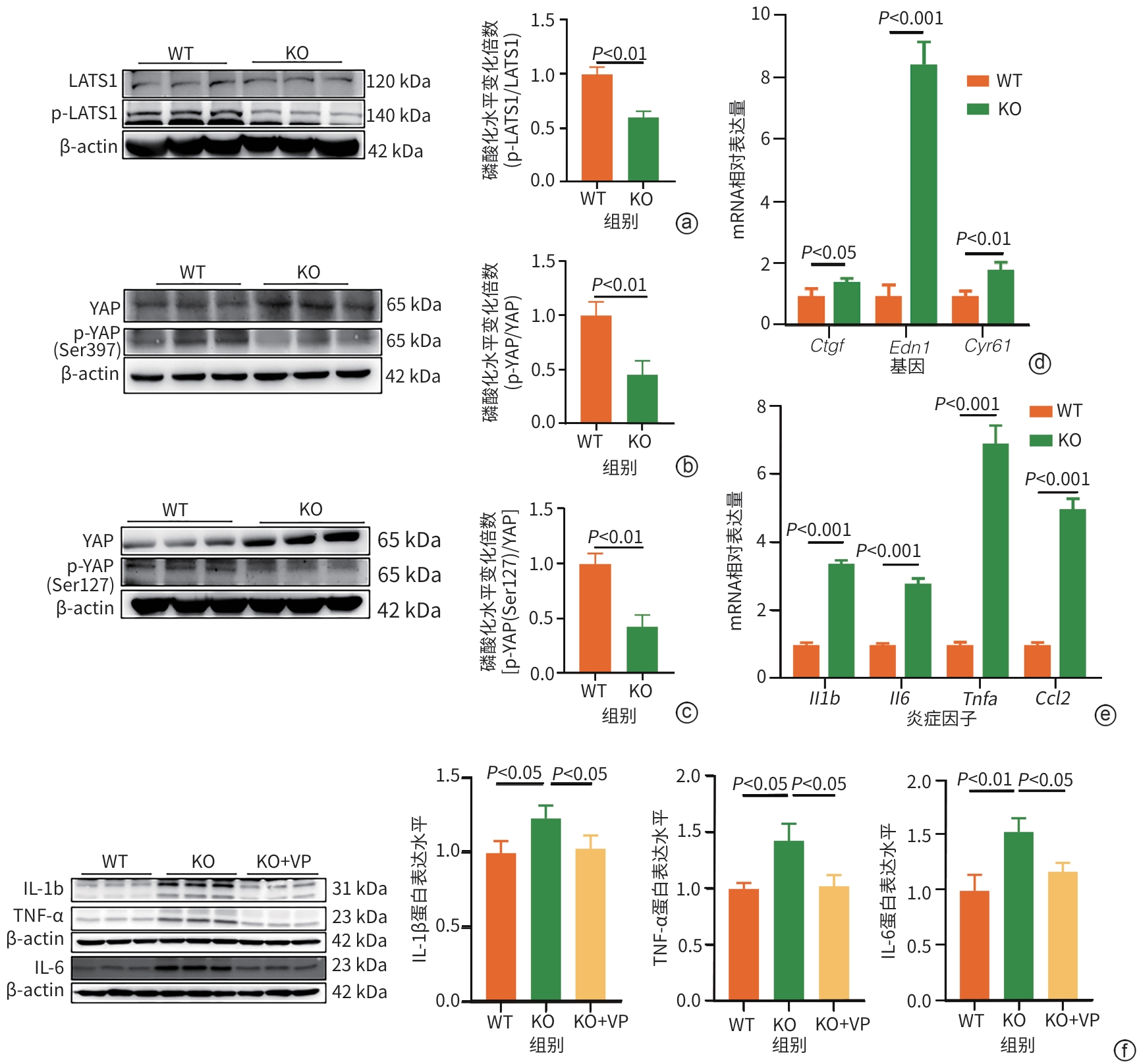
注: a,蛋白质印迹法检测小鼠肝巨噬细胞中LATS1、p-LATS1蛋白表达水平;b,蛋白质印迹法检测小鼠肝巨噬细胞中YAP、p-YAP(Ser397)蛋白表达水平;c,蛋白质印迹法检测小鼠肝巨噬细胞中YAP、p-YAP(Ser127)蛋白表达水平;d,qRT-PCR检测小鼠肝巨噬细胞中YAP下游靶基因Edn1、Cyr61及Ctgf的mRNA表达;e,qRT-PCR检测小鼠肝巨噬细胞中炎症因子Il1β、Tnfα、Il6及Ccl2的mRNA表达水平;f,VP处理后,蛋白质印迹法检测小鼠肝巨噬细胞中IL-1β、TNF-α及IL-6蛋白表达水平。LATS1,大肿瘤抑制激酶1;p-LATS1,磷酸化大肿瘤抑制激酶1;YAP,Yes相关蛋白;p-YAP,磷酸化Yes相关蛋白;Cyr61,富含半胱氨酸的血管生成诱导因子61;Edn1,内皮素1;Ctgf,结缔组织生长因子;Il1β,白细胞介素1β;Tnfα,肿瘤坏死因子α;Il6,白细胞介素6;Ccl2,C-C基序趋化因子配体2;LSR,脂解刺激脂蛋白受体;VP,维替泊芬。
图 9 LSR缺失通过激活YAP通路促进肝巨噬细胞炎症反应
Figure 9. Deficiency of LSR promotes the inflammatory response in hepatic macrophages by activating the YAP pathway
表 1 qRT-PCR引物序列
Table 1. Sequences of the primers for qRT-PCR
基因 引物序列 基因 引物序列 F4/80 上游 5'-TGACTCACCTTGTGGTCCTAA-3'
下游 5'- CTTCCCAGAATCCAGTCTTTCC-3'Stard4 上游 5'-AGCCCGTGGTCACAGATTG-3'
下游 5'-GCAACTCGCCACTCATCTTC-3'Ctgf 上游 5'-GGGCCTCTTCTGCGATTTC-3'
下游 5'- ATCCAGGCAAGTGCATTGGTA-3'Abcg5 上游 5'-AGGGCCTCACATCAACAGAG-3'
下游 5'- GCTGACGCTGTAGGACACAT-3'Il6 上游 5'-TAGTCCTTCCTACCCCAATTTCC-3'
下游 5'- TTGGTCCTTAGCCACTCCTTC-3'Acsl1 上游 5'-TGCCAGAGCTGATTGACATTC-3'
下游 5'- GGCATACCAGAAGGTGGTGAG-3'Il1β 上游 5'-GACCTTCCAGGATGAGGACA-3'
下游 5'- AGCTCATATGGGTCCGACAG-3'Aldh3a2 上游5'-GCGGAGGATGGTGCAAGAG-3'
下游 5'-CATTGAGTTCACTTTTGCTCAGG-3'Ccl2 上游 5'-TTAAAAACCTGGATCGGAACCAA-3'
下游 5'-GCATTAGCTTCAGATTTACGGGT-3'Edn1 上游 5'-GCACCGGAGCTGAGAATGG-3'
下游 5'-GTGGCAGAAGTAGACACACTC-3'Lsr 上游 5'-CCCCCTGTGCGAGGAAAAT-3'
下游 5'-CTCCCAAGTATCCAGAGAGTAGG-3'Cyr61 上游 5'-CTGCGCTAAACAACTCAACGA-3'
下游 5'-GCAGATCCCTTTCAGAGCGG-3'Tnfα 上游 5'-CGTCGTAGCAAACCACCAAG-3'
下游 5'-TTGAAGAGAACCTGGGAGTAGACA-3'β-actin 上游 5'-GGCTGTATTCCCCTCCATCG-3'
下游 5'-CCAGTTGGTAACAATGCCATGT-3'Timp1 上游 5'- GCAACTCGGACCTGGTCATAA-3'
下游 5'-CGGCCCGTGATGAGAAACT-3'Collagen Ⅰ 上游 5'-GCTCCTCTTAGGGGCCACT-3'
下游 5'-CCACGTCTCACCATTGGGG-3'α-SMA 上游 5'-GTCCCAGACATCAGGGAGTAA-3'
下游 5'-TCGGATACTTCAGCGTCAGGA-3'Fn 上游 5'-ATGTGGACCCCTCCTGATAGT-3'
下游 5'-GCCCAGTGATTTCAGCAAAGG-3'Fasn 上游 5'-GGAGGTGGTGATAGCCGGTAT-3'
下游 5'-TGGGTAATCCATAGAGCCCAG-3'Tgfβ1 上游 5'-CTCCCGTGGCTTCTAGTGC-3'
下游 5'-GCCTTAGTTTGGACAG-GATCTG-3'Cpt1α 上游 5'-CTCCGCCTGAGCCATGAAG-3'
下游 5'-CACCAGTGATGATGCCATTCT-3'Col4a1 上游 5'-CTGGCACAAAAGGGACGAG-3'
下游 5'-ACGTGGCCGAGAATTTCACC-3'Cyp7a1 上游 5'-GGGATTGCTGTGGTAGTGAGC-3'
下游 5'- GGTATGGAATCAACCCGTTGTC-3'Ly6c 上游 5'-GCAGTGCTACGAGTGCTATGG-3'
下游 5'-ACTGACGGGTCTTTAGTTTCCTT-3'Scd1 上游 5'-TTCTTGCGATACACTCTGGTGC-3'
下游 5'- CGGGATTGAATGTTCTTGTCGT-3'Ccl5 上游 5'-GCTGCTTTGCCTACCTCTCC-3'
下游 5'-TCGAGTGACAAACACGACTGC-3'注:F4/80,EGF样模块黏蛋白样激素受体样序列1;Ctgf,结缔组织生长因子;Il6,白细胞介素6;Il1β,白细胞介素1β;Ccl2,C-C基序趋化因子配体2;Lsr,脂解刺激脂蛋白受体;Tnfα,肿瘤坏死因子α;Timp1,金属蛋白酶组织抑制因子1;α-SMA,平滑肌肌动蛋白α;Fasn,脂肪酸合成酶;Cpt1a,肉碱棕榈酰转移酶1α;Cyp7a1,细胞色素P450家族7亚家族A成员1;Scd1,硬脂酰辅酶A去饱和酶1;Stard4,StAR相关脂质转移结构域蛋白4;Abcg5,三磷酸腺苷结合盒转运蛋白G5;Acsl1,长链脂酰辅酶A合成酶1;Aldh3a2,醛脱氢酶家族3亚家族A成员2;Cyr61,富含半胱氨酸的血管生成诱导因子61;Edn1,内皮素1;Collagen Ⅰ,Ⅰ型胶原蛋白;Fn,纤维连接蛋白;Tgfβ1,转化生长因子β1;Col4a1,Ⅳ型胶原α1链;Ccl5,C-C基序趋化因子配体5;Ly6c,淋巴细胞抗原6家族成员C;qRT-PCR,实时荧光定量逆转录聚合酶链式反应。
 下载: 导出CSV
表 2 38 周普通饮食各组小鼠Stard4、Scd1、Acsl1、Tnfα、Ccl2、F4/80、α-SMA、Collagen Ⅰ及Timp1的mRNA相对表达量
Table 2. mRNA expression levels of Stard4, Scd1, Acsl1, Tnfα, Ccl2, F4/80, α-SMA, Collagen Ⅰ and Timp1 in mice of each group fed with normal chow diet at 38 weeks of age
基因名称 WT组(n=6) KO组(n=6) 统计值 P值 Stard4 1.000±0.202 1.181±0.284 t=1.272 0.232 Scd1 1.000±0.917 0.972±0.620 t=0.062 0.952 Acsl1 1.000±0.447 1.297±0.401 t=1.213 0.253 Tnfα 0.895(0.587~1.477) 1.181(0.988~1.676) Z=0.801 0.485 Ccl2 0.730(0.678~1.549) 0.835(0.696~1.188) Z=0.160 0.937 F4/80 0.876(0.797~1.194) 1.153(0.967~1.387) Z=1.601 0.132 α-SMA 1.000±0.470 1.192±0.742 t=0.535 0.604 Collagen Ⅰ 1.000±0.287 0.964±0.140 t=0.279 0.785 Timp1 0.655(0.423~1.559) 0.626(0.370~0.779) Z=0.801 0.485 注:Stard4,StAR相关脂质转移结构域蛋白4;Scd1,硬脂酰辅酶A去饱和酶1;Acsl1,长链脂酰辅酶A合成酶1;Tnfα,肿瘤坏死因子α;Ccl2,C-C基序趋化因子配体2;F4/80,EGF样模块黏蛋白样激素受体样1序列;α-SMA,平滑肌肌动蛋白α;Collagen Ⅰ,Ⅰ型胶原蛋白;Timp1,金属蛋白酶组织抑制剂1。
下载: 导出CSV
表 3 HFD各组小鼠Fasn、Scd1、Cpt1a、Cyp7a1、Ccl2、Il1β、F4/80、α-SMA及Timp1的mRNA相对表达量
Table 3. mRNA expression levels of Fasn, Scd1, Cpt1a, Cyp7a1, Ccl2, Il1β, F4/80, α-SMA and Timp1 in each group of high-fat diet-induced mouse model
基因名称 WT组(n=12) KO组(n=12) 统计值 P值 Fasn 0.868(0.510~1.556) 1.667(1.624~2.602) Z=2.192 0.030 Scd1 1.000±0.461 1.996±0.749 t=2.351 0.047 Cpt1a 1.000±0.380 0.576±0.246 t=2.361 0.040 Cyp7a1 1.000±0.470 0.252±0.165 t=4.194 0.002 Ccl2 1.000±0.316 1.684±0.735 t=3.093 0.004 Il1β 0.812(0.597~1.210) 1.592(1.033~2.488) Z=2.288 0.022 F4/80 1.000±0.328 1.516±0.395 t=2.433 0.033 α-SMA 1.000±0.409 2.832±1.581 t=2.499 0.031 Timp1 1.000±0.437 1.666±0.997 t=2.119 0.046 注:Fasn,脂肪酸合成酶;Scd1,硬脂酰辅酶A去饱和酶1;Cpt1a,肉碱棕榈酰转移酶1α;Cyp7a1,细胞色素P450家族7亚家族A成员1;Ccl2,C-C基序趋化因子配体2;Il1β,白细胞介素1β;F4/80,EGF样模块黏蛋白样激素受体样序列1;α-SMA,平滑肌肌动蛋白α;Timp1,金属蛋白酶组织抑制剂1。
下载: 导出CSV
表 4 HFHC各组小鼠Stard4、Abcg5、Aldh3a2、Acsl1、Ly6c、Ccl2、Il6、Ccl5、α-SMA、Collagen Ⅰ、Timp1及Ctgf的mRNA相对表达量
Table 4. mRNA expression levels of Stard4, Abcg5, Aldh3a2, Acsl1, Ly6c, Ccl2, Il6, Ccl5, α-SMA, Collagen Ⅰ, Timp1 and Ctgf in each group of high-fat and high-cholesterol diet-induced mouse model
基因名称 WT组(n=12) KO组(n=12) 统计值 P值 Stard4 1.000±0.540 0.628±0.173 t=2.274 0.033 Abcg5 1.000±0.605 0.469±0.334 t=2.746 0.012 Aldh3a2 1.000±0.482 1.738±0.920 t=2.461 0.022 Acsl1 0.900(0.718~1.099) 1.850(0.962~2.568) Z=2.230 0.026 Ly6c 1.000±0.561 1.775±0.777 t=2.598 0.018 Ccl2 0.694(0.468~1.404) 1.478(0.855~3.180) Z=2.285 0.022 Il6 1.000±0.807 2.279±1.497 t=2.495 0.022 Ccl5 1.000±0.483 1.519±0.612 t=2.305 0.031 α-SMA 1.000±0.487 1.985±1.197 t=2.186 0.043 Collagen Ⅰ 1.000±0.516 1.886±0.886 t=2.759 0.013 Timp1 1.000±0.743 2.156±1.407 t=2.409 0.026 Ctgf 0.735(0.517~1.310) 1.604(1.168~1.985) Z=2.216 0.027 注:Stard4,StAR相关脂质转移结构域蛋白4;Abcg5,三磷酸腺苷结合盒转运蛋白G5;Aldh3a2,醛脱氢酶家族3亚家族A成员2;Acsl1,长链脂酰辅酶A合成酶1;Ccl5,C-C基序趋化因子配体5;Il6,白细胞介素6;Ccl2,C-C基序趋化因子配体2;Ly6c,淋巴细胞抗原6家族成员C;α-SMA,平滑肌肌动蛋白α;Collagen Ⅰ,Ⅰ型胶原蛋白;Timp1,金属蛋白酶组织抑制剂1;Ctgf,结缔组织生长因子。
下载: 导出CSV
表 5 骨髓移植后HFHC各组小鼠Stard4、Abcg5、Tnfα、Ccl2、Ly6c、Il1β、α-SMA、Timp1、Fn、Col4a1及Tgfβ1 mRNA相对表达量
Table 5. mRNA expression levels of Stard4, Abcg5, Tnfα, Ccl2, Ly6c, Il1β, α-SMA, Timp1, Fn, Col4a1 and Tgfβ1 in mice of each group in the high-fat and high-cholesterol diet mouse model after bone marrow transplantation model
基因名称 KO→KO组(n=10) WT→KO组(n=10) 统计值 P值 Stard4 1.000±0.600 1.551±0.567 t=2.196 0.040 Abcg5 1.000±0.385 1.558±0.733 t=2.290 0.033 Tnfα 1.107(0.470~1.440) 0.380(0.314~0.466) Z=2.123 0.035 Ccl2 0.955(0.626~1.284) 0.269(0.145~0.346) Z=2.571 0.009 Ly6c 0.949(0.550~1.252) 0.420(0.261~0.689) Z=2.531 0.010 Il1β 0.927(0.687~1.369) 0.423(0.224~0.730) Z=2.570 0.008 α-SMA 1.000±0.646 0.458±0.277 t=2.436 0.026 Timp1 1.000±0.387 0.555±0.396 t=2.401 0.029 Fn 0.984(0.753~1.260) 0.631(0.494~1.032) Z=2.116 0.036 Col4a1 1.000±0.355 0.607±0.303 t=2.712 0.014 Tgfβ1 0.869(0.674~2.053) 0.443(0.236~0.785) Z=2.205 0.028 注:Stard4,StAR相关脂质转移结构域蛋白4;Abcg5,三磷酸腺苷结合盒转运蛋白G5;Tnfα,肿瘤坏死因子α;Il1β,白细胞介素1β;Ccl2,C-C基序趋化因子配体2;Ly6c,淋巴细胞抗原6家族成员C;α-SMA,平滑肌肌动蛋白α;Timp1,金属蛋白酶组织抑制剂1;Fn,纤维连接蛋白;Col4a1,Ⅳ型胶原α1链;Tgfβ1,转化生长因子β1;WT→KO组,野生型供体→巨噬细胞Lsr敲除受体;KO→KO组,巨噬细胞Lsr敲除供体→敲除受体。
下载: 导出CSV
-
[1] Rinella M E, Lazarus J V, Ratziu V, et al. A multisociety Delphi consensus statement on new fatty liver disease nomenclature[J]. Hepatology, 2023, 78( 6): 1966- 1986. DOI: 10.1097/HEP.0000000000000520. [2] Younossi Z M, Golabi P, Paik J M, et al. The global epidemiology of nonalcoholic fatty liver disease(NAFLD) and nonalcoholic steatohepatitis(NASH): A systematic review[J]. Hepatology, 2023, 77( 4): 1335- 1347. DOI: 10.1097/HEP.0000000000000004. [3] European Association for the Study of the Liver(EASL), European Association for the Study of Diabetes(EASD), European Association for the Study of Obesity(EASO). EASL-EASD-EASO clinical practice guidelines on the management of metabolic dysfunction-associated steatotic liver disease(MASLD)[J]. Obes Facts, 2024, 17( 4): 374- 444. DOI: 10.1159/000539371. [4] Chinese Society of Hepatology, Chinese Medical Association. Guidelines for the prevention and treatment of metabolic dysfunction-associated(non-alcoholic) fatty liver disease(version 2024)[J]. J Prac Hepatol, 2024, 27( 4): 494- 510. DOI: 10.3760/cma.j.cn501113-20240327-00163.中华医学会肝病学分会. 代谢相关(非酒精性)脂肪性肝病防治指南(2024年版)[J]. 实用肝脏病杂志, 2024, 27( 4): 494- 510. DOI: 10.3760/cma.j.cn501113-20240327-00163. [5] Yen F T, Masson M, Clossais-Besnard N, et al. Molecular cloning of a lipolysis-stimulated remnant receptor expressed in the liver[J]. J Biol Chem, 1999, 274( 19): 13390- 13398. DOI: 10.1074/jbc.274.19.13390. [6] Narvekar P, Berriel Diaz M, Krones-Herzig A, et al. Liver-specific loss of lipolysis-stimulated lipoprotein receptor triggers systemic hyperlipidemia in mice[J]. Diabetes, 2009, 58( 5): 1040- 1049. DOI: 10.2337/db08-1184. [7] Masuda S, Oda Y, Sasaki H, et al. LSR defines cell corners for tricellular tight junction formation in epithelial cells[J]. J Cell Sci, 2011, 124( Pt 4): 548- 555. DOI: 10.1242/jcs.072058. [8] Sohet F, Lin C, Munji R N, et al. LSR/angulin-1 is a tricellular tight junction protein involved in blood-brain barrier formation[J]. J Cell Biol, 2015, 208( 6): 703- 711. DOI: 10.1083/jcb.201410131. [9] Park S J, Garcia Diaz J, Um E, et al. Major roles of Kupffer cells and macrophages in NAFLD development[J]. Front Endocrinol, 2023, 14: 1150118. DOI: 10.3389/fendo.2023.1150118. [10] Xu G X, Wei S, Yu C, et al. Activation of Kupffer cells in NAFLD and NASH: Mechanisms and therapeutic interventions[J]. Front Cell Dev Biol, 2023, 11: 1199519. DOI: 10.3389/fcell.2023.1199519. [11] Wiszniewski M, Mori D, Sanchez Puch S I, et al. Divergent hepatic and adipose tissue effects of Kupffer cell depletion in a male rat model of metabolic-associated steatohepatitis[J]. Biology, 2025, 14( 8): 1058. DOI: 10.3390/biology14081058. [12] Tian Y X, Ni Y M, Zhang T, et al. Targeting hepatic macrophages for non-alcoholic fatty liver disease therapy[J]. Front Cell Dev Biol, 2024, 12: 1444198. DOI: 10.3389/fcell.2024.1444198. [13] Meli V S, Veerasubramanian P K, Downing T L, et al. Mechanosensation to inflammation: Roles for YAP/TAZ in innate immune cells[J]. Sci Signal, 2023, 16( 783): eadc9656. DOI: 10.1126/scisignal.adc9656. [14] Zhang J Q, Chen W N, Song K, et al. YAP activation in liver macrophages via depletion of MST1/MST2 enhances liver inflammation and fibrosis in MASLD[J]. FASEB J, 2024, 38( 17): e70026. DOI: 10.1096/fj.202400813RR. [15] Lee N Y, Choi M G, Lee E J, et al. Interplay between YAP/TAZ and metabolic dysfunction-associated steatotic liver disease progression[J]. Arch Pharmacal Res, 2024, 47( 6): 558- 570. DOI: 10.1007/s12272-024-01501-5. [16] Cohen J. Statistical power analysis for the behavioral sciences[M]. 2nd ed. Hillsdale: Lawrence Erlbaum Associates, 1988. [17] Sawilowsky S S. New effect size rules of thumb[J]. J Mod App Stat Meth, 2009, 8( 2): 597- 599. DOI: 10.22237/jmasm/1257035100. [18] Zhang X L, Hartmann P. How to calculate sample size in animal and human studies[J]. Front Med, 2023, 10: 1215927. DOI: 10.3389/fmed.2023.1215927. [19] Do A, Zahrawi F, Mehal W Z. Therapeutic landscape of metabolic dysfunction-associated steatohepatitis(MASH)[J]. Nat Rev Drug Discov, 2025, 24( 3): 171- 189. DOI: 10.1038/s41573-024-01084-2. [20] Steinberg G R, Carpentier A C, Wang D D. MASH: The nexus of metabolism, inflammation, and fibrosis[J]. J Clin Invest, 2025, 135( 18): e186420. DOI: 10.1172/JCI186420. [21] Jiang Z H, Qian B L, Xu T J, et al. Immune microenvironment on the molecular mechanisms and therapeutic targets of MAFLD[J]. ImmunoTargets Ther, 2025, 14: 719- 733. DOI: 10.2147/ITT.S530451. [22] Tsomidis I, Tsakou A, Voumvouraki A, et al. The role of Kupffer cells and liver macrophages in the pathogenesis of metabolic dysfunction-associated steatotic liver disease[J]. Biomedicines, 2026, 14( 1): 151. DOI: 10.3390/biomedicines14010151. [23] Matsuda M, Seki E. Hepatic stellate cell-macrophage crosstalk in liver fibrosis and carcinogenesis[J]. Semin Liver Dis, 2020, 40( 3): 307- 320. DOI: 10.1055/s-0040-1708876. [24] Yen F T, Roitel O, Bonnard L, et al. Lipolysis stimulated lipoprotein receptor: A novel molecular link between hyperlipidemia, weight gain, and atherosclerosis in mice[J]. J Biol Chem, 2008, 283( 37): 25650- 25659. DOI: 10.1074/jbc.M801027200. [25] Taru V, Szabo G, Mehal W, et al. Inflammasomes in chronic liver disease: Hepatic injury, fibrosis progression and systemic inflammation[J]. J Hepatol, 2024, 81( 5): 895- 910. DOI: 10.1016/j.jhep.2024.06.016. [26] Dong X, Zhang X B, Liu P, et al. Lipolysis-stimulated lipoprotein receptor impairs hepatocellular carcinoma and inhibits the oncogenic activity of YAP1 via PPPY motif[J]. Front Oncol, 2022, 12: 896412. DOI: 10.3389/fonc.2022.896412. [27] An Y N, Wang C, Fan B Z, et al. LSR targets YAP to modulate intestinal Paneth cell differentiation[J]. Cell Rep, 2023, 42( 9): 113118. DOI: 10.1016/j.celrep.2023.113118. [28] Song K, Kwon H, Han C, et al. Yes-associated protein in Kupffer cells enhances the production of proinflammatory cytokines and promotes the development of nonalcoholic steatohepatitis[J]. Hepatology, 2020, 72( 1): 72- 87. DOI: 10.1002/hep.30990. [29] Liang J J, Fraser I D C, Bryant C E. Lipid regulation of NLRP3 inflammasome activity through organelle stress[J]. Trends Immunol, 2021, 42( 9): 807- 823. DOI: 10.1016/j.it.2021.07.005. [30] Zhou Y, Tong Z Z, Jiang S H, et al. The roles of endoplasmic reticulum in NLRP3 inflammasome activation[J]. Cells, 2020, 9( 5): 1219. DOI: 10.3390/cells9051219. [31] Liu Y Q, An Y N, Li G B, et al. Regulatory mechanism of macrophage polarization based on Hippo pathway[J]. Front Immunol, 2023, 14: 1279591. DOI: 10.3389/fimmu.2023.1279591. [32] Zhang J, Wu H P, Ren X X, et al. Hippo/YAP signaling’s multifaceted crosstalk in cancer[J]. Front Cell Dev Biol, 2025, 13: 1595362. DOI: 10.3389/fcell.2025.1595362. [33] Clausen B E, Burkhardt C, Reith W, et al. Conditional gene targeting in macrophages and granulocytes using LysMcre mice[J]. Transgenic Res, 1999, 8( 4): 265- 277. DOI: 10.1023/a: 1008942828960. [34] Wang Y, Yu F X. Angiomotin family proteins in the Hippo signaling pathway[J]. Bioessays, 2024, 46( 8): e2400076. DOI: 10.1002/bies.202400076. -

 本文二维码
计量
- 文章访问数: 15
- HTML全文浏览量: 11
- PDF下载量: 0
- 被引次数: 0