PDF下载 ( 16384 KB)

骨髓间充质干细胞来源的外泌体中微RNA-21-5p对血管生成的调控作用
DOI: 10.12449/JCH260921
Regulatory effect of miR-21-5p in bone marrow mesenchymal stem cell-derived exosome on angiogenesis
-
摘要:
目的 验证骨髓间充质干细胞来源外泌体(BMSC-Exo)中微RNA(microRNA,miR)-21-5p通过磷脂酰肌醇3-激酶(PI3K)/蛋白激酶B(Akt)信号通路对血管生成的调控作用。 方法 分离并培养大鼠骨髓间充质干细胞(BMSC),将其分为阴性对照组(NC组)与抑制剂组(inhibitor组),分别转染miR-21-5p阴性对照与miR-21-5p inhibitor,提取外泌体并进行鉴定。将外泌体与人脐静脉内皮细胞(HUVEC)共培养,采用MTT法检测细胞增殖(24、48和72 h),采用管腔形成实验检测血管形成能力,采用蛋白质印迹法(Western blot)检测磷酸化Akt(p-Akt)、总Akt及血管内皮生长因子(VEGF)的蛋白表达水平。使用PI3K抑制剂LY294002处理后观察HUVEC血管形成能力的变化。计量资料两组间比较采用独立样本t检验,多组间比较采用单因素方差分析,进一步两两比较采用LSD-t检验。 结果 流式细胞术结果显示,第3代BMSC高表达CD29(98.18%)和CD90(97.85%),低表达CD45(0.22%)。实时荧光定量逆转录聚合酶链式反应结果显示,与NC组比较,转染miR-21-5pinhibitor后BMSC中miR-21-5p的相对表达量显著降低(0.38±0.05 vs 1.00±0.08,t=11.38,P<0.01)。MTT结果显示,与对照组比较,25、50、100和200 μg/mL外泌体处理组的HUVEC增殖活性均显著增强(P值均<0.05),其中100 μg/mL外泌体处理72 h时促增殖效果最显著,400 μg/mL外泌体处理组细胞增殖活性显著降低(P<0.01)。管腔形成实验结果显示,与对照组和NC-Exo组比较,inhibitor-Exo组血管形成能力显著下降(P值均<0.05);与inhibitor-Exo组比较,加入PI3K抑制剂LY294002后血管形成能力进一步显著下降(P<0.01)。Western blot结果显示,与对照组和NC-Exo组比较,inhibitor-Exo组p-Akt及VEGF蛋白表达水平显著降低(P值均<0.05);与inhibitor-Exo组比较,加入LY294002后p-Akt及VEGF蛋白表达水平进一步显著降低(P值均<0.01)。 结论 BMSC-Exo通过其携带的miR-21-5p靶向激活PI3K/Akt信号通路,上调VEGF表达,促进血管内皮细胞增殖与血管形成。 Abstract:Objective To investigate the regulatory effect of miR-21-5p in bone marrow mesenchymal stem cell-derived exosome (BMSC-Exo) on angiogenesis through the phosphatidylinositol 3-kinase (PI3K)/protein kinase B (Akt) signaling pathway. Methods Bone marrow mesenchymal stem cells (BMSCs) were isolated from rats, cultured, and divided into negative control group (NC group) and inhibitor group, which were transfected with miR-21-5p negative control and miR-21-5p inhibitor, respectively, and then exosomes were extracted and identified. Exosomes were co-cultured with human umbilical vein endothelial cells (HUVECs). MTT assay was used to measure cell proliferation (24, 48, and 72 hours), tube formation assay was used to assess angiogenesis ability, and Western blot was used to measure the protein expression levels of phosphorylated Akt (p-Akt), total Akt, and vascular endothelial growth factor (VEGF). The change in the angiogenesis ability of HUVECs was observed after treatment with the PI3K inhibitor LY294002. The independent samples t-test was used for comparison of continuous data between two groups, and a one-way analysis of variance was used for comparison between multiple groups, while the least significant difference t-test was used for further comparison between two groups. Results Flow cytometry showed that the third-generation BMSCs showed high expression levels of CD29 (98.18%) and CD90 (97.85%) and a low expression level of CD45 (0.22%). Quantitative real-time reverse transcription-polymerase chain reaction showed that compared with the NC group, there was a significant reduction in the relative expression level of miR-21-5p in BMSCs after transfection with miR-21-5p inhibitor (0.38±0.05 vs 1.00±0.08, t=11.38, P<0.01). MTT assay showed that compared with the control group, the 25, 50, 100, and 200 μg/mL exosome treatment groups had a significant increase in the proliferative activity of HUVECs (all P<0.05), among which 100 μg/mL exosome treatment for 72 hours showed the most significant pro-proliferative effect, while the 400 μg/mL exosome treatment group showed a significant reduction in the proliferative activity of HUVECs (P<0.01). Tube formation assay showed that compared with the control group and the NC-Exo group, the inhibitor-Exo group showed a significant reduction in angiogenesis ability (both P<0.05), and compared with the inhibitor-Exo group, there was a further significant reduction in angiogenesis ability after addition of the PI3K inhibitor LY294002 (P<0.01). Western blot showed that compared with the control group and the NC-Exo group, the inhibitor-Exo group had significant reduction in the protein expression levels of p-Akt and VEGF (all P<0.05), and compared with the inhibitor-Exo group, the protein expression levels of p-Akt and VEGF further decreased significantly after the addition of LY294002 (both P<0.01). Conclusion BMSC-Exo targets and activates the PI3K/Akt signaling pathway through miR-21-5p, upregulates the expression of VEGF, and promotes vascular endothelial cell proliferation and angiogenesis. -
Key words:
- Liver Failure, Acute /
- Exosomes /
- MicroRNAs /
- Mesenchymal Stem Cells
-
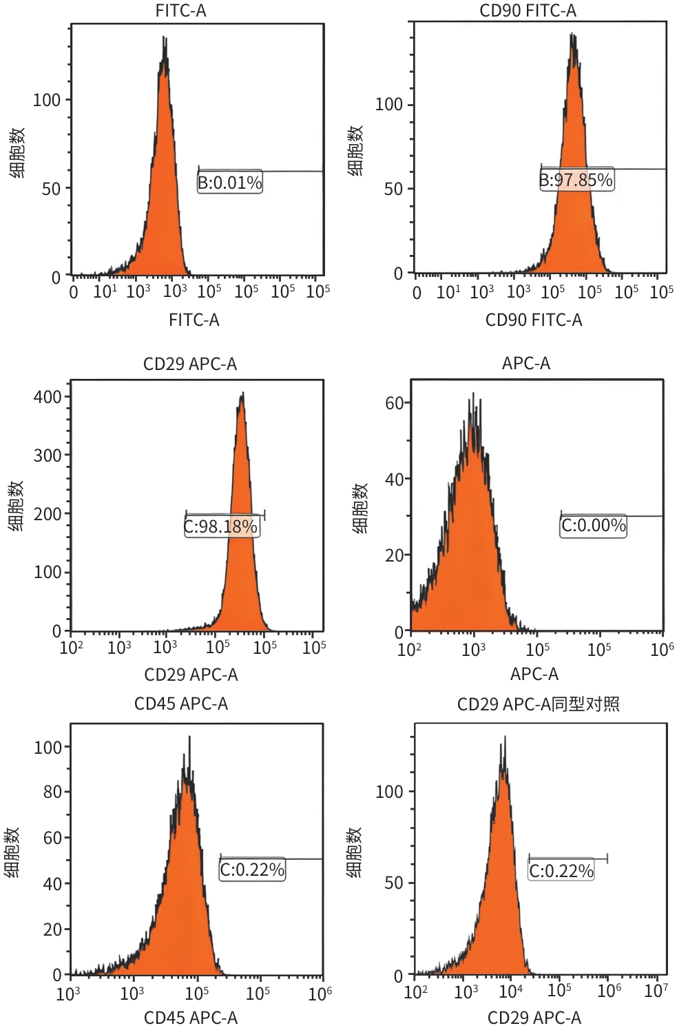
注: FITC,异硫氰酸荧光素;APC,别藻蓝蛋白。
图 1 大鼠骨髓间充质干细胞表面特异性标志物CD29、CD90和CD45的表达情况
Figure 1. Expression profile of surface-specific markers CD29, CD90, and CD45 in rat bone marrow mesenchymal stem cells
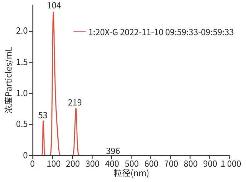
图 2 纳米颗粒跟踪分析仪测定大鼠骨髓间充质干细胞来源外泌体粒径分布
Figure 2. Particle size distribution of rat bone marrow mesenchymal stem cell-derived exosomes determined by nanoparticle tracking analysis
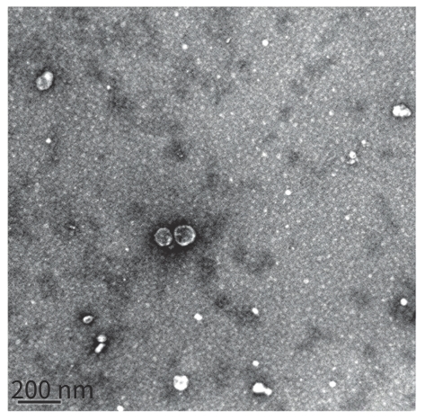
图 3 透射电镜观察大鼠骨髓间充质干细胞来源外泌体形态
Figure 3. Morphological observation of rat bone marrow mesenchymal stem cell-derived exosomes using transmission electron microscopy
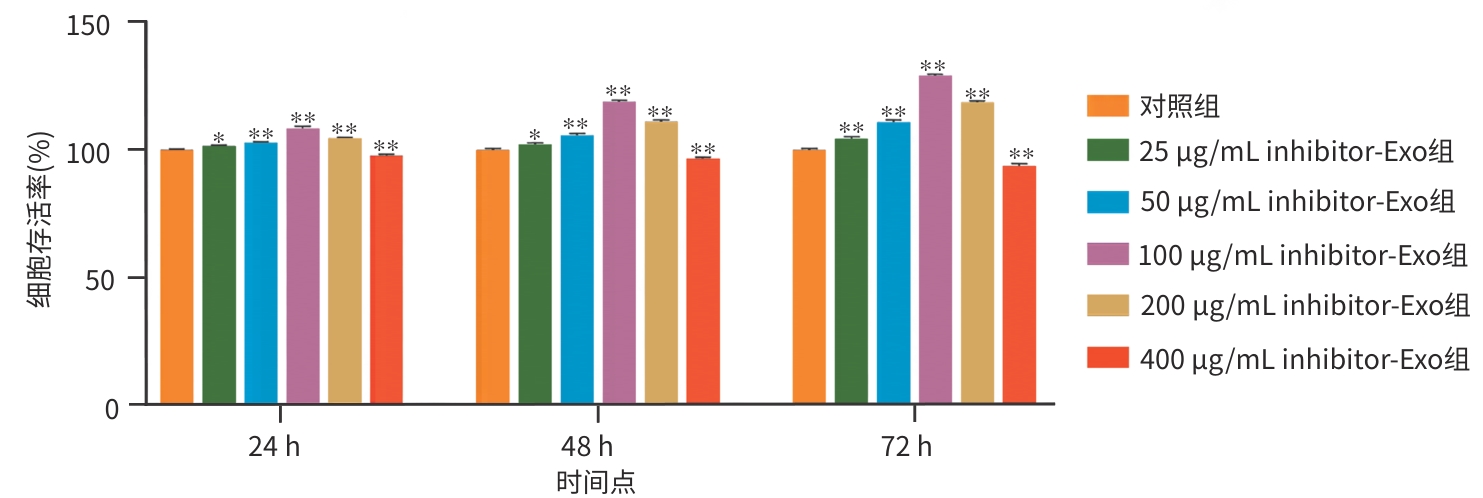
注: 与对照组比较,*P<0.05,**P<0.01。HUVEC,人脐静脉内皮细胞。
图 4 不同浓度外泌体对HUVEC增殖的影响
Figure 4. Effect of different concentrations of exosomes on the proliferation of HUVEC
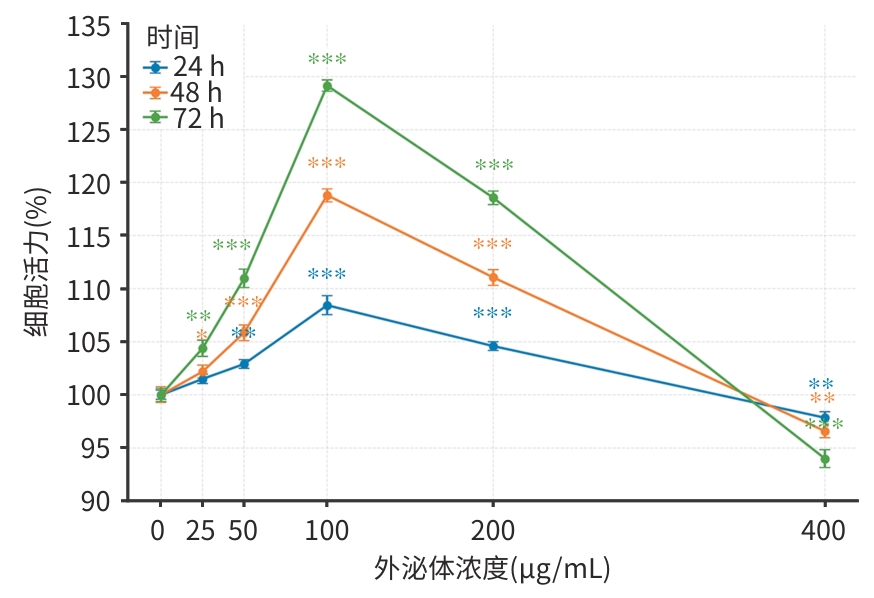
注: 采用MTT法检测细胞活力,以对照组活力为100%进行归一化处理。与对照组比较,*P<0.05,**P<0.01,***P<0.001。HUVEC,人脐静脉内皮细胞。
图 5 不同浓度miR-21-5p inhibitor外泌体对HUVEC活力的影响
Figure 5. Effect of different concentrations of miR-21-5p inhibitor-loaded exosomes on HUVEC viability
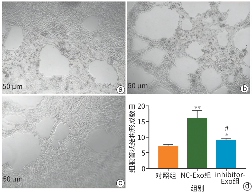
注: a,对照组;b,NC-Exo组;c,inhibitor-Exo组;d,各组管腔形成数目定量分析。与对照组比较,*P<0.05,**P<0.01;与NC-Exo组比较,#P<0.01。HUVEC,人脐静脉内皮细胞。
图 6 抑制外泌体中miR-21-5p后对HUVEC血管形成能力的影响
Figure 6. Effect of miR-21-5p inhibition in exosomes on the angiogenic capacity of HUVEC
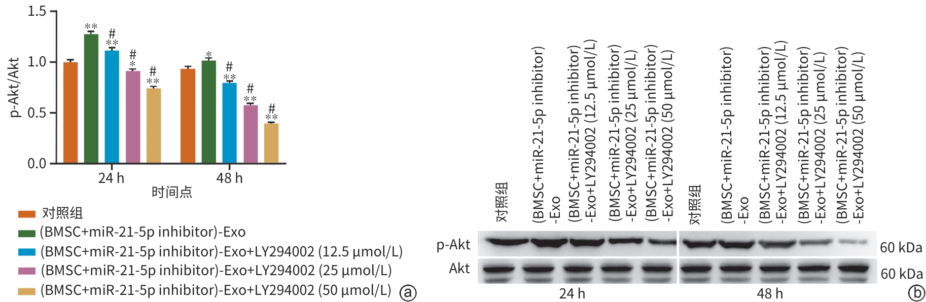
注: a,p-Akt/Akt蛋白相对表达量定量分析;b,p-Akt及总Akt蛋白条带。与对照组比较,*P<0.05,**P<0.01;与(BMSC+miR-21-5p inhibitor)-Exo组比较,#P<0.01。HUVEC,人脐静脉内皮细胞;PI3K,磷脂酰肌醇3-激酶;p-Akt,磷酸化蛋白激酶B。
图 7 蛋白质印迹法测定不同浓度PI3K抑制剂LY294002对p-Akt蛋白表达的影响
Figure 7. Effect of different concentrations of the PI3K inhibitor LY294002 on p-Akt protein expression as determined by Western blot
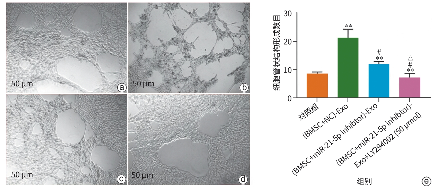
注: a,对照组;b,(BMSC+NC)-Exo组;c,(BMSC+miR-21-5p inhibitor)-Exo组;d,(BMSC+miR-21-5p inhibitor)-Exo+LY294002(50 μmol/L)组;e,各组管腔形成数目定量分析。与对照组比较,
**P<0.01;与(BMSC+NC)-Exo组比较,#P<0.01;与(BMSC+miR-21-5p inhibitor)-Exo组比较,△P<0.05。BMSC,骨髓间充质干细胞;HUVEC,人脐静脉内皮细胞;PI3K,磷脂酰肌醇3-激酶。 图 8 PI3K抑制剂LY294002处理对HUVEC血管形成能力的影响
Figure 8. Effect of treatment with the PI3K inhibitor LY294002 on the angiogenic capacity of HUVEC
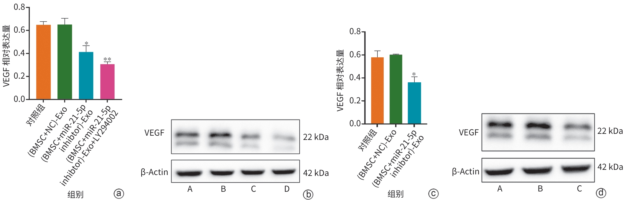
注: a,VEGF蛋白相对表达量定量分析;b,VEGF及β-Actin蛋白条带图[A:对照组;B:(BMSC+NC)-Exo组;C:(BMSC+miR-21-5p inhibitor)-Exo组;D:(BMSC+miR-21-5p inhibitor)-Exo+LY294002组];c,VEGF蛋白相对表达量定量分析(重复验证);d,VEGF及β-Actin蛋白条带图[重复验证,A:对照组;B:(BMSC+NC)-Exo组;C:(BMSC+miR-21-5p inhibitor)-Exo组]。与对照组比较:*P<0.05,**P<0.01。VEGF,血管内皮生长因子。
图 9 蛋白质印迹法检测各组细胞中VEGF蛋白表达水平
Figure 9. Western blot detection of VEGF protein expression levels in each group of cells
-
[1] Liver Failure and Artificial Liver Group, Chinese Society of Infectious Diseases, Chinese Medical Association; Severe Liver Disease and Artificial Liver Group, Chinese Society of Hepatology, Chinese Medical Association. Guideline for diagnosis and treatment of liver failure(2024 version)[J]. J Clin Hepatol, 2024, 40( 12): 2371- 2387. DOI: 10.12449/JCH241206.中华医学会感染病学分会肝衰竭与人工肝学组, 中华医学会肝病学分会重型肝病与人工肝学组. 肝衰竭诊治指南(2024年版)[J]. 临床肝胆病杂志, 2024, 40( 12): 2371- 2387. DOI: 10.12449/JCH241206. [2] Maiwall R, Kulkarni A V, Arab J P, et al. Acute liver failure[J]. Lancet, 2024, 404( 10454): 789- 802. DOI: 10.1016/S0140-6736(24)00693-7. [3] Naved S A, Ali Parpia S S. Anaesthetic management of liver transplantation in patients with fulminant hepatic failure[J]. J Pak Med Assoc, 2025, 75( 1): 111- 114. DOI: 10.47391/JPMA.9055. [4] Sorbini M, Carradori T, Patrono D, et al. Circulating cell-free DNA in liver transplantation: A pre- and post-transplant biomarker of graft dysfunction[J]. Artif Organs, 2025, 49( 4): 649- 662. DOI: 10.1111/aor.14910. [5] Khanam A, Kottilil S. Acute-on-chronic liver failure: Pathophysiological mechanisms and management[J]. Front Med, 2021, 8: 752875. DOI: 10.3389/fmed.2021.752875. [6] Sarin S K, Choudhury A, Kumar A, et al. Acute-on-chronic liver failure: Pathophysiological mechanisms and clinical management[J]. Nat Rev Gastroenterol Hepatol, 2026, 23( 5): 411- 431. DOI: 10.1038/s41575-025-01159-4. [7] Furuta T, Furuya K, Zheng Y W, et al. Novel alternative transplantation therapy for orthotopic liver transplantation in liver failure: A systematic review[J]. World J Transplant, 2020, 10( 3): 64- 78. DOI: 10.5500/wjt.v10.i3.64. [8] Psaraki A, Ntari L, Karakostas C, et al. Extracellular vesicles derived from mesenchymal stem/stromal cells: The regenerative impact in liver diseases[J]. Hepatology, 2022, 75( 6): 1590- 1603. DOI: 10.1002/hep.32129. [9] Maldonado V V, Patel N H, Smith E E, et al. Clinical utility of mesenchymal stem/stromal cells in regenerative medicine and cellular therapy[J]. J Biol Eng, 2023, 17( 1): 44. DOI: 10.1186/s13036-023-00361-9. [10] Luo Xiaofan, Luo Yehao, Ting Lyu, et al. Regulating mechanism of mesenchymal stem cells on the progression of liver cirrhosis[J]. J Liaoning Univ Tradit Chin Med, 2022, 24( 8): 65- 69. DOI: 10.13194/j.issn.1673-842x.2022.08.015.罗筱凡, 罗业浩, 吕挺, 等. 间充质干细胞对肝硬化进展的调控机制[J]. 辽宁中医药大学学报, 2022, 24( 8): 65- 69. DOI: 10.13194/j.issn.1673-842x.2022.08.015. [11] Mittal A, Jakhmola V R, Baweja S. Bioengineered extracellular vesicles: The path to precision medicine in liver diseases[J]. Liver Res, 2025, 9( 1): 17- 28. DOI: 10.1016/j.livres.2025.02.002. [12] Youssef E, Palmer D, Fletcher B, et al. Exosomes in precision oncology and beyond: From bench to bedside in diagnostics and therapeutics[J]. Cancers, 2025, 17( 6): 940. DOI: 10.3390/cancers17060940. [13] Wang X Q, Munson M J, Friis K, et al. Hybrid extracellular vesicles for efficient loading and functional delivery of mRNA[J]. J Extracell Vesicles, 2025, 14( 12): e70201. DOI: 10.1002/jev2.70201. [14] Mathieu M, Martin-Jaular L, Lavieu G, et al. Specificities of secretion and uptake of exosomes and other extracellular vesicles for cell-to-cell communication[J]. Nat Cell Biol, 2019, 21( 1): 9- 17. DOI: 10.1038/s41556-018-0250-9. [15] Shokravi S, Borisov V, Zaman B A, et al. Mesenchymal stromal cells(MSCs) and their exosome in acute liver failure(ALF): A comprehensive review[J]. Stem Cell Res Ther, 2022, 13( 1): 192. DOI: 10.1186/s13287-022-02825-z. [16] Wang J L, Huang D Q, Ren H Z, et al. Biomimic trained immunity-MSCs delivery microcarriers for acute liver failure regeneration[J]. Small, 2022, 18( 36): e2200858. DOI: 10.1002/smll.202200858. [17] Sun Lieqian, Gu Mengyu, Yang Jie, et al. Effect of bone marrow-derived mesenchymal stem cell transplantation on mitochondrial autophagy in rats with vascular dementia through ROS/Nrf2 signaling and its mechanism[J]. J Jilin Univ Med Ed, 2025, 51( 3): 610- 620. DOI: 10.13481/j.1671-587X.20250306.孙烈乾, 顾梦宇, 杨杰, 等. 骨髓来源的间充质干细胞移植通过ROS/Nrf2信号对血管性痴呆大鼠线粒体自噬的影响及其机制[J]. 吉林大学学报(医学版), 2025, 51( 3): 610- 620. DOI: 10.13481/j.1671-587X.20250306. [18] Zhang L, Song Y L, Chen L, et al. miR-20a-containing exosomes from umbilical cord mesenchymal stem cells alleviates liver ischemia/reperfusion injury[J]. J Cell Physiol, 2020, 235( 4): 3698- 3710. DOI: 10.1002/jcp.29264. [19] Kim S H, Kim C H, Lee C H, et al. Glycoengineered stem cell-derived extracellular vesicles for targeted therapy of acute kidney injury[J]. Biomaterials, 2025, 318: 123165. DOI: 10.1016/j.biomaterials.2025.123165. [20] Wang Z R, Ling J, You S L, et al. Exosomes derived from liver failure patients’ plasma stimulated mesenchymal stem cells alleviate acute liver failure[J]. Stem Cell Res Ther, 2025, 16( 1): 48. DOI: 10.1186/s13287-025-04163-2. [21] Li Rui, Wang Dianxiang, Liang Zhaowei, et al. Current research status and application prospects of mesenchymal stem cell-derived exosomes in islet transplantation[J]. Organ Transplantation, 2025, 16( 1): 163- 168. DOI: 10.12464/j.issn.1674-7445.2024194.李睿, 王殿相, 梁兆伟, 等. 间充质干细胞来源的外泌体在胰岛移植中的作用研究现状及应用前景[J]. 器官移植, 2025, 16( 1): 163- 168. DOI: 10.12464/j.issn.1674-7445.2024194. [22] Zheng Rongjiong, Deng Zerun, Han Dan, et al. Mechanism underlying rat hepatocyte apoptosis regulated by exosomes derived from bone marrow mesenchymal stem cells[J]. Chin J Tissue Eng Res, 2024, 28( 1): 44- 49. DOI: 10.12307/2023.916.郑嵘炅, 邓泽润, 韩丹, 等. 骨髓间充质干细胞来源外泌体调节大鼠肝细胞凋亡的机制[J]. 中国组织工程研究, 2024, 28( 1): 44- 49. DOI: 10.12307/2023.916. [23] Maher S Z, Schreibman I R. The clinical spectrum and manifestations of acute liver failure[J]. Clin Liver Dis, 2018, 22( 2): 361- 374. DOI: 10.1016/j.cld.2018.01.012. [24] Wang Y J, Xie X L, Liu H Q, et al. LR12 promotes liver repair by improving the resolution of inflammation and liver regeneration in mice with thioacetamide-(TAA-) induced acute liver failure[J]. Mediators Inflamm, 2021, 2021: 2327721. DOI: 10.1155/2021/2327721. [25] Li M J, Wang Z F, Yan X X, et al. Luopan mountain pig bone marrow mesenchymal stem cells promote liver regeneration in D-galactosamine-induced acute liver failure rats by regulating the PTEN-PI3K/Akt/mTOR pathway[J]. Biology, 2025, 14( 10): 1363. DOI: 10.3390/biology14101363. [26] Stravitz R T, Fontana R J, Karvellas C, et al. Future directions in acute liver failure[J]. Hepatology, 2023, 78( 4): 1266- 1289. DOI: 10.1097/HEP.0000000000000458. [27] Luo S, Wu F, Yang X J. Single-cell transcriptome atlas reveals mitophagy dynamics in acute chemical injury model and the role of MSCs transplantation[J]. Stem Cell Res Ther, 2025, 16( 1): 350. DOI: 10.1186/s13287-025-04491-3. [28] Xie D, Yu L N, Wang Z Y, et al. Exosomes derived from human umbilical cord mesenchymal stem cells inhibit hepatocyte pyroptosis via miR-423-5p/ZBP1 in acute liver failure[J]. Hum Cell, 2025, 38( 5): 124. DOI: 10.1007/s13577-025-01248-1. [29] Tang H X, Lu W, Yang Q, et al. Exosomes derived from 3D-cultured hUCMSCs exhibit superior hepatoprotection against ALF[J]. iScience, 2025, 28( 9): 113419. DOI: 10.1016/j.isci.2025.113419. [30] Wu J, Chen L H, Sun S Y, et al. Mesenchymal stem cell-derived exosomes: The dawn of diabetic wound healing[J]. World J Diabetes, 2022, 13( 12): 1066- 1095. DOI: 10.4239/wjd.v13.i12.1066. [31] Vadhan A, Gupta T, Hsu W L. Mesenchymal stem cell-derived exosomes as a treatment option for osteoarthritis[J]. Int J Mol Sci, 2024, 25( 17): 9149. DOI: 10.3390/ijms25179149. [32] Yao Shun, Yang Yongqi, Liu Cuicui, et al. Advances in the druggability and clinical translation of platelets[J]. Chin J Cell Biol, 2026, 48( 1): 116- 128. DOI: 10.11844/cjcb.2026.01.0008.姚舜, 杨咏琪, 刘翠翠, 等. 血小板临床转化研究进展[J]. 中国细胞生物学学报, 2026, 48( 1): 116- 128. DOI: 10.11844/cjcb.2026.01.0008. [33] Su Qiumei, Zhou Ou, Tan Wen, et al. Progress of mechanism of mesenchymal stem cell-derived extracellular vesicles in the treatment of bronchopulmonary dysplasia[J]. J Pediatr Pharm, 2026, 32( 1): 52- 56. DOI: 10.13407/j.cnki.jpp.1672-108X.2026.01.014.粟秋梅, 周欧, 谭雯, 等. 间充质干细胞来源的细胞外囊泡治疗支气管肺发育不良机制研究进展[J]. 儿科药学杂志, 2026, 32( 1): 52- 56. DOI: 10.13407/j.cnki.jpp.1672-108X.2026.01.014. [34] He L L, Zhang H W, Zhao N B, et al. A novel approach in biomedical engineering: The use of polyvinyl alcohol hydrogel encapsulating human umbilical cord mesenchymal stem cell-derived exosomes for enhanced osteogenic differentiation and angiogenesis in bone regeneration[J]. Int J Biol Macromol, 2024, 270: 132116. DOI: 10.1016/j.ijbiomac.2024.132116. [35] Tan W, Ma L, Li Y L, et al. Glycoside components promote endothelial progenitor cell-derived exosomes repairing damaged vascular endothelium via the PI3K/AKT signaling pathway[J]. Phytomedicine, 2025, 141: 156694. DOI: 10.1016/j.phymed.2025.156694. [36] He P P, Zeng W R, Li J, et al. ATF4 regulates PI3K/AKT signaling axis to promote angiogenesis after myocardial infarction[J]. Vitro Cell Dev Biol Anim, 2025, 61( 8): 909- 920. DOI: 10.1007/s11626-025-01085-4. [37] Peng C H, Chen H M, Li Y W, et al. LRIG3 suppresses angiogenesis by regulating the PI3K/AKT/VEGFA signaling pathway in glioma[J]. Front Oncol, 2021, 11: 621154. DOI: 10.3389/fonc.2021.621154. [38] Li J, Shi R D, Li Q, et al. miRNA-21-5p targeting PTEN to regulate PI3K/Akt/mTOR pathway in retinal pigment epithelial cell photodamage[J]. Int J Ophthalmol, 2025, 18( 4): 575- 581. DOI: 10.18240/ijo.2025.04.02. [39] Cheng Y, Guo Q Y, Cheng Y L, et al. Endostatin-expressing endometrial mesenchymal stem cells inhibit angiogenesis in endometriosis through the miRNA-21-5p/TIMP3/PI3K/Akt/mTOR pathway[J]. Stem Cells Transl Med, 2025, 14( 2): szae079. DOI: 10.1093/stcltm/szae079. -

 下载:
 本文二维码
计量
- 文章访问数: 17
- HTML全文浏览量: 7
- PDF下载量: 0
- 被引次数: 0