PDF下载 ( 1655 KB)

肝巨噬细胞在代谢相关脂肪性肝病中的作用机制及相关靶向治疗策略
DOI: 10.12449/JCH260924
Mechanism of action of hepatic macrophages in metabolic dysfunction-associated fatty liver disease and related targeted therapeutic strategies
-
摘要: 代谢相关脂肪性肝病(MAFLD)是目前全球最常见的慢性肝病,其疾病谱包括单纯性脂肪肝、代谢相关脂肪性肝炎、肝纤维化、肝硬化和肝细胞癌。近年来研究揭示,肝巨噬细胞在MAFLD发生发展中发挥重要调控作用。肝巨噬细胞主要包括定居肝脏的库普弗细胞和单核来源的巨噬细胞,通过调节炎症反应、脂质代谢、纤维化进程和细胞死亡途径,参与疾病的病理生理过程。本文综述了肝巨噬细胞在MAFLD中的类型多样性、功能可塑性及其与肝细胞、肠道微生物之间的相互作用,重点阐述了肝巨噬细胞及相关信号通路在MAFLD进展中的核心机制,并探讨了以肝巨噬细胞为靶点的新型治疗策略。深入理解肝巨噬细胞在MAFLD中的作用机制,可为开发MAFLD的精准治疗方法提供重要理论基础。Abstract: Metabolic dysfunction-associated fatty liver disease (MAFLD) is currently the most prevalent chronic liver disease worldwide, and its disease spectrum encompasses simple steatosis, metabolic-associated steatohepatitis, hepatic fibrosis, liver cirrhosis, and hepatocellular carcinoma. Recent studies have shown that hepatic macrophages play an important regulatory role in the development and progression of MAFLD. Hepatic macrophages mainly consist of resident Kupffer cells and monocyte-derived macrophages, which participate in the pathophysiological processes of the disease by regulating inflammatory response, lipid metabolism, fibrogenic process, and cell death pathways. This article summarizes the diversity and functional plasticity of hepatic macrophages in MAFLD, reviews their interactions with hepatocytes and gut microbiota, elaborates on the core mechanism of hepatic macrophages and related signaling pathways in the progression of MAFLD, and further discusses novel therapeutic strategies targeting hepatic macrophages. An in-depth understanding of the mechanism of action of hepatic macrophages in MAFLD may provide a crucial theoretical basis for developing precise therapeutic approaches for this disease.
-
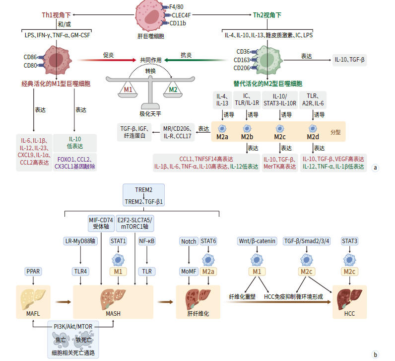
注: a,肝巨噬细胞M1/M2极化平衡及亚型分化调控示意图;b,下游信号通路调控机制示意图。CLEC4F,C型凝集素结构域家族4成员F;CD11b,分化簇11b;F4/80,F4/80分子;Th1,辅助性T细胞1;Th2,辅助性T细胞2;IL,白细胞介素;IC,免疫复合物;LPS,脂多糖;IFN-γ,干扰素γ;GM-CSF,粒细胞-巨噬细胞集落刺激因子;CD163,分化簇163;CD86,分化簇86;CD36,分化簇36;CD206,分化簇206;CD80,分化簇80;STAT,信号转导与转录激活因子;IL-1R,白细胞介素1受体;TLR,Toll样受体;A2R,A2腺苷受体;TGF-β,转化生长因子β;MR,甘露糖受体;CCL17,CC亚族趋化因子配体17;CCL2,CC亚族趋化因子配体2;CCL1,CC亚族趋化因子配体1;CXCL9,C-X-C基序趋化因子配体9;CX3CL1,C-X3-C基序趋化因子配体1;FOXO1,叉头框蛋白O1;IGF,胰岛素样生长因子;TNFSF14,肿瘤坏死因子超家族成员14;MerTK,Mer受体酪氨酸激酶;VEGF,血管内皮生长因子;NF-κB,核因子κB;Wnt/β-catenin,无翼样糖蛋白/β-联蛋白通路;PI3K/Akt/mTOR,磷脂酰肌醇3-激酶/蛋白激酶B/哺乳动物雷帕霉素靶蛋白通路;MIF-CD74,巨噬细胞迁移抑制因子-分化簇74轴;E2F2-SLC7A5-mTORC1,叉头框蛋白E2-溶质载体家族7成员5-哺乳动物雷帕霉素靶蛋白复合物1轴;PPAR,过氧化物酶体增殖物激活受体;MyD88,髓样分化因子88;TNF-α,肿瘤坏死因子α;MoMF,骨髓来源的单核巨噬细胞;TREM,髓样细胞触发受体;Smad,母系抗十五表态蛋白同源物;MAFLD,代谢相关脂肪性肝病;MAFL,代谢相关单纯性脂肪肝;MASH,代谢相关脂肪性肝炎;HCC,肝细胞癌。
图 1 肝巨噬细胞及相关信号通路在MAFLD进展中的作用机制
Figure 1. Roles and mechanisms of hepatic macrophages and associated signaling pathways in MAFLD progression
表 1 2024—2025年肝巨噬细胞靶向治疗MAFLD的策略
Table 1. Hepatic macrophage-related therapeutic strategies for MAFLD (2024—2025)
疾病领域 策略 核心机制 研发阶段 MASH M2巨噬细胞仿生纳米
递药平台(O@OLGA@M)利用M2膜炎症趋化性靶向递送奥贝胆酸,重编程M1→
M2,协同调节脂代谢、抗炎和抗纤维化,并调控肠道
菌群[15]临床前(2025-09-25报道) HCC 仿生靶向铁死亡纳米平台
(Sv@PM-M2p)同时靶向肝癌细胞与M2型TAM,诱导双重铁死亡,逆转
免疫抑制,重塑肿瘤微环境[58]临床前(2025-11-24报道) 肝纤维化/
肝硬化CAR-Ms(靶向uPAR) CAR-Ms特异性归巢肝脏,调节免疫微环境,促进中性粒
细胞和M2型巨噬细胞浸润,激活抗纤维化T细胞反应,减
轻纤维化[63]临床前(2024-10-02报道) 自体工程巨噬细胞
(RTX001)(国外)自体巨噬细胞经工程化改造,具有抗炎、抗纤维化功能,
用于终末期肝病患者的细胞疗法[61]Ⅰ/Ⅱ期临床(2025-10-02
报道)自体工程巨噬细胞
(MACF-001)(国内)采用自主知识产权的抗纤维化Payloads组合与递送系统,
维持巨噬细胞免疫修复表型[62]早期临床研究中 非工程巨噬细胞
(MATCH实验)单剂量自体巨噬细胞输注后,67%患者的肝纤维化评分
改善≥1级,MELD评分显著降低,无严重不良反应[64]Ⅱ期临床(2025-03-01
报道)自体单核细胞来源
巨噬细胞输注在代偿期肝硬化患者中显示安全性,并呈现抗炎潜力(降
低促炎因子、提升抗炎因子)Ⅱ期临床(2025-01-13
报道)注:MAFLD,代谢相关脂肪性肝病;MASH,代谢相关脂肪性肝炎;HCC,肝细胞癌;CAR-Ms,嵌合抗原受体修饰巨噬细胞;uPAR,尿激酶型纤溶酶原激活物受体;RTX001,自体工程巨噬细胞(国外);MACF-001,自体工程巨噬细胞(国内);MELD,终末期肝病模型;O@OLGA@M,M2巨噬细胞仿生纳米递药平台;Sv@PM-M2p,仿生靶向铁死亡纳米平台;TAM,肿瘤相关巨噬细胞。
 下载: 导出CSV
-
[1] Estes C, Chan H L Y, Chien R N, et al. Modelling NAFLD disease burden in four Asian regions: 2019-2030[J]. Aliment Pharmacol Ther, 2020, 51( 8): 801- 811. DOI: 10.1111/apt.15673. [2] Sandireddy R, Sakthivel S, Gupta P, et al. Systemic impacts of metabolic dysfunction-associated steatotic liver disease(MASLD) and metabolic dysfunction-associated steatohepatitis(MASH) on heart, muscle, and kidney related diseases[J]. Front Cell Dev Biol, 2024, 12: 1433857. DOI: 10.3389/fcell.2024.1433857. [3] González-Serrano S, Onoiu A I, Camps J, et al. Immunometabolic integration in therapeutic strategies for managing MASLD[J]. Front Pharmacol, 2025, 16: 1693753. DOI: 10.3389/fphar.2025.1693753. [4] Krenkel O, Hundertmark J, Abdallah A T, et al. Myeloid cells in liver and bone marrow acquire a functionally distinct inflammatory phenotype during obesity-related steatohepatitis[J]. Gut, 2020, 69( 3): 551- 563. DOI: 10.1136/gutjnl-2019-318382. [5] Orci L A, Kreutzfeldt M, Goossens N, et al. Tolerogenic properties of liver macrophages in non-alcoholic steatohepatitis[J]. Liver Int, 2020, 40( 3): 609- 621. DOI: 10.1111/liv.14336. [6] Scott C L, Zheng F, de Baetselier P, et al. Bone marrow-derived monocytes give rise to self-renewing and fully differentiated Kupffer cells[J]. Nat Commun, 2016, 7: 10321. DOI: 10.1038/ncomms10321. [7] Sierro F, Evrard M, Rizzetto S, et al. A liver capsular network of monocyte-derived macrophages restricts hepatic dissemination of intraperitoneal bacteria by neutrophil recruitment[J]. Immunity, 2017, 47( 2): 374- 388. e 6. DOI: 10.1016/j.immuni.2017.07.018. [8] Gomez Perdiguero E, Klapproth K, Schulz C, et al. Tissue-resident macrophages originate from yolk-sac-derived erythro-myeloid progenitors[J]. Nature, 2015, 518( 7540): 547- 551. DOI: 10.1038/nature13989. [9] He Y, Chen Y F, Qian S Y, et al. Immunopathogenic mechanisms and immunoregulatory therapies in MASLD[J]. Cell Mol Immunol, 2025, 22( 10): 1159- 1177. DOI: 10.1038/s41423-025-01307-5. [10] Devisscher L, Scott C L, Lefere S, et al. Non-alcoholic steatohepatitis induces transient changes within the liver macrophage pool[J]. Cell Immunol, 2017, 322: 74- 83. DOI: 10.1016/j.cellimm.2017.10.006. [11] Viola A, Munari F, Sánchez-Rodríguez R, et al. The metabolic signature of macrophage responses[J]. Front Immunol, 2019, 10: 1462. DOI: 10.3389/fimmu.2019.01462. [12] Wang L X, Zhang S X, Wu H J, et al. M2b macrophage polarization and its roles in diseases[J]. J Leukoc Biol, 2019, 106( 2): 345- 358. DOI: 10.1002/JLB.3RU1018-378RR. [13] Cui C, Gao S, Shi J F, et al. Gut-liver axis: The role of intestinal microbiota and their metabolites in the progression of metabolic dysfunction-associated steatotic liver disease[J]. Gut Liver, 2025, 19( 4): 479- 507. DOI: 10.5009/gnl240539. [14] Miyamoto Y, Kikuta J, Matsui T, et al. Periportal macrophages protect against commensal-driven liver inflammation[J]. Nature, 2024, 629( 8013): 901- 909. DOI: 10.1038/s41586-024-07372-6. [15] Pan X E, Chen X Y, Ma Y, et al. Chemotaxis-guided nanoplatform for non-alcoholic steatohepatitis therapy via macrophage reprogramming, hepatoprotection and gut microbiome modulation[J]. Biomaterials, 2026, 326: 123694. DOI: 10.1016/j.biomaterials.2025.123694. [16] de Muynck K, Vanderborght B, van Vlierberghe H, et al. The gut-liver axis in chronic liver disease: A macrophage perspective[J]. Cells, 2021, 10( 11): 2959. DOI: 10.3390/cells10112959. [17] Balmer M L, Slack E, de Gottardi A, et al. The liver may act as a firewall mediating mutualism between the host and its gut commensal microbiota[J]. Sci Transl Med, 2014, 6( 237): 237ra66. DOI: 10.1126/scitranslmed.3008618. [18] di Ciaula A, Baj J, Garruti G, et al. Liver steatosis, gut-liver axis, microbiome and environmental factors. A never-ending bidirectional cross-talk[J]. J Clin Med, 2020, 9( 8): 2648. DOI: 10.3390/jcm9082648. [19] Yi Z F, Fan H, Liu X X, et al. Adrenomedullin improves intestinal epithelial barrier function by downregulating myosin light chain phosphorylation in ulcerative colitis rats[J]. Mol Med Rep, 2015, 12( 3): 3615- 3620. DOI: 10.3892/mmr.2015.3887. [20] Rizvi F, Everton E, Smith A R, et al. Murine liver repair via transient activation of regenerative pathways in hepatocytes using lipid nanoparticle-complexed nucleoside-modified mRNA[J]. Nat Commun, 2021, 12: 613. DOI: 10.1038/s41467-021-20903-3. [21] Hendrikx T, Porsch F, Kiss M G, et al. Soluble TREM2 levels reflect the recruitment and expansion of TREM2(+) macrophages that localize to fibrotic areas and limit NASH[J]. J Hepatol, 2022, 77( 5): 1373- 1385. DOI: 10.1016/j.jhep.2022.06.004. [22] Li L, Cui L, Lin P, et al. Kupffer-cell-derived IL-6 is repurposed for hepatocyte dedifferentiation via activating progenitor genes from injury-specific enhancers[J]. Cell Stem Cell, 2023, 30( 3): 283- 299.e9. DOI: 10.1016/j.stem.2023.01.009. [23] del Mar Rigual M, Angulo-Aguado M, Zagorac S, et al. Macrophages harness hepatocyte glutamate to boost liver regeneration[J]. Nature, 2025, 641( 8064): 1005- 1016. DOI: 10.1038/s41586-025-08778-6. [24] Wang H Y, Poulain S, Cao W X, et al. Palmitic acid induced the onset of lipotoxicity in a HepaSH-on-chip model with raised of H2O2 and IL-6, and altered p38/MAPK& JAK/STAT pathways[J]. Toxicology, 2026, 520: 154345. DOI: 10.1016/j.tox.2025.154345. [25] Xu X, Li H G, Lin H, et al. MIF-mediated crosstalk between THRSP+ hepatocytes and CD74+lipid-associated macrophages in hepatic periportal zone drives MASH[J]. Hepatology, 2026, 83( 4): 868- 887. DOI: 10.1097/HEP.0000000000001429. [26] Xu R N, Vujić N, Bianco V, et al. Lipid-associated macrophages between aggravation and alleviation of metabolic diseases[J]. Trends Endocrinol Metab, 2024, 35( 11): 981- 995. DOI: 10.1016/j.tem.2024.04.009. [27] Tian Y X, Ni Y M, Zhang T, et al. Targeting hepatic macrophages for non-alcoholic fatty liver disease therapy[J]. Front Cell Dev Biol, 2024, 12: 1444198. DOI: 10.3389/fcell.2024.1444198. [28] Lumeng C N, Bodzin J L, Saltiel A R. Obesity induces a phenotypic switch in adipose tissue macrophage polarization[J]. J Clin Invest, 2007, 117( 1): 175- 184. DOI: 10.1172/JCI29881. [29] Wang J J, Cai Y, Feng Y X, et al. Diabetic kidney disease macrophage cholesterol efflux: A revolution from metabolism to immune[J]. Front Endocrinol, 2025, 16: 1714167. DOI: 10.3389/fendo.2025.1714167. [30] Shi S Y, Zhou Y Y, Zhang H M, et al. TREM2 in MASH: Integrating lipid metabolism and immune response[J]. Front Immunol, 2025, 16: 1604837. DOI: 10.3389/fimmu.2025.1604837. [31] Perugorria M J, Esparza-Baquer A, Oakley F, et al. Non-parenchymal TREM-2 protects the liver from immune-mediated hepatocellular damage[J]. Gut, 2019, 68( 3): 533- 546. DOI: 10.1136/gutjnl-2017-314107. [32] Indira Chandran V, Wernberg C W, Lauridsen M M, et al. Circulating TREM2 as a noninvasive diagnostic biomarker for NASH in patients with elevated liver stiffness[J]. Hepatology, 2023, 77( 2): 558- 572. DOI: 10.1002/hep.32620. [33] Song K, Kwon H, Han C, et al. Yes-associated protein in kupffer cells enhances the production of proinflammatory cytokines and promotes the development of nonalcoholic steatohepatitis[J]. Hepatology, 2020, 72( 1): 72- 87. DOI: 10.1002/hep.30990. [34] Zou Xuchen, Chen Qian, Shi Chunxia, et al. Effect of macrophage migration inhibitory factor on liver inflammation in mice with acute liver failure[J]. J Med Res, 2024, 53( 5): 38- 41, 47. DOI: 10.11969/j.issn.1673-548X.2024.05.009.邹旭晨, 陈倩, 石春霞, 等. 巨噬细胞移动抑制因子在急性肝衰竭小鼠肝脏炎症中的作用研究[J]. 医学研究杂志, 2024, 53( 5): 38- 41, 47. DOI: 10.11969/j.issn.1673-548X.2024.05.009. [35] Tacke F. Targeting hepatic macrophages to treat liver diseases[J]. J Hepatol, 2017, 66( 6): 1300- 1312. DOI: 10.1016/j.jhep.2017.02.026. [36] Zhang J, Wang Y, Fan M Y, et al. Reactive oxygen species regulation by NCF1 governs ferroptosis susceptibility of Kupffer cells to MASH[J]. Cell Metab, 2024, 36( 8): 1745- 1763. e 6. DOI: 10.1016/j.cmet.2024.05.008. [37] Valenti L, Moscatiello S, Vanni E, et al. Venesection for non-alcoholic fatty liver disease unresponsive to lifestyle counselling: A propensity score-adjusted observational study[J]. QJM, 2011, 104( 2): 141- 149. DOI: 10.1093/qjmed/hcq170. [38] Tsurusaki S, Tsuchiya Y, Koumura T, et al. Hepatic ferroptosis plays an important role as the trigger for initiating inflammation in nonalcoholic steatohepatitis[J]. Cell Death Dis, 2019, 10: 449. DOI: 10.1038/s41419-019-1678-y. [39] Pierantonelli I, Svegliati-Baroni G. Nonalcoholic fatty liver disease: Basic pathogenetic mechanisms in the progression from NAFLD to NASH[J]. Transplantation, 2019, 103( 1): e1- e13. DOI: 10.1097/TP.0000000000002480. [40] Yi Z H, Li S Q, Ke J Y, et al. Baicalein relieves ferroptosis-mediated phagocytosis inhibition of macrophages in ovarian endometriosis[J]. Curr Issues Mol Biol, 2022, 44( 12): 6189- 6204. DOI: 10.3390/cimb44120422. [41] Kohlhepp M S, Liu H Y, Tacke F, et al. The contradictory roles of macrophages in non-alcoholic fatty liver disease and primary liver cancer-Challenges and opportunities[J]. Front Mol Biosci, 2023, 10: 1129831. DOI: 10.3389/fmolb.2023.1129831. [42] Guillot A, Winkler M, Silva Afonso M, et al. Mapping the hepatic immune landscape identifies monocytic macrophages as key drivers of steatohepatitis and cholangiopathy progression[J]. Hepatology, 2023, 78( 1): 150- 166. DOI: 10.1097/HEP.0000000000000270. [43] Guo W, Li Z Y, Anagnostopoulos G, et al. Notch signaling regulates macrophage-mediated inflammation in metabolic dysfunction-associated steatotic liver disease[J]. Immunity, 2024, 57( 10): 2310- 2327. e 6. DOI: 10.1016/j.immuni.2024.08.016. [44] Guillot A, Tacke F. Spatial dimension of macrophage heterogeneity in liver diseases[J]. eGastroenterology, 2023, 1( 1): e000003. DOI: 10.1136/egastro-2023-000003. [45] Ma Simin, Zhu Hanxiao, Li Yuqing, et al. Research progress of THR-β agonists in the treatment of MASH[J]. Acta Pharm Sin, 2025, 60( 9): 2690- 2701. DOI: 10.16438/j.0513-4870.2025-0137.马思敏, 朱含笑, 李昱晴, 等. THR-β激动剂在非酒精性脂肪肝炎治疗方面的研究进展[J]. 药学学报, 2025, 60( 9): 2690- 2701. DOI: 10.16438/j.0513-4870.2025-0137. [46] Kholodenko I V, Yarygin K N. Hepatic macrophages as targets for the MSC-based cell therapy in non-alcoholic steatohepatitis[J]. Biomedicines, 2023, 11( 11): 3056. DOI: 10.3390/biomedicines11113056. [47] Obata Y, Abe K, Miyazaki M, et al. The transfer of the hepatocyte growth factor gene by macrophages ameliorates the progression of peritoneal fibrosis in mice[J]. Int J Mol Sci, 2023, 24( 8): 6951. DOI: 10.3390/ijms24086951. [48] Peiseler M, Schwabe R, Hampe J, et al. Immune mechanisms linking metabolic injury to inflammation and fibrosis in fatty liver disease-novel insights into cellular communication circuits[J]. J Hepatol, 2022, 77( 4): 1136- 1160. DOI: 10.1016/j.jhep.2022.06.012. [49] Niu Yuanyuan, Wang Longde, Xu Wenjuan, et al. Research progress in the role of M1/M2 polarization of macrophages in different liver diseases[J]. J Shanghai Jiao Tong Univ Med Sci, 2024, 44( 4): 509- 517. DOI: 10.3969/j.issn.1674-8115.2024.04.012.牛媛媛, 汪龙德, 胥文娟, 等. 巨噬细胞M1/M2型极化在不同肝病中的作用研究进展[J]. 上海交通大学学报(医学版), 2024, 44( 4): 509- 517. DOI: 10.3969/j.issn.1674-8115.2024.04.012. [50] Wang Yuhang, Chen Zhiming, Yin Yucheng, et al. Radiotherapy sensitivity regulation of tumor-associated macrophages[J]. Trauma and Crit Medicine, 2024, 12( 3): 182- 185, 190. DOI: 10.16048/j.issn.2095-5561.2024.03.13.王禹杭, 陈志明, 殷雨成, 等. 肿瘤相关巨噬细胞放射治疗敏感性调节机制与应用进展[J]. 创伤与急危重病医学, 2024, 12( 3): 182- 185,190. DOI: 10.16048/j.issn.2095-5561.2024.03.13. [51] Xia Anliang, Sun Beicheng. Research progress of macrophages in non-alcoholic fatty liver diseases[J]. J Nanjing Med Univ Nat Sci, 2023, 43( 10): 1456- 1463. DOI: 10.7655/NYDXBNS20231020.夏安亮, 孙倍成. 巨噬细胞参与非酒精性脂肪性肝病的研究进展[J]. 南京医科大学学报(自然科学版), 2023, 43( 10): 1456- 1463. DOI: 10.7655/NYDXBNS20231020. [52] Nair B, Nath L R. Inevitable role of TGF-β1 in progression of nonalcoholic fatty liver disease[J]. J Recept Signal Transduct Res, 2020, 40( 3): 195- 200. DOI: 10.1080/10799893.2020.1726952. [53] Liu Z, Wang H, Liang Y, et al. E2F2 reprograms macrophage function by modulating material and energy metabolism in the progression of metabolic dysfunction-associated steatohepatitis[J]. Adv Sci, 2024, 11( 48): 2410880. DOI: 10.1002/advs.202410880. [54] Wree A, Eguchi A, McGeough M D, et al. NLRP3 inflammasome activation results in hepatocyte pyroptosis, liver inflammation, and fibrosis in mice[J]. Hepatology, 2014, 59( 3): 898- 910. DOI: 10.1002/hep.26592. [55] Liu Aifang, Luo Lei, Yang Wenlong. Advances of clinical trials related to Resmetirom as an approved new drug for metabolic dysfunction-associated steatohepatitis[J]. J Clin Hepatol, 2025, 41( 1): 145- 150. DOI: 10.12449/JCH250122.刘爱芳, 罗磊, 杨文龙. 瑞美替罗(Resmetirom)治疗代谢相关脂肪性肝炎的临床试验进展[J]. 临床肝胆病杂志, 2025, 41( 1): 145- 150. DOI: 10.12449/JCH250122. [56] Lai J Q, Zhao L L, Hong C, et al. Baicalein triggers ferroptosis in colorectal cancer cells via blocking the JAK2/STAT3/GPX4 axis[J]. Acta Pharmacol Sin, 2024, 45( 8): 1715- 1726. DOI: 10.1038/s41401-024-01258-z. [57] Niu Chunyan, Zhao Xiangyang. Hepatic macrophage-targeted therapy for fatty liver disease: Opportunities and challenges[J]. J Clin Hepatol, 2020, 36( 6): 1393- 1397. DOI: 10.3969/j.issn.1001-5256.2020.06.045.牛春燕, 赵向阳. 肝巨噬细胞靶向治疗脂肪性肝病及相关肝癌的机遇与挑战[J]. 临床肝胆病杂志, 2020, 36( 6): 1393- 1397. DOI: 10.3969/j.issn.1001-5256.2020.06.045. [58] Tang C Y, He C, Wang D C, et al. Co-delivery of sorafenib and an FSP1 inhibitor triggers dual ferroptosis in tumor cells and immunosuppressive macrophages for enhanced immunotherapy in mouse models of hepatocellular carcinoma[J]. Nat Commun, 2025, 16( 1): 10096. DOI: 10.1038/s41467-025-65056-9. [59] Chen Y N, Hu M R, Wang L, et al. Macrophage M1/M2 polarization[J]. Eur J Pharmacol, 2020, 877: 173090. DOI: 10.1016/j.ejphar.2020.173090. [60] Zhao Y, Ye W L, Wang Y D, et al. HGF/c-met: A key promoter in liver regeneration[J]. Front Pharmacol, 2022, 13: 808855. DOI: 10.3389/fphar.2022.808855. [61] Brass C E A. RTX001:An engineered autologous macrophage cell therapy for end-stage liver disease[C]// EASL Congress 2024. Milan, 2024. [62] Nanjing Biomedical Valley. Yuanmai Cell, the first domestically engineered macrophage therapy for liver cirrhosis, has achieved clinical breakthroughs[EB/OL].( 2025-10-17)[ 2025-10-25]. https://www.njbpv.cn/index.php?m=content&c=index&a=show&catid=15&id=7970. https://www.njbpv.cn/index.php?m=content&c=index&a=show&catid=15&id=7970南京生物医药谷. 元迈细胞国内首个工程化巨噬细胞治疗肝硬化临床获突破[EB/OL].( 2025-10-17)[ 2025-10-25]. https://www.njbpv.cn/index.php?m=content&c=index&a=show&catid=15&id=7970. https://www.njbpv.cn/index.php?m=content&c=index&a=show&catid=15&id=7970 [63] Dai H R, Zhu C, Huai Q, et al. Chimeric antigen receptor-modified macrophages ameliorate liver fibrosis in preclinical models[J]. J Hepatol, 2024, 80( 6): 913- 927. DOI: 10.1016/j.jhep.2024.01.034. [64] Brennan P N, MacMillan M, Manship T, et al. Autologous macrophage therapy for liver cirrhosis: A phase 2 open-label randomized controlled trial[J]. Nat Med, 2025, 31( 3): 979- 987. DOI: 10.1038/s41591-024-03406-8. -

 本文二维码
计量
- 文章访问数: 16
- HTML全文浏览量: 6
- PDF下载量: 1
- 被引次数: 0