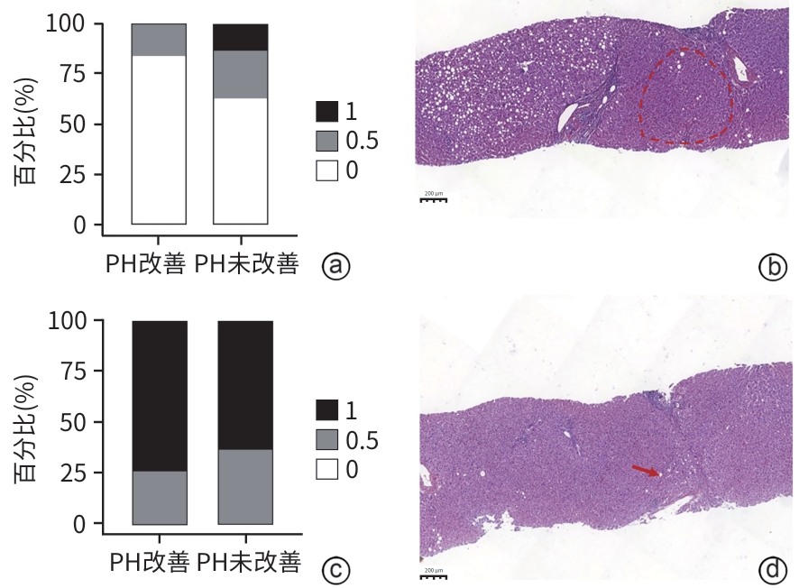
中文English
ISSN 1001-5256 (Print)
ISSN 2097-3497 (Online)
CN 22-1108/R
Volume 41 Issue 8
Aug.  2025
Turn off MathJax
Article Contents

Histological factors for improving portal hypertension in patients with chronic hepatitis B cirrhosis

DOI: 10.12449/JCH250815
Research funding:

National Science and Technology Major Project (2014ZX10005001);

National Natural Science Foundation of China (82274305);

National Natural Science Foundation of China (82305200);

Shanghai Key Specialty of Traditional Chinese Clinical Medicine (shslczdzk01201);

Science and technology development project of Shanghai Academy of Traditional Chinese Medicine (24YJS03)

More Information
  • Corresponding author: LIU Chenghai, chenghailiu@hotmail.com (ORCID: 0000-0002-1696-6008)
  • Received Date: 2025-01-15
  • Accepted Date: 2025-02-13
  • Published Date: 2025-08-25
  •   Objective  To investigate the histological and cellular bases for the improvement of portal hypertension (PH) by observing liver histopathological changes after treatment in patients with cirrhotic portal hypertension, and to provide a basis for clinical drug development.  Methods  A total of 322 patients with hepatitis B cirrhosis who completed 48 weeks of antiviral therapy or combined anti-fibrotic treatment in 20 hospitals across 12 provinces in China from September 2014 to October 2018 were enrolled, and the noninvasive diagnostic criteria for clinically significant portal hypertension (CSPH) from Baveno Ⅶ were used to assess the severity of PH; 43 patients with a confirmed diagnosis of CSPH were identified based on liver stiffness measurement (LSM) ≥25 kPa before treatment, and according to whether the severity of PH was reduced by ≥2 grades after treatment, the patients were divided into PH improvement (n=19) group and PH non-improvement group(n=24). Related data were collected, including demographic data, laboratory tests. Liver fibrosis were assessed, including HE staining and reticular fiber staining; liver microvascular lesions were assessed, including obliterative portal venopathy (OPV), nodular regenerative hyperplasia (NRH), and incomplete septal fibrosis (ISF). Single immunohistochemical staining was performed for von Willebrand factor (vWF), and fibronectin; multiplex immunohistochemical staining was performed for fibrinogen, CD32b, CD31, alpha-smooth muscle actin (α-SMA). The independent-samples t test or the Mann-Whitney U test was used for comparison of continuous data between two groups, and the chi-square test was used for comparison of categorical data between two groups.  Results  After 48 weeks of treatment, 43 patients had significant improvements in red blood cell count, alanine aminotransferase, aspartate aminotransferase, aspartate aminotransferase-to-platelet ratio index score, liver fibrosis grade, and PH grade (all P<0.05), among whom 19 patients showed a reduction in PH severity by ≥2 grades (PH improvement group), while the remaining patients were enrolled as the PH non-improvement group. There was no significant difference in the outcome of liver fibrosis between the two groups (χ²=3.380, P=0.066). Microvascular lesion assessment showed that compared with the PH non-improvement group, the PH improvement group had significantly lower OPV severity, microvascular density (the expression level of vWF), and expression of fibronectin (all P<0.05), while there were no significant differences in NRH severity, ISF severity, and the expression level of fibrinogen between the two groups (all P>0.05). Cytological evaluation showed no significant differences in the expression levels of CD32b, CD31, and α-SMA between the two groups before and after treatment (all P>0.05), and comparison of the expression levels before and after treatment showed that the PH improvement group had a significant increase in the expression level of CD32b (t=-2.007, P=0.045) and a significant reduction in the expression level of α-SMA (t=2.628, P=0.013).  Conclusion  The pathological features of PH improvement are associated with liver fibrosis regression and the improvement in liver microvascular lesions, and at the cellular level, PH improvement is associated with the dedifferentiation of liver sinusoidal endothelial cells and the activated phenotype of hepatic stellate cells.


  • loading
  • [1]
    BOSCH J, GROSZMANN RJ, SHAH VH. Evolution in the understanding of the pathophysiological basis of portal hypertension: How changes in paradigm are leading to successful new treatments[J]. J Hepatol, 2015, 62( 1 Suppl): S121- S130. DOI: 10.1016/j.jhep.2015.01.003.
    [2]
    GUIXÉ‍-MUNTET S, QUESADA-VÁZQUEZ S, GRACIA-SANCHO J. Pathophysiology and therapeutic options for cirrhotic portal hypertension[J]. Lancet Gastroenterol Hepatol, 2024, 9( 7): 646- 663. DOI: 10.1016/S2468-1253(23)00438-7.
    [3]
    GUO YN, LYU J, LIU CH. Medicinal treatment of portal hypertension in liver cirrhosis[J]. Chin J Integr Tradit West Med Liver Dis, 2021, 31( 11): 961- 964.

    郭亚楠, 吕靖, 刘成海. 肝硬化门静脉高压症的药物治疗[J]. 中西医结合肝病杂志, 2021, 31( 11): 961- 964.
    [4]
    SUN X, LIU CH. Consensus interpretation of Baveno Ⅶ in the diagnosis and treatment of portal hypertension in cirrhosis: A physician’s perspective[J]. Chin J Integr Tradit West Med Dig, 2023, 31( 2): 79- 84.

    孙鑫, 刘成海. 肝硬化门静脉高压诊治Baveno Ⅶ共识解读: 内科视角[J]. 中国中西医结合消化杂志, 2023, 31( 2): 79- 84.
    [5]
    de FRANCHIS R, BOSCH J, GARCIA-TSAO G, et al. Baveno VII-Renewing consensus in portal hypertension[J]. J Hepatol, 2022, 76( 4): 959- 974. DOI: 10.1016/j.jhep.2021.12.022.
    [6]
    CAMPRECIÓS G, VILASECA M, TRIPATHI DM, et al. Interspecies transcriptomic comparison identifies a potential Porto-sinusoidal vascular disorder rat model suitable for in vivo drug testing[J]. Liver Int, 2024, 44( 1): 180- 190. DOI: 10.1111/liv.15765.
    [7]
    WEIDNER N, SEMPLE JP, WELCH WR, et al. Tumor angiogenesis and metastasis: Correlation in invasive breast carcinoma[J]. N Engl J Med, 1991, 324( 1): 1- 8. DOI: 10.1056/NEJM199101033240101.
    [8]
    SELICEAN S, WANG C, GUIXÉ-MUNTET S, et al. Regression of portal hypertension: Underlying mechanisms and therapeutic strategies[J]. Hepatol Int, 2021, 15( 1): 36- 50. DOI: 10.1007/s12072-021-10135-4.
    [9]
    ABBEY P, SHALIMAR. Nodular regenerative hyperplasia[J]. Curr Hepatol Rep, 2023, 22( 3): 182- 192. DOI: 10.1007/s11901-023-00613-8.
    [0]
    WANLESS IR, NAKASHIMA E, SHERMAN M. Regression of human cirrhosis. Morphologic features and the genesis of incomplete septal cirrhosis[J]. Arch Pathol Lab Med, 2000, 124( 11): 1599- 1607. DOI: 10.5858/2000-124-1599-ROHC.
    [11]
    GRACIA-SANCHO J, MARRONE G, FERNÁNDEZ-IGLESIAS A. Hepatic microcirculation and mechanisms of portal hypertension[J]. Nat Rev Gastroenterol Hepatol, 2019, 16( 4): 221- 234. DOI: 10.1038/s41575-018-0097-3.
    [12]
    Chinese Society of Hepatology, Chinese Society of Gastroenterology, and Chinese Society of Digestive Endoscopology of Chinese Medical Association. Guidelines on the management of esophagogastric variceal bleeding in cirrhotic portal hypertension[J]. J Clin Hepatol, 2023, 39( 3): 527- 538. DOI: 10.3969/j.issn.1001-5256.2023.03.znygf.

    中华医学会肝病学分会, 中华医学会消化病学分会, 中华医学会消化内镜学分会. 肝硬化门静脉高压食管胃静脉曲张出血的防治指南[J]. 临床肝胆病杂志, 2023, 39( 3): 527- 538. DOI: 10.3969/j.issn.1001-5256.2023.03.znygf.
    [13]
    Beijing Society of Portal Hypertension, Beijing Medical Association; Portal Hypertension Expert Committee, Liver Disease Committee of Chinese Research Hospital Association; Liver Disease Committee of Chinese Research Hospital Association. Expert consensus on multidisciplinary diagnosis and treatment of cirrhotic portal hypertension(based on hepatic venous pressure gradient)[J]. J Clin Hepatol, 2021, 37( 9): 2037- 2044. DOI: 10.3969/j.issn.1001-5256.2021.09.008.

    北京医师协会门静脉高压专科医师分会, 中国研究型医院学会肝病专业委员会门静脉高压学组, 中国研究型医院学会肝病专业委员会. 肝硬化门静脉高压症多学科诊治(基于肝静脉压力梯度)专家共识[J]. 临床肝胆病杂志, 2021, 37( 9): 2037- 2044. DOI: 10.3969/j.issn.1001-5256.2021.09.008.
    [14]
    GRACIA-SANCHO J, MAESO-DÍAZ R, FERNÁNDEZ-IGLESIAS A, et al. New cellular and molecular targets for the treatment of portal hypertension[J]. Hepatol Int, 2015, 9( 2): 183- 191. DOI: 10.1007/s12072-015-9613-5.
    [15]
    GARCIA-TSAO G, FRIEDMAN S, IREDALE J, et al. Now there are many(stages) where before there was one: In search of a pathophysiological classification of cirrhosis[J]. Hepatology, 2010, 51( 4): 1445- 1449. DOI: 10.1002/hep.23478.
    [16]
    DAI WM, LU LG, CAI XB. Association between liver sinusoidal endothelial cells and liver fibrosis[J]. J Clin Hepatol, 2023, 39( 2): 419- 423. DOI: 10.3969/j.issn.1001-5256.2023.02.027.

    戴伟明, 陆伦根, 蔡晓波. 肝窦内皮细胞与肝纤维化的关系[J]. 临床肝胆病杂志, 2023, 39( 2): 419- 423. DOI: 10.3969/j.issn.1001-5256.2023.02.027.
    [17]
    LI MB, LI JY, FENG DP. Research advances in the reversal of liver fibrosis[J]. J Clin Hepatol, 2023, 39( 1): 193- 198. DOI: 10.3969/j.issn.1001-5256.2023.01.030.

    李满彪, 李金玉, 冯对平. 肝纤维化逆转的研究进展[J]. 临床肝胆病杂志, 2023, 39( 1): 193- 198. DOI: 10.3969/j.issn.1001-5256.2023.01.030.
    [18]
    IWAKIRI Y, TREBICKA J. Portal hypertension in cirrhosis: Pathophysiological mechanisms and therapy[J]. JHEP Rep, 2021, 3( 4): 100316. DOI: 10.1016/j.jhepr.2021.100316.
    [19]
    NAKASHIMA E, KAGE M, WANLESS IR. Idiopathic portal hypertension: histologic evidence that some cases may be regressed cirrhosis with portal vein thrombosis[J]. Hepatology, 1999, 30: 218A.
    [20]
    GUIDO M, SARCOGNATO S, RUSSO FP, et al. Focus on histological abnormalities of intrahepatic vasculature in chronic viral hepatitis[J]. Liver Int, 2018, 38( 10): 1770- 1776. DOI: 10.1111/liv.13718.
    [21]
    FERNANDEZ M. Molecular pathophysiology of portal hypertension[J]. Hepatology, 2015, 61( 4): 1406- 1415. DOI: 10.1002/hep.27343.
  • 加载中

Catalog

    通讯作者: 陈斌, bchen63@163.com
    • 1. 

      沈阳化工大学材料科学与工程学院 沈阳 110142

    1. 本站搜索
    2. 百度学术搜索
    3. 万方数据库搜索
    4. CNKI搜索

    Figures(3)  / Tables(4)

    Article Metrics

    Article views (298) PDF downloads(33) Cited by()
    Proportional views
    Related

    /

    DownLoad:  Full-Size Img  PowerPoint
    Return
    Return