
 PDF下载 ( 917 KB)

超声内镜引导下置入弹簧圈联合组织胶注射治疗合并自发性分流的胃静脉曲张的效果分析
DOI: 10.12449/JCH240416
Efficacy of endoscopic ultrasound-guided coil placement combined with tissue adhesive injection in treatment of gastric varices with spontaneous shunt
-
摘要:
目的 评估超声内镜(EUS)引导下置入弹簧圈联合组织胶注射(ECI)治疗合并自发性分流的胃静脉曲张的有效性、安全性和经济性。 方法 回顾性分析2019年3月—2022年9月因急性胃静脉曲张出血合并自发性门体分流在襄阳市中心医院住院并接受改良球囊封堵逆行静脉闭塞术(BRTO)联合内镜下ECI或EUS引导下置入弹簧圈联合ECI治疗的患者。统计分析两种手术方式的疗效(技术成功率,5天、1年再出血率,再出血时间)、安全性(异位栓塞发生率、组织胶用量、聚桂醇用量)和经济性(住院费用和时间)差异。符合正态分布的计量资料两组间比较采用成组t检验,不符合正态分布的计量资料两组间比较采用Mann-Whitney U检验。采用Kaplan-Meier法对再出血情况进行评估和比较。计数资料两组间比较采用χ2检验。 结果 25例患者在EUS引导下成功放置弹簧圈并注射组织胶,技术成功率100%,中位组织胶用量为2.5 mL,中位聚桂醇用量为11.0 mL,平均住院时间为(14.88±3.21)d,平均住院费用为(32 660.00±4 602.07)元,5天再出血率为0;2例失访,23例完整随访患者中住院期间异位栓塞发生率为0,中位再出血时间为689 d。14例患者接受改良BRTO联合内镜下ECI,技术成功率100%,术中中位组织胶用量为5.0 mL,高于EUS组(U=39.000,P<0.001),中位聚桂醇用量为10.5 mL;平均住院时间为(15.38±4.94)d;平均住院费用为(57 583.47±18 955.40)元,高于EUS组(t=-6.310,P<0.001);5天再出血率为0;无失访,14例完整随访患者中住院期间异位栓塞发生率为0,中位再出血时间为244.50 d。Kaplan-Meier生存曲线分析表明,2组患者再出血风险无明显差异(χ2=1.448,P=0.229)。 结论 EUS引导置入弹簧圈联合ECI是一种相对安全有效的胃静脉曲张出血治疗技术,技术成功率高,严重不良事件发生率低,疗效与BRTO手术无明显差异,但安全性和经济性更高。 -
关键词:
- 食管和胃静脉曲张 /
- 自发性门体分流 /
- 超声内镜 /
- 球囊封堵逆行静脉闭塞术
Abstract:Objective To investigate the efficacy, safety, and cost-effectiveness of endoscopic ultrasound (EUS)-guided coil placement combined with tissue adhesive injection in the treatment of gastric varices with spontaneous shunt. Methods A retrospective analysis was performed for the patients with acute gastric variceal bleeding and spontaneous portosystemic shunt who were hospitalized and received balloon-occluded retrograde transvenous obliteration (BRTO) combined with endoscopic tissue adhesive injection or EUS-guided coil placement combined with tissue adhesive injection in Xiangyang Central Hospital from March 2019 to September 2022. The two surgical procedures were compared in terms of efficacy (technical success rate, 5-day rebleeding rate, 1-year rebleeding rate, and time to rebleeding), safety (the incidence rate of ectopic embolism, the amount of tissue adhesive used, and the amount of lauromacrogol used), and cost-effectiveness (hospital costs and length of hospital stay). The t-test was used for comparison of normally distributed continuous data between two groups, and the Mann-Whitney U test was used for comparison of non-normally distributed continuous data between two groups. The Kaplan-Meier method was used to estimate the rebleeding. The chi-square test was used for comparison of categorical data between two groups. Results A total of 25 patients received successful EUS-guided coil placement and tissue adhesive injection, with a technical success rate of 100%, a median amount of 2.5 mL tissue adhesive used, a median amount of 11.0 mL lauromacrogol used, a mean length of hospital stay of 14.88±3.21 days, a mean hospital cost of 32 660.00±4 602.07 yuan, and a 5-day rebleeding rate of 0%; among these patients, 2 were lost to follow-up, and 23 patients with complete follow-up data had an incidence rate of ectopic embolism of 0% and a median time to rebleeding of 689 days. A total of 14 patients underwent modified BRTO combined with endoscopic tissue adhesive injection, with a technical success rate of 100%; a median amount of 5.0 mL tissue adhesive used during surgery, which was significantly higher than that used in EUS (U=39.000, P<0.001); a median amount of 10.5 mL lauromacrogol used during surgery; a mean length of hospital stay of 15.38±4.94 days; a mean hospital cost of 57 583.47±18 955.40 yuan, which was significantly higher than that used in EUS (t=-6.310, P<0.001); a 5-day rebleeding rate of 0%. No patient was lost to follow-up, and all 14 patients had an incidence rate of ectopic embolism of 0% and a median time to rebleeding of 244.50 days, with no significant difference between the two groups (χ2=1.448, P=0.229). Conclusion EUS-guided coil placement combined with tissue adhesive injection is a relatively safe and effective technique for the treatment of gastric variceal bleeding and has a high technical success rate, a low incidence rate of serious adverse events, and similar efficacy to BRTO, with higher safety and cost-effectiveness. -
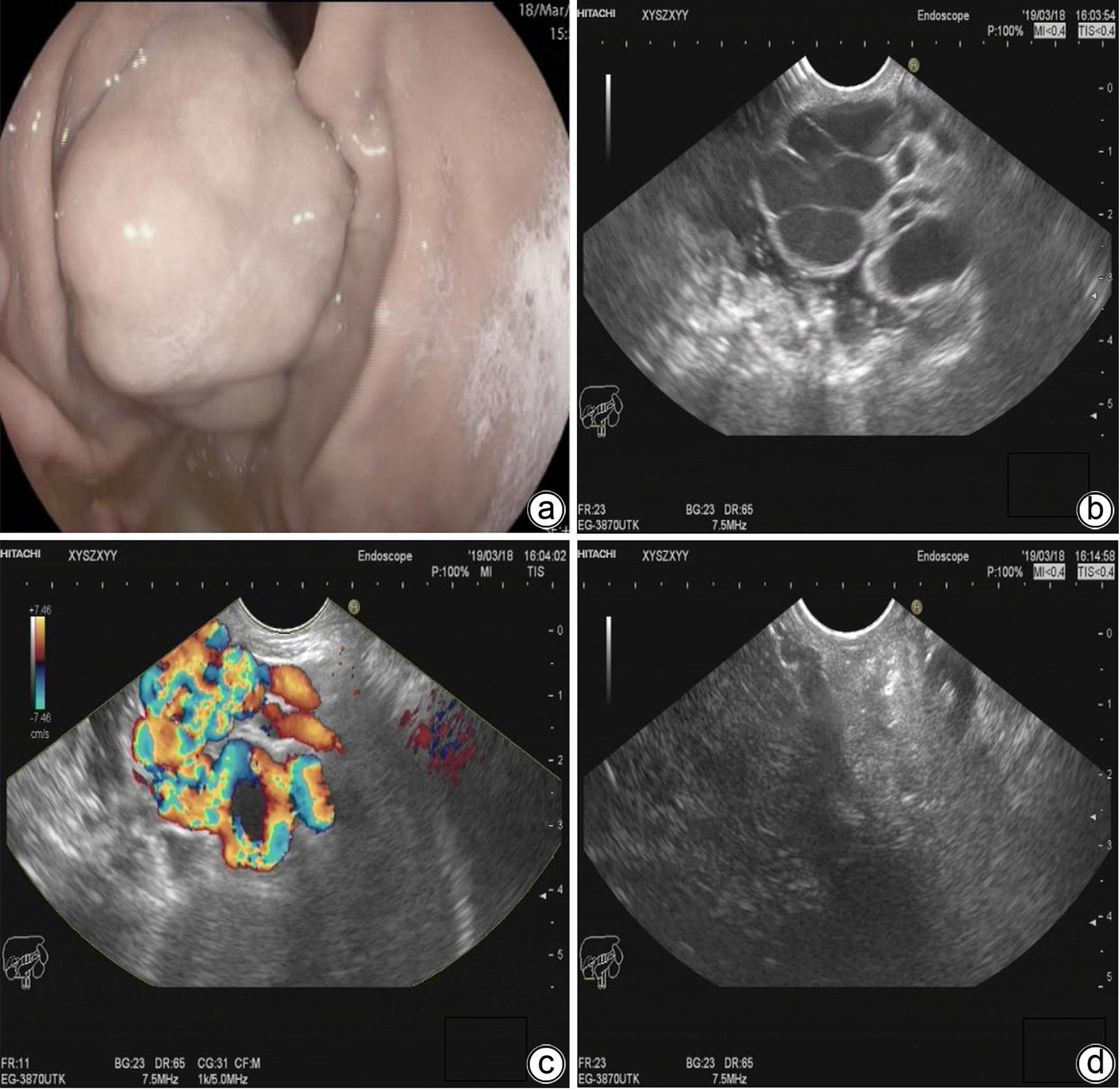
注: a,曲张静脉团块;b,EUS扇扫可见无回声区;c,超声动态监测可见血流信号;d,EUS引导下置入弹簧圈联合ECI治疗后可见血流信号消失。
图 1 EUS组手术过程
Figure 1. Surgical procedure of EUS group
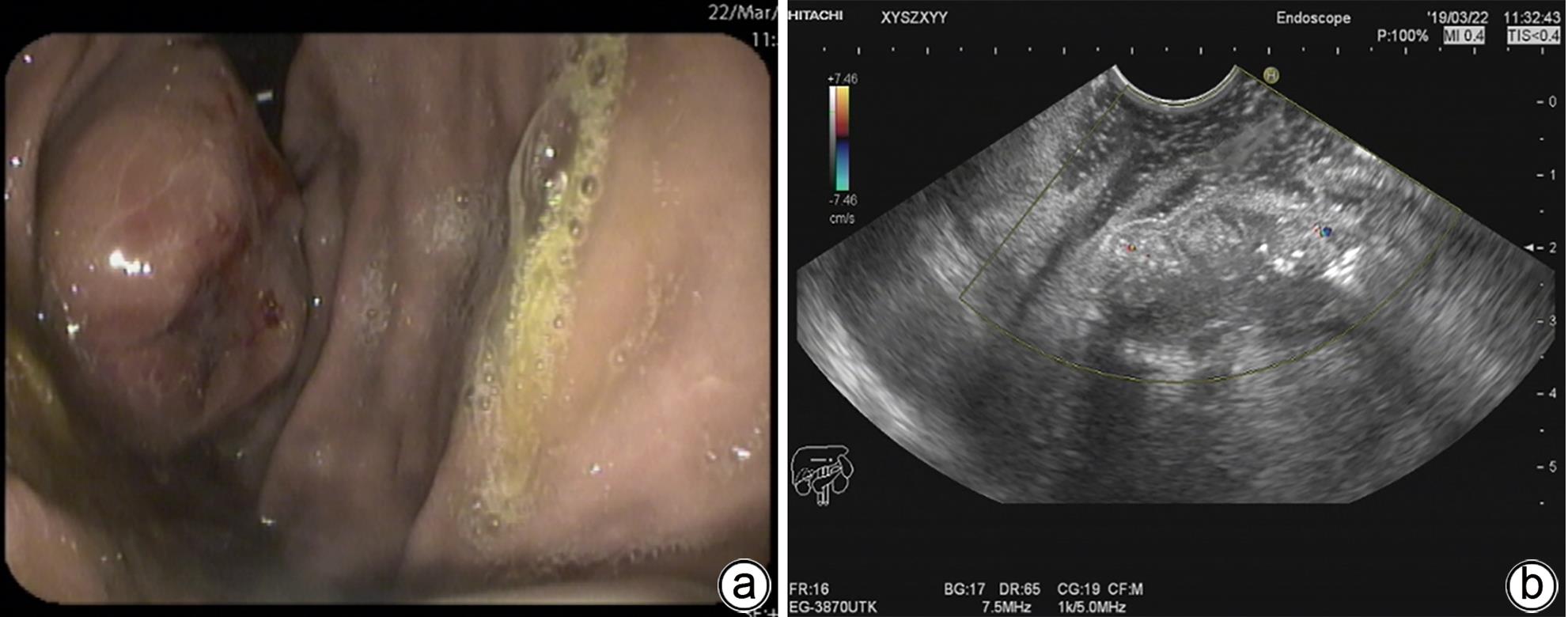
注: a,术后5天胃镜可见曲张静脉硬化;b,术后5天超声可见曲张静脉血流信号消失。
图 2 术后影像学复查结果
Figure 2. Postoperative images
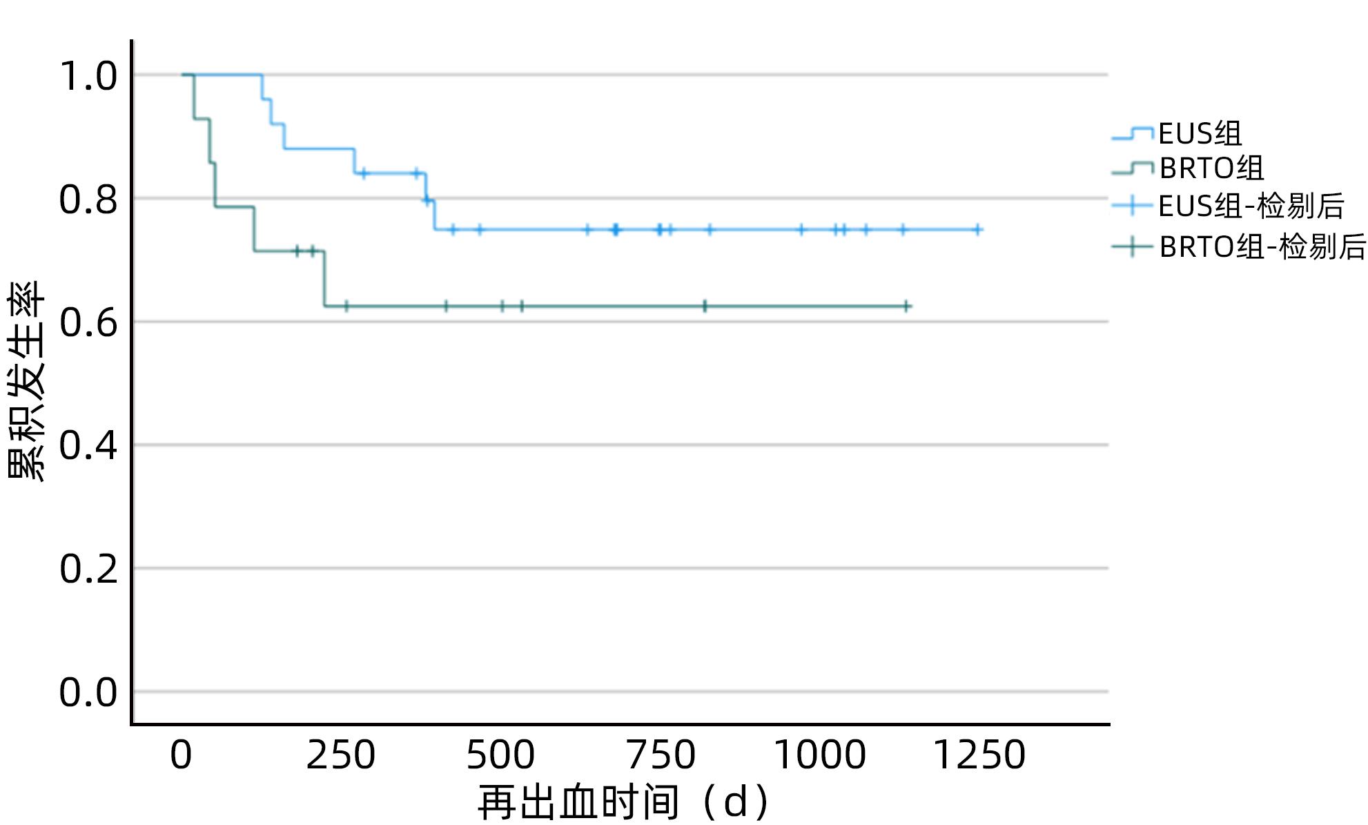
图 3 EUS组与BRTO组再出血时间分析
Figure 3. The Kaplan-Meier curve of the EUS group and BRTO group
表 1 EUS组和BRTO组患者一般资料
Table 1. General information of patients with gastric varices and spontaneous portosystemic shunt
项目 EUS组(n=25) BRTO组(n=14) 男/女(例) 13/12 6/8 年龄(岁) 60.88 57.64±10.82 静脉曲张类型[例(%)] IGV1型 4(16.00) 11(78.57) GOV1型 2(8.00) 0(0.00) GOV2型 19(76.00) 3(21.43) 分流类型[例(%)] 胃肾分流 17(68.00) 10(71.43) 脾肾分流 5(20.00) 2(14.29) 胃肾+脾肾分流 3(12.00) 2(14.29) 术前胃静脉曲张直径(cm) 1.44±0.39 1.48±0.34 分流道最大直径(cm) 11.05±2.80 11.45±1.84 分流道最小直径(cm) 6.17±1.47 6.57±1.34 术前Child评分(分) 8.32±1.46 8.50±1.34 术前MELD评分(分) 6.47±3.38 5.82±1.78 注:IGV1,孤立性胃静脉曲张1型;GOV1,食管胃静脉曲张1型;GOV2,食管胃静脉曲张2型。  下载: 导出CSV
表 2 EUS组与BRTO组治疗有效性、安全性及经济性分析
Table 2. Effectiveness, safety and economic analysis between theEUS group and theBRTO group
项目 EUS组(n=25) BRTO组(n=14) 统计值 P值 有效性指标[例(%)] 技术成功率 25(100.00) 14(100.00) χ2=0.000 >0.05 5天再出血率 0(0.00) 0(0.00) χ2=0.000 >0.05 1年再出血率 4(17.39) 5(35.71) χ2=6.466 0.011 安全性指标(mL) 组织胶用量 2.5(1.5~3.5) 5.0(3.4~6.8) U=39.000 <0.001 聚桂醇用量 11.0(7.0~14.0) 10.5(6.8~20.0) U=151.000 0.496 经济性指标 住院时间(d) 14.88±3.21 15.38±4.94 t=-0.33 0.743 住院费用(元) 32 660.00±4 602.07 57 583.47±18 955.40 t=-6.31 <0.001 注:BRTO组住院时间存在1个异常值,追溯原因后选择剔除。
下载: 导出CSV
-
[1] LESMANA CRA, RAHARJO M, GANI RA. Managing liver cirrhotic complications: Overview of esophageal and gastric varices[J]. Clin Mol Hepatol, 2020, 26( 4): 444- 460. DOI: 10.3350/cmh.2020.0022. [2] LUO R, GAO J, GAN W, et al. Clinical-radiomics nomogram for predicting esophagogastric variceal bleeding risk noninvasively in patients with cirrhosis[J]. World J Gastroenterol, 2023, 29( 6): 1076- 1089. DOI: 10.3748/wjg.v29.i6.1076. [3] LUO XF, HERNÁNDEZ-GEA V. Update on the management of gastric varices[J]. Liver Int, 2022, 42( 6): 1250- 1258. DOI: 10.1111/liv.15181. [4] KHOURY T, NADELLA D, WILES A, et al. A review article on gastric varices with focus on the emerging role of endoscopic ultrasound-guided angiotherapy[J]. Eur J Gastroenterol Hepatol, 2018, 30( 12): 1411- 1415. DOI: 10.1097/MEG.0000000000001200. [5] GARCIA-TSAO G, SANYAL AJ, GRACE ND, et al. Prevention and management of gastroesophageal varices and variceal hemorrhage in cirrhosis[J]. Hepatology, 2007, 46( 3): 922- 938. DOI: 10.1002/hep.21907. [6] de FRANCHIS R, FACULTY BV. Revising consensus in portal hypertension: Report of the Baveno V consensus workshop on methodology of diagnosis and therapy in portal hypertension[J]. J Hepatol, 2010, 53( 4): 762- 768. DOI: 10.1016/j.jhep.2010.06.004. [7] TAN YY, GONG J, CHU Y, et al. Current status of diagnosis and treatment for gastric varices in liver cirrhosis with spontaneous portalsystemic shunt[J]. Chin J Dig Endosc, 2023, 40( 1): 78- 81. DOI: 10.3760/cma.j.cn321463-20220222-00029.谭玉勇, 龚箭, 楚毅, 等. 肝硬化胃静脉曲张伴自发性门体分流的诊治现状[J]. 中华消化内镜杂志, 2023, 40( 1): 78- 81. DOI: 10.3760/cma.j.cn321463-20220222-00029. [8] WU Q, JIANG H, LINGHU EQ, et al. BRTO assisted endoscopic Histoacryl injection in treating gastric varices with gastrorenal shunt[J]. Minim Invasive Ther Allied Technol, 2016, 25( 6): 337- 344. DOI: 10.1080/13645706.2016.1192552. [9] LO GH, LIN CW, TAI CM, et al. A prospective, randomized trial of thrombin versus cyanoacrylate injection in the control of acute gastric variceal hemorrhage[J]. Endoscopy, 2020, 52( 7): 548- 555. DOI: 10.1055/a-1127-3170. [10] CHEN MK, DING Z, XIAO Y, et al. Preliminary study on the treatment of obvious splenorenal shunt with huge gastric varices by ultrasonic endoscope combined with gastroscope[J]. Chin J Dig Endosc, 2016, 33( 10): 707- 710. DOI: 10.3760/cma.j.issn.1007-5232.2016.10.013.陈明锴, 丁震, 肖勇, 等. 超声内镜联合胃镜治疗明显脾肾分流合并巨大胃静脉曲张的初步探讨[J]. 中华消化内镜杂志, 2016, 33( 10): 707- 710. DOI: 10.3760/cma.j.issn.1007-5232.2016.10.013. [11] MCCARTY TR, BAZARBASHI AN, HATHORN KE, et al. Combination therapy versus monotherapy for EUS-guided management of gastric varices: A systematic review and meta-analysis[J]. Endosc Ultrasound, 2020, 9( 1): 6- 15. DOI: 10.4103/eus.eus_37_19. [12] Endoscopic Diagnosis and Treatment of Esophagogastric Varices Group of Chinese Society of Digestive Endoscopology. Chinese expert consensus on endoscopic cyanoacrylate injection for gastrointestinal varices with portal hypertension induced by liver cirrhosis(2022, Changsha)[J]. Chin J Dig Endosc, 2023, 40( 1): 12- 23. DOI: 10.3760/cma.j.cn321463-20221016-00530.中华医学会消化内镜学分会食管胃静脉曲张内镜诊断与治疗学组. 肝硬化门静脉高压消化道静脉曲张内镜下组织胶注射治疗专家共识(2022, 长沙)[J]. 中华消化内镜杂志, 2023, 40( 1): 12- 23. DOI: 10.3760/cma.j.cn321463-20221016-00530. [13] HUANG ZY, ZHANG WH, LV FJ, et al. Efficacy and safety of EUS-guided coil embolization combined with endoscopic cyanoacrylate injection versus balloon-occluded retrograde transvenous obliteration for gastric varices with high-risk ectopic embolism: A multicenter and retrospective cohort study[J]. Endosc Ultrasound, 2023, 12( 1): 74- 83. DOI: 10.4103/EUS-D-21-00260. [14] KIM DJ, DARCY MD, MANI NB, et al. Modified balloon-occluded retrograde transvenous obliteration(BRTO) techniques for the treatment of gastric varices: Vascular plug-assisted retrograde transvenous obliteration(PARTO)/coil-assisted retrograde transvenous obliteration(CARTO)/balloon-occluded antegrade transvenous obliteration(BATO)[J]. Cardiovasc Intervent Radiol, 2018, 41( 6): 835- 847. DOI: 10.1007/s00270-018-1896-1. [15] ROBLES-MEDRANDA C, VALERO M, NEBEL JA, et al. Endoscopic-ultrasound-guided coil and cyanoacrylate embolization for gastric varices and the roles of endoscopic Doppler and endosonographic varicealography in vascular targeting[J]. Dig Endosc, 2019, 31( 3): 283- 290. DOI: 10.1111/den.13305. [16] TANG L, LI X, CUI J, et al. EUS-guided coil placement and cyanoacrylate glue injection for gastric variceal bleeding with obvious spontaneous portosystemic shunts[J]. Endosc Ultrasound, 2023, 12( 1): 84- 89. DOI: 10.4103/EUS-D-22-00006. [17] NAKAMURA K, OKAMOTO T, SHIRATORI Y, et al. Endoscopic ultrasound-guided coil deployment with sclerotherapy for gastric varices[J]. Clin J Gastroenterol, 2021, 14( 1): 76- 83. DOI: 10.1007/s12328-020-01259-5. [18] WANG XM, YU S, CHEN X, et al. Endoscopic ultrasound-guided injection of coils and cyanoacrylate glue for the treatment of gastric fundal varices with abnormal shunts: A series of case reports[J]. J Int Med Res, 2019, 47( 4): 1802- 1809. DOI: 10.1177/0300060519830207. [19] ZHANG J, ZHU LR, YAO LW, et al. Deep learning-based pancreas segmentation and station recognition system in EUS: Development and validation of a useful training tool(with video)[J]. Gastrointest Endosc, 2020, 92( 4): 874- 885.e3. DOI: 10.1016/j.gie.2020.04.071. [20] BAYANI A, HOSSEINI A, ASADI F, et al. Identifying predictors of varices grading in patients with cirrhosis using ensemble learning[J]. Clin Chem Lab Med, 2022, 60( 12): 1938- 1945. DOI: 10.1515/cclm-2022-0508. [21] LEE CM, LEE SS, CHOI WM, et al. An index based on deep learning-measured spleen volume on CT for the assessment of high-risk varix in B-viral compensated cirrhosis[J]. Eur Radiol, 2021, 31( 5): 3355- 3365. DOI: 10.1007/s00330-020-07430-3. -

 本文二维码
本文二维码
计量
- 文章访问数: 1128
- HTML全文浏览量: 617
- PDF下载量: 81
- 被引次数: 0

